Introduction
Fowl cholera is an infectious disease caused by the bacterium Pasteurella multocida. This species is named “multocida”, which may be interpreted as a bacterium that "kills" (cida) "many" (multo). In 1879, Pasteur was able to cultivate this bacterium; this was the first time that disease-causing bacteria were grown in culture media, outside the animal host. Pasteur inadvertently found that hens were protected from P. multocida challenge by inoculating an old broth cultures that have been attenuated. These were the first documented trials that were carried out with bacterial vaccines.
Production losses
Fowl cholera causes very high economic losses in chicken and turkey breeders, especially in broiler breeders, due to high mortality, low production of hatching eggs and reduction of fertility. Furthermore, fowl cholera was also described in broilers, turkeys and layers. Cases of complicated infectious coryza (Avibacterium paragallinarum) mainly associated with simultaneous infections of P. multocida have been described in chicken egg layer farms.
Pasteurella multocida
Pasteurella multocida is a Gram negative, very short characteristic cocobacillus; it also may form a few long strands of which coccoid forms dissociate free from its ends and are arranged into short chains (photo No. 1). Furthermore, in fresh smears or tissue imprints, stained with methylene blue, bacilli with a typical bipolar staining may be observed. In slide smears of bacterial suspensions, carried out with the addition of Chinese ink, the presence of negatively stained bacilli, due to the presence of polysaccharide capsules, are frequently observed in most P. multocida strains.
Four subspecies, which differ by sugar fermentation tests, are recognized as follows: gallicida, septica, multocida and tigris. The subspecies gallicida is recognized as the causative agent of fowl cholera but has also been isolated from cattle. The subspecies septica has been isolated from dogs, cats, birds and humans. The subspecies multocida causes various diseases of importance in different species of domestic animals and humans. The subspecies tigris has only been described in human wounds caused by tiger bites.
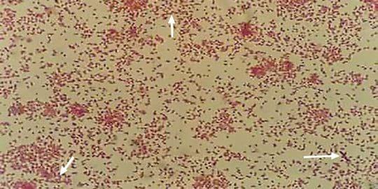
Photo N°1 This photo shows a Gram-Hucker stained smear obtained from a pure culture in Columbia blood agar plate after incubation for 24 hours at 37°C. At 1000X it can be seen that
Pasteurella multocida is a Gram negative bacterium (stained red) with pleomorphic forms, coccobacilli or coccoid of 0.2-2 microns, grouped loose, in pairs or in few short chains with some filaments (arrows).
Lipopolysaccharides and somatic serotypingThe specificity of the somatic serotypes is determined by the type of lipopolysaccharide (LPS). The Heddleston precipitin test uses antisera prepared in chickens and thermostable antigens extracted from saline suspensions of formalinized bacteria. To date, 16 somatic serotypes have been isolated from poultry, cattle, pigs and humans.
LPS combined with the carrier protein elicit the production of antibodies that protect birds against fowl cholera. Unlike other bacteria, in P. multocida there are two LPS, designated A and B, which are very similar and only show minor differences in their internal structure. LPS A is responsible for the pathophysiological properties of endotoxins. Furthermore, some strains of P. multocida produce a third LPS, designated C. It is believed that this simultaneous expression of several LPS improves survival of P. multocida in the bird.
Capsular typing
Capsules are highly hydrated polysaccharides, externally located and adherent to the bacterial cell wall. Many P. multocida strains express a capsule on their surfaces.
The ability of P. multocida to invade and reproduce in the host is increased by the presence of the capsule. By passive hemagglutination test, which is performed using sensitized glutaraldehyde-fixed sheep erythrocytes, 5 capsular types (A, B, D, E and F) have been described. Currently, PCR tests are available to determine these capsular types.
Capsule A mainly consists of hyaluronic acid, capsule D of heparin and capsule F of chondroitin whereas the exact chemical structure of the polysaccharide of types B and E capsules are not yet elucidated. The capsule is related to the pathogenesis and host predilection: strains with type A capsule are related to the majority of fowl cholera outbreaks, type B and E to bovine haemorrhagic septicaemia and type D to porcine atrophic rhinitis.
Strains of P. multocida causing fowl cholera outbreaks
Outbreaks of fowl cholera are usually associated with strains of P. multocida of serotype 1, 3 and 4. Many of these strains belong to the subspecies gallicida or multocida and belong to capsular type A. Some outbreaks in turkeys are associated with capsular type F.
Virulence factors of Pasteurella multocida
The mechanisms by which these bacteria invade the mucosa, evade innate immunity and cause systemic disease are slowly being elucidated. As P. multocida may be part of the microbiota in the upper respiratory tract , it may behave as a primary invasive pathogen or as a secondary pathogen , according to how virulence factors are expressed and act, counteracting the immune response of the host. Furthermore, some virulence factors are critical in determining the pathogenicity of certain strains in some hosts but not in others. Among the virulence factors it may be worth mentioning the lipopolysaccharide capsule, the iron acquisition system and some adhesins. The capsule is clearly involved in bacterial avoidance of phagocytosis and resistance to complement, while complete lipopolysaccharide is critical for bacterial survival in the host. The protean toxins of P. multocida strains occur in capsular type A and D. It is believed that the type A capsule, composed of hyaluronic acid, might serve as a mask against the immune system since the hyaluronic acid structure is indistinguishable from the one that constitutes the tissue structure of the bird.
Reservoirs of infection
Poultry,domestic and wild birds, including seabirds, may become infected and ill or even die from fowl cholera. All kinds of animals including humans may maintain the infection as carriers for a long period of time. Furthermore, this bacterium is able to survive for a long period of time in the environment.
Pathogenicity differences between strains
Certain strains are fully adapted to produce a specific disease in a given animal species but the same strains may be incapable of infecting a different animal species. Poultry are affected with different degrees of morbidity and mortality, which may vary according to the species of bird, health status, environmental factors or management as well as pathogenic differences between P. multocida strains. Some strains, very virulent in primo-isolation, often lose their virulence when they are successively cultivated in artificial media (in vitro passages attenuation). On the other hand, successive passages in susceptible birds (in vivo) make these same strains reacquire their initial pathogenicity.
Susceptibility of poultry
The relationship between birds and fowl cholera is complex and variable, depending on the type of bird, variations of each strain or individual variations of birds within the flock. Adult birds are more susceptible to suffer acute cholera as compared to very young poultry birds that are protected by trans-ovo maternal immunity.
Furthermore, the diversity of conditions that have the different production systems, the quantity and density of birds in each house and the variable application of biosecurity measures should also be considered. Due to the stress caused by the high performance demands of the poultry industry, birds are raised in intensive production systems. Consequently, these birds are very sensitive and often suffer infections that require the use of an appropriate health management. Generally, the reduction of associated risk factors may reduce pathogen multiplication and dissemination.
Routes of infection and persistence of the pathogen on farms
The disease is spread by ingestion through drinking water, food or litter contaminated with droppings from sick or carrier birds. The respiratory route is another common way of infection, either directly by sneezing among birds or by indirectly inhaling contaminated dust. Wounds or skin lesions may also be a source of infection.
Symptoms and lesions
Fowl cholera has three clinical presentations: hyperacute, acute and chronic. The hyperacute form appears with the sudden death of birds without expressing the slightest symptom or detectable pathognomonic injury. The acute presentation course lasts for 1-2 days, during which the birds show anorexia, fever, intense thirst, sleepiness (photo No. 2A), prostration, profuse diarrhoea and sometimes bloody stained faeces, respiratory distress with abundant mucus and violet colour of the combs and wattles (photo No. 2B) due to intense cyanosis. During the acute course of fowl cholera the lesions have the characteristic signs of a haemorrhagic septicaemia with petechiae and generalized haemorrhages in organs and skin, hepatomegaly and hepatic congestion (photo No. 3), oedematous lungs which sometimes have small purulent grey areas, and a congestive spleen but without showing evident splenomegaly. The chronically diseased birds usually survive for a long time and become cachectic; commonly these birds have noticeable swelling of the combs and wattles, which is more evident in male breeders. The incision of the combs and wattles shows a purulent content or yellowing caseous lesions from which oozes a purulent fluid (photo No. 4), sometimes these lesions may spread as subcutaneous abscesses. Yellowish cheesy-like caseum may be found, either in small pieces or big masses, which may be located in the air sacs (photo No. 3) and / or peritoneum (photo No. 5). Petechiae may be found in the heart and gizzard. Hepatomegaly and congestion of the liver is frequently found, with or without white spots of necrosis. Cardiac dilation and arthritis may also be found in a few birds of the flock. In the last phase of the disease, septicaemia occurs and P. multocida freely multiplies in the blood stream affecting the entire circulatory system.
As has been mentioned above, mortality and morbidity are variable. Hyperacute forms are infrequent in industrial poultry thanks to the implementation of immunization plans, as well as the use of strict application of good management and biosecurity measures. Therefore, most acute cases evolve into chronic. On the other hand, if the outbreak occurs in an unimmunized poultry flock, high mortality may be reached, as frequently happens in small scale or family poultry farms.
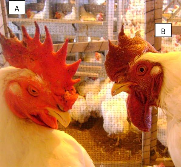 Photo N°2
 This photo shows two chickens suffering from an acute case of fowl cholera. The rooster on the left is depressed and sleepy (A). The hen on the right shows cyanotic comb and wattles (B).
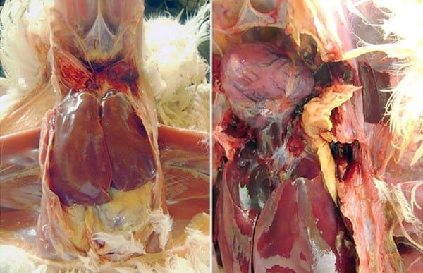 Photo N°3
 The picture on the left shows a broiler breeder liver with remarkable congestion and hepatomegaly, collection of fibrin over the Glisson’s capsule and a haemorrhagic organ at the right side edge.
In the picture on the right it is shown another chicken with evident hepatomegaly, collection of fibrin over the Glisson’s capsule and a great cheesy-like bright yellow caseum mass over one of the air sacs.
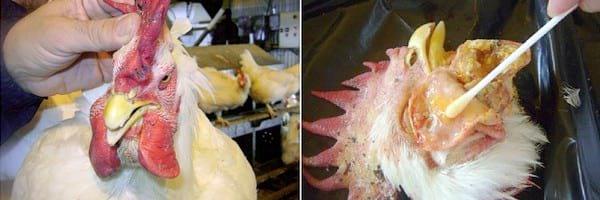
Photo N°4 This photo shows a chronic clinical case of fowl cholera in a rooster. Note the severe swelling of the comb and both wattles. The incision of the wattles displays abundant yellow caseum material from which oozes a purulent fluid. This fluid may be sampled with a cotton swab directly cultured onto an agar plate.
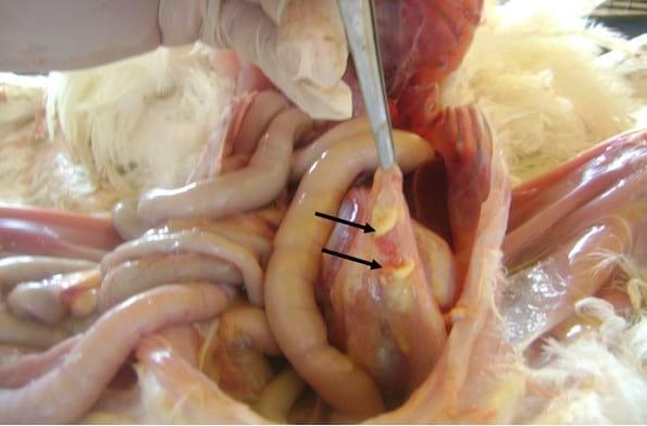
Photo N°5 Two lumps of yellow caseum material are found in the peritoneal omentum.
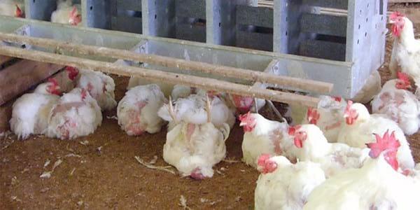
PhotoN°6 In an outbreak of fowl cholera, the weakened sick birds are usually hidden under the nests to protect themselves from pecking . These birds, dying or recently dead, are suitable for sampling and culturing of
Pasteurella multocida.
Isolation of Pasteurella multocidaIsolation is very important, not only for aetiological diagnosis but also to obtain strains from the farms. These strains may be useful for further development of inactivated vaccines (bacterins) that include local or regional antigens. These updated bacterins may be used to immunize future batches of birds. Also, by vaccinating a diseased flock with strains isolated from the same affected flock it is possible to prevent fowl cholera in those birds that have not yet become sick.
Isolation of P. multocida is more difficult in chronic cases and selection of birds in affected poultry houses must be done with great care. Recently dead birds and infected birds showing clinical signs and lesions compatible with fowl cholera should be selected for samplings. Ideally, birds with congested, swollen, warm wattles should be selected. If dead or sick birds are not found, as often happens in subclinical cases, very weak birds, drowsy or cachectic, which are usually hidden in corners or under the nest boxes should be looked for (Photo 6).
According to the pathological lesions the following samples may be cultivated: congested livers, spleens and lungs and/or with purulent or necrotic lesions; free caseous masses in the peritoneum or air sacs; synovial content or caseous material from the interior of affected joints; and indurated caseum or purulent contents within swollen wattles.
P. multocida grows in rich culture media, such as agar base plus 0.1% decomplemented equine serum, dextrose starch agar, brain-heart agar or Columbia blood agar. Plates are incubated for 18 to 24 hours at 37°C. In acute cases, non-haemolytic colonies growing onto Columbia blood agar are smaller than 2-3 mm in diameter, grey and only sometimes adherent to the medium. In contrast, in acute cases the colonies grow larger and are mucoid (Photo7A). The aforementioned agar base with serum allows easy differentiation of typical P. multocida colonies from contaminants when transverse oblique illumination from a lamp is applied; when obliquely illuminated, the translucent colonies of P. multocida take on a bluish or iridescent hue, while colonies of other contaminating bacterial species are opaque, yellowish or whitish (photo No. 7B). Simultaneous plating of samples onto agars with and without blood, allows easy isolation from organs, fluids or swabs obtained from sick birds. Final identification is performed using pure cross sub-culturing of individual colonies from both agars. To prevent loss of immune-protective antigens of the selected strains, intended for the production of bacterins, primo-isolates should be kept frozen immediately after isolation.
 Photo N°7
 A pure culture of
Pasteurella multocida colonies colonies grown onto Columbia agar with the addition of 7% desfibrinated bovine whole blood (A).
A contaminated culture of
Pasteurella multocida developed onto agar base with the addition of 0.1% decomplemented horse serum (B). Note the bluish translucent colonies of
Pasteurella multocida (arrows 1) in the last strokes of the plate mingled with contaminant colonies, which are opaque and yellowish (arrow 2).
Prophylaxis and vaccinationManagement measures and disinfection, along with vaccination are practical to prevent and control both acute and chronic fowl cholera. Effective bacterins must contain virulent strains of avian origin. Antigenicity and protective power should be experimentally demonstrated by challenge trials. On the other hand, vaccines should be prepared from primo-isolates cultures, using a new aliquot each time the vaccine is prepared. The diversity of strains belonging to each geographical region requires adaptation and formulation of bacterins in order to contain representative strains that reflect epidemiological situation at the time of vaccination. In our experience, the best adjuvant is aluminium hydroxide gel; oily adjuvants should be carefully selected as some may cause severe necrotic side-effects in the application site due to the synergy of P. multocida antigens with the oils. It is desirable to vaccinate before 20 weeks of age, administering two doses of bacterin separated by a 3 to 4 week interval. In areas that are very exposed to the disease, it is advisable to apply the first dose from the 5th week of life onwards. Bacterins may be administered subcutaneously behind the neck or intramuscularly into the breast. In infected flocks, exposed to severe P. multocida challenge, revaccination should be carried out every 6 months.
The live vaccine based on the attenuated strain of the University of Clemenson is available for oral vaccination of turkeys before 14 weeks of age and intradermal vaccination of 6 to 12 weeks old chickens by introducing a lancet into the skin of the alar fold. Vaccination of older birds is contraindicated because this strain retains some virulence. In Australia commercial vaccines have been developed based on auxotrophic mutants ring-A designated pMP1 (serotype 1) and pMP3 (serotype 3). Recently, it has been shown that turkeys reach adequate protection through the administration of inactivated vaccines based on a peptide (rFB2). Furthermore, chickens can be protected using a novel vaccine based on DNA encoding outer membrane proteins genes OmpH and OmpA.
Despite vaccines provide good protection, outbreaks are often reported in vaccinated flocks. The lack of protection could be explained by the absence of cross-protection between various strains and serotypes acting on a farm, the constant evolution of indigenous strains in the same farm or other factors related to biosecurity measures, health condition of the flock or other causes. The pathogenicity of P. multocida is very variable as it quickly adapts to environmental changes and to the poultry host.
Therefore, it is advisable that the vaccines should include regional or local strains. These strains should carry antigens of the acting serotypes and should be selected considering other factors such as capsular type, virulence genes, pattern of antibiotic resistance and / or phylogenetic characterization.
Antibiotic treatment
It must be considered that all birds in an infected flock are lifelong carriers of P. multocida. In these chronically infected flocks it is common to administer treatments with antibiotics and, although mortality may be temporarily stopped, the flock continues to be infected. Treatments usually give variable results, depending mainly on the drug used and the virulence of the acting strain in the outbreak. P. multocida often develops resistance to antibiotics that are commonly used. Regardless of its therapeutic action, the inappropriate use of antibiotics is of concern, on one hand, due to the residues in meat and eggs and, on the other hand, by an increase in resistance to antibiotics or chemotherapeutic agents and the danger that these resistance gens be transmitted to humans. Globally, there are more and more prohibitions to the preventive routine use of antibiotics at sub-therapeutic doses. These restrictions significantly reduce the available tools to treat the birds. Therefore, it is best to apply preventive immunization of poultry and limit to a minimum the use of antibiotics. Whenever possible, it is recommended to perform sensitivity tests (susceptibility) before treatment. However, sometimes it is necessary to act very fast and immediately treat the birds with antibiotics while waiting for the laboratory results to be ready. In these cases, the rapid treatment of choice could be made using florfenicol, trimethoprim-sulfamethoxazole or tetracycline. Secondly, depending on availability, ampicillin, kanamycin, colistin and enrofloxacin may be used. Generally, it is not advisable to administer streptomycin, gentamicin or neomycin.