1. INTRODUCTION:
Gallibacterium anatis is now considered to be an important bacterial disease responsible for decreased egg production in commercial layers, since it causes pathological changes in the reproductive tract and causes also respiratory manifestations in commercial broilers (Bojesen et al., 2003).
Gallibacterium was recently established as a new genus within the family Pasteurellaceae Pohl 1981 (Christensen et al., 2003a). Bacteria belonging to this genus had previously been reported as Pasteurella anatis, avian Pasteurella haemolyticalike organisms or Actinobacillus salpingitidis.
The genus Gallibacterium contains 5 named species; Gallibacterium anatis Biovar haemolytica and biovar anatis, G. melopsittaci, G. trehalosifermentans, G. salpingitidis, G. genomospecies; 1, 2, 3 and un-named taxon (group 5) (Christensen et al., 2003b; Bojesen, 2003; Bisgaard et al., 2009 and Janda, 2011).
The pathogenic potential reported for Gallibacterium is highly variable. These organisms have been isolated from clinically healthy birds, in which they have been suggested to constitute a part of the upper respiratory tract and lower genital tract flora (Bisgaard, 1977; Mushin et al., 1980; Bojesen et al., 2003). However, others have isolated Gallibacterium in pure culture from diseased birds affected with salpingitis, oophoritis, peritonitis, septicaemia, pericarditis, hepatitis and upper respiratory tract lesions, indicating that at least some strains of Gallibacterium possess a pathogenic potential (Shaw et al., 1990; Mirle et al., 1991; Suzuki et al., 1996 and 1997).
G. anatis isolates have also been shown to possess decreased susceptibility toward many antimicrobial agents (Bojesen et al., 2011).
On account of the economic importance of this disease, this study was designed for identification and examination of the pathogenicity of G. anatis from diseased chickens in Egypt.
2. MATERIALS AND METHODS:
Samples:
Tracheal, oviductal, as well as ovarian samples, were collected from different 64 chicken flocks (53 broiler, 6 layer and 5 breeder flocks) in El-behira and Alexandria governorates for detection of Gallibacterium anatis infection.
2.1. Bacterial isolation and identification:
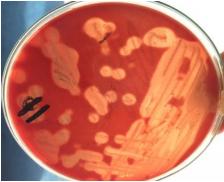
Tracheal, oviductal swabs as well as ovarian samples were cultivated onto a plate of blood agar base (Oxoid), supplemented with 5% citrated bovine blood and incubated overnight at 37 oC under micro-aerophilic condition. The colonies of G. anatis on blood agar appeared smooth and shiny, grayish, semitransparent, circular raised colonies with an entire margine and a butyrous consistency, 1-2 mm in diameter and surrounded by a wide β- haemolytic zone (1-2 mm) after 24 h incubation as defined by (Bisgaard, 1982 and Christensen et al, 2003a).
Microscopic examination revealed Gram negative, rod-shaped or pleomorphic, nonmotile. Biochemical identification of G. anatis isolates showed catalase and oxidase positive, indole and urease negative (Christensen et al., 2003a and Cruickshank et al., 1975).
2.2. Molecular characterization (PCR):
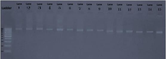
Oligonucleotide primers were designed, based on 16S rRNA sequences from GenBank (Bojesen et al., 2007a), representing all Gallibacterium and related members of Pasteurellaceae. The forward primer 1133fgal (5'- TATTCTTTGTTACCARCGG) was predicted to be specific for Gallibacterium, for the reverse amplification, the general 23S rRNA gene sequence primer 114r (5'- GGTTTCCCCATTCGG) was chosen (Lane, 1991) which gives predicted amplicons of 1080 bp.
2.3. Extraction of DNA were done using a thermo-scientific extraction kits (00248760):
PCR: DNA samples were amplified in a total volume of 50 μl of the following reaction mixture: 25 µl of PCR master mix, 2 µl of both of forward and reverse primer for G.anatis gene, 11 µl of nuclease free water, and 10 µl of the template DNA. The samples were denatured at 95°C for 4 min and the PCR was run for 35 cycles with 95°C denaturation for 30 s, 55°C annealing for 1 min and 72°C extension for 2 min. The PCR was terminated at 72°C extension for 10 min (Bojesen et al., 2007a).
2.4. Detection of PCR products:
Aliquots of amplified PCR products were visualized by electrophoresis on a 1.5% (w/v) agarose gel in the presence of 100-bp DNA ladder supplied from QIAGEN. The agarose gel was supplemented with ethidium bromide under UV illumination.
2.4. Antimicrobial susceptibility testing:
Antimicrobials susceptibility testing of G. anatis isolates was performed using disc diffusion test (Oxoid, UK). The antimicrobials used were Cefotaxime, Florfenicol, Norfloxacin, Ciprofloxacin, Gentamycin, Erythromycin, Ampicillin, Amoxicillin, Cephradine, Doxycycline, Oxytetracycline, Sulphamethoxazole + Trimethoprim, Streptomycin, Lincomycin, and Spectinomycin. All isolates were grown overnight on 5% citrated sheep blood agar at 37°C in microaerophilic condition, then cultures were suspended in 0.85% NaCl to an optical density equivalent to that of McFarland 0.5 standards. Each isolate was then inoculated onto Mueller Hinton agar medium (Oxoid, UK), then 15 minutes later, the antimicrobial discs were applied. Plates were incubated anaerobically at 37°C for 24 hours and the interpretation was performed according to the manufacturer.
2.6. Experimental infection with G. anatis isolates in commercial broiler chickens:
A total of 110 commercial broiler chickens, one day-old were used for experimental infection. All birds were reared in separate units and kept under daily observation for 5 weeks. They were divided into 11 groups (10 birds each), and birds in all groups were supplied with clean drinking water and commercial feed, ad-libitum. At day 22 of age, all chickens (except the control negative group) were infected intranasally (IN) with 10 different G. anatis isolates at a dose of 0.5 ml/bird of 109 CFU/ml. Experimentally infected birds were observed for 10 days post infection (dpi).
2.7. Parameters used to evaluate the pathogenicity in experimental infection:
A. The clinical sign, postmortem (PM) lesion and mortality rates were recorded daily during the experimental infection.
B. Re-isolation of G. anatis from collected samples which were collected from trachea, lung and liver 2, 5 and 10 dpi (at the age of 24, 27 and 32 days).
C. Histopathological examination from collected samples which were collected from trachea, lung and liver 2, 5 and 10 dpi (at the age of 24, 27 and 32 days).
3. RESULTS AND DISCUSSION:
During this study, G. anatis was isolated from 15 out of 64 chicken flocks (23.43%) mostly all these isolates were from broilers (13/15) with 1 isolate from layers and 1 isolate from breeders (Fig. 1) (Table, 1) and this is the first time of isolation of G. anatis from commercial broiler chickens in Egypt. The previous survey in 2014 done by (Elbestawy, 2014) who investigated 38 chicken flocks (26 layer, 3 broiler breeder and 9 broiler flocks) and obtained 7 G. anatis isolates from only 5 positive commercial layer flocks (5/38) (13.1%) while the samples from broilers and broiler breeders were negative.
The Molecular characterization demonstrated that all 15 of the examined field isolates were G. anatis at 1080 bp (Fig. 2). This was similar to findings of Bojesen et al. (2007a) who reported that the same PCR amplicon of approx. 1080 bp was present in all 25 examined reference strains.
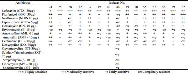
The in-vitro sensitivity of G. anatis isolates to 15 different antimicrobials revealed that these isolates were highly sensitive to cefotaxime, moderately to doxycycline, norfloxacin, florfenicol and ciprofloxacin, fairly sensitive to gentamycin, amoxicillin and ampicillin, as well as resistant to erythromycin, cephradin, oxytetracyclin, sulpha.trimethoprim, streptomycin, lincomycin, spectinomycin. The results of the in-vitro antibiotic sensitivity testing of the G. anatis strains agreed with Bojesen et al. (2011), who noted that G. anatis were resistant to tetracycline and sulfamethoxazoletrimethoprime in 92% and 97%, respectively for the field strains, While on the other hand Bojesen et al. (2007b) and Guo (2011) reported that all G. anatis isolates were susceptible to amoxicillin-clavulanic acid, apramycin, cefpodoxime, ceftifur, cephalotin, chloramphenicol, colistin, florfenicol, gentamycin, spectinomycin, streptomycin, erythromycin, tiamulin and tilmicosin but their isolates were resistant to tetracycline, sulfamethoxazole, ciprofloxacin and nalidixic acid.
Chuan-qing et al. (2008) said that all G. anatis isolates were highly sensitive to the third generation cephalosporin in antimicrobial resistance testing. Also, Dewhirst et al. (1993); Janda (2011) and Jones et al. (2013) agreed that G. anatis is a phylogenetically diverse organism from the family Pasteurellaceae and this can be supported by the fact that all isolates were resistant to oxytetracyclin, sulphamethoxazol-trimethoprime and streptomycin which are the main antibiotics involved in the treatment of Pasteurellacae infections in chickens, turkeys and ducks in Egypt.
Also, Elbestawy, (2014) recorded a complete resistance of 7 Egyptian G. anatis isolates against 5 different antimicrobials to oxytetracycline, sulphamethoxazol +trimethoprim, streptomycin, lincomycin and spectinomycin and the isolates were sensitive to chloramphenicol.
The decrease in antimicrobial susceptibility may be due to inappropriate use of antimicrobials in Egypt which represents a risk to control this bacterial infection.
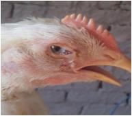
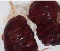
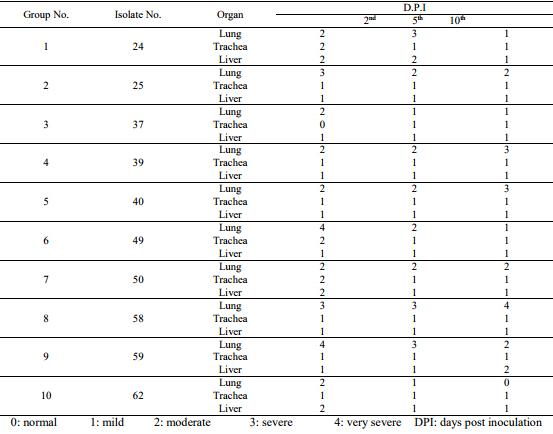
After IN experimental infection of commercial broiler chickens with 10 G. anatis isolates at 22 days of age, all chickens showed depression, respiratory signs as rales, coughing, sneezing, conjunctivitis and teary eyes at 2nd dpi with a decreased feed and water intake at 4th dpi. During the PM examination, the tracheal and lung lesions varied from severe tracheitis and pneumonia in chickens infected with isolates No. 2, 4, 5, 8, and 9 at 2nd, 5th and 10th dpi to mild tracheitis and congested lungs in other infected chicken groups. Air sacculitis was only seen in chickens experimentally infected with isolates No 2 and 4, these data matched with Bojesen et al. (2004) and Shaw et al. (1990) mentioned that at least some strains of Gallibacterium possess a pathogenic potential.
However, there was no any mortalities appeared in any chicken group indicating the importance of stress factors predisposing this type of bacterial infection like impaired host immunity, co-infections and environmental factors as bad ventilation, overcrowding and climatic changes leading to the penetration of Gallibacterium organism into the mucosa of the respiratory tract then into the systemic circulation causing mortalities as supported by Bojesen (2003) and Neubauer et al. (2009).
Re-isolation of G. anatis from the experimental chickens, all was positive from tracheal swabs till 10 dpi as the results shown before with Paudel et al. (2013) mentioned that the bacterial re-isolation was possible following IN infection, mainly from the trachea, choana, lung from 3 until 10 dpi.
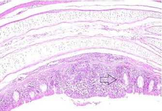
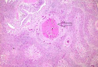
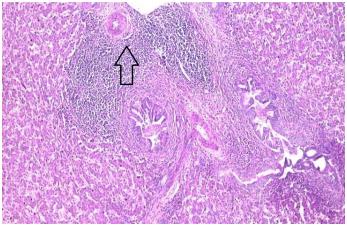
All the histopathological lesions appeared on 2nd dpi as in tracheal samples showed catarrhal tracheitis with hyperplasia of mucous secreting cells (goblet cells), inflammatory cell infiltration which persisted along the experiment in all chicken groups (Fig. 5). Lungs showed interstitial pneumonia with granulomatous lesions and also persisted along the experiment (Fig. 6). In the liver, there were hyperplasia of bile ductules with focal perivascular granulocytes with mononuclear cells infiltration and these lesions persisted for 10 dpi (Fig. 7). The same results were typically shown before by Elbestawy, (2014).
The severity of histopathological lesions were recorded (Table, 3) in which the tracheal and lung lesions varied from severe tracheitis and granulomatous pneumonia in chickens infected with isolates No. 2, 4, 5, 8, and 9 at 2nd, 5th and 10th dpi, however, hepatitis decreased gradually till 10th dpi which had the mildest lesion even in these 5 isolates. This finding is supported by Paudel et al. (2013) who evaluated the pathogenicity of 3 different isolates of G. anatis from three different geographical locations (Mexico, China and Austria) and reported that the Mexican isolate showed a somewhat higher pathogenicity than the other strains after I/N infection which showed a longlasting bacteraemia, targeting the respiratory tract proved by histopathological and bacterial counting from organs.1).
Fig. 1: Haemolytic colonies of G. anatis on bovine blood agar
Fig. 2: PCR of the examined isolates (1080 bp)
Fig. 3: Occular discharge of experimentally infected birds group 5 at 5 dpi
Fig. 4: Pneumonia of experimentally infected birds group 5 at 5 dpi
Table (1): Incidence of G. anatis among chicken flocks:
Fig. 5: Trachea experimentally infected birds group 4 at 10 dpi showed focal thickening of mucosal layer with focal loss of columnar lining epithelium, submucosal mononuclear cells infiltration with activation of mucous glands (Goblet cells) (H&E X100).
Fig. 6: Lung of experimentally infected birds of group 2 at 5 dpi showed interstitial and bronchial pneumonia with granulomatous lesions (H&E X50).
Fig. 7: liver of experimentally infected birds of group 5 at 2 dpi showed hyperplasia of bile ductules with focal perivascular granulocytes with mononuclear cells infiltration (H&E X100).
Table (2): Sensitivity test for G. anatis isolates:
Table (3): Histopathological score lesions of lung, trachea and liver from experimentally infected commercial broiler chickens with 10 G. anatis isolates:
4. CONCLUSION:
From the data obtained in this work, we proved the first isolation and molecular characterization of G. anatis isolates from broiler chickens in Egypt, and upon the experimental infection of commercial broiler chickens concerning the gross lesions, histopathological examination and bacterial reisolation there was a pathogenicity difference between the examined 10 G. anatis isolates as 5 isolates No., 2, 4, 5, 8, and 9 were more pathogenic than the other 5 isolates. Also, these isolates were sensitive to cefotaxime, norfloxacin, doxycycline and florfenicol antibiotics.
This article was originally published in Alexandria Journal of Veterinary Sciences 2016, Apr. 49 (2): 42-49 ISSN 1110-2047, www.alexjvs.com. DOI: 10.5455/ajvs.224857. This is an Open Access article licensed under the terms of the Creative Commons Attribution Non-Commercial License (http://creativecommons.org/licenses/by-sa/3.0/). 5. REFERENCES:
Bisgaard, M. 1982. Isolation and characterization of some previously unreported taxa from poultry with phenotypical characters related to Actinobacillus and Pasteurella species. Acta Pathologica Microbiolgica et Immunologica Scandinavica (B), 90: 59-67.
Bisgaard, M. 1977. Incidence of Pasteurella haemolytica in the respiratory tract of apparently healthy chickens and chickens with infectious bronchitis. Characterisation of 213 strains. Avian Pathol. 6 : 285-292.
Bisgaard, M., Korczak, B.M., Busse, H.J., Kuhnert, P., Bojesen, A.M., Christensen, H. 2009. Classification of the taxon 2 and taxon 3 complex of Bisgaard within Gallibacterium and description of Gallibacterium melopsittaci sp. nov., Gallibacterium trehalosifermentans sp. nov. and Gallibacterium salpingitidis sp. nov. Int. J. Syst. Evol. Microbiol. 59: 735-744.
Bojesen, A.M. 2003. Gallibacterium infection in chickens. PhD, Thesis, Dep. Vet. Microb., Royal Vet.& Agricul. Univ.Frederiksberg, Denmark.
Bojesen, A.M., Nielsen, S.S., Bisgaard, M. 2003. Prevalence and transmission of haemolytic Gallibacterium species in chicken production systems with different biosecurity levels. Avian Pathol. 32: 503-510.
Bojesen, A. M., Nielsen, O. L., Christensen, J. P., Bisgaard, M. 2004. In vivo studies of Gallibacterium anatis infection in chickens. Avian Pathol. 33: 145-152.
Bojesen A.M., Vazquez, M.E., Robles, F., Gonzalez C., Soriano, E.V., Olsen J.E., Christensen H. 2007a. Specific identification of Gallibacterium by a PCR using primers targeting the 16S rRNA and 23S rRNA genes. Vet. Microbiol. 123: 262-268.
Bojesen, A.M., Vazquez, M.E., Gonzalez, C., Aarestrup, F.M. 2007b. Antimicrobial susceptibility of Gallibacterium from chickens in Denmark and Mexico. XVth WVPA Congress, China, pp-243.
Bojesen, A.M., Vazquez, M.E., Bager, R.J., Ifrah, D., Gonzalez, C., Aarestrup, F.M. 2011. Antimicrobial susceptibility and tetracycline resistance determinant genotyping of Gallibacterium anatis. Vet. Microbiol., 148 : 105:110
Christensen, H., Bisgaard, M., Bojesen, A.M., Mutters, R., Olsen, J.E. 2003a. Genetic relationships among strains of biovars of avian isolates classified as ‘Pasteurella haemolytica ’, ‘Actinobacillus salpingitidi’ or Pasteurella anatis with proposal of Gallibacterium anatis gen. nov., comb. nov and description of additional genomospecies within Gallibacterium gen. nov. Inter. J. Syst. Evol. Microbiol. 53: 275-287.
Christensen, H.G., Foster, J.P., Christensen, T., Pennycott, J.E., Olsen, E., Bisgaard, M. 2003b. Phylogenetic analysis by 16S rRNA sequence comparison of avian taxa of Bisgaard and characterization and description of two new taxa of Pasteurellaceae. J. Appl. Microbiol. 95:354-363.
Chuan-qing, w., Lu, C., Xia, Y., He-ping, H., Hong-ying, L., Zhi-feng, P.,Lu-ping, Z., Xue, X., Hiu-min, L., Ren-yi, F. and Lun-tao, G. 2008. A Premilinary Study of Gallibacterium anatis Infection in Laying Hens. J. Henan Agricul. Sci.
Cruickshank, R, Marmion, J.P. and Swain, R.H.A. 1975. Med. Microbiol., 12th Ed., Living stone Ltd, Edinburgh, London, New York.
Dewhirst, R.E., Paster, B.J., Olsen, I., Fraser, G. J. 1993. Phylogeny of the Pasteurellaceae as determined by comparison of 16S ribosomal ribonucleic acid sequences. Zentralblat für Bakteriologie 279: 35-44.
El-Bestawy, A.R. 2014. Studies on Gallibacterium anatis infection in chickens. Ph.D thesis in Poultry Diseases, Alexandria University.
Guo, L.T. 2011. Studies on drug resistance and resistant genes of Gallibacterium anatis strains isolated from chickens in different localities. Master Thesis in Vet. Med., China (abstract).
Janda, W.M. 2011. Update on Family Pasteurellaceae and the Status of Genus Pasteurella and Genus Actinobacillus. Clin. Microbiol. Newsletter, Vol. 33, No. 18, P 135-144.
Jones, K.H., Thornton, J.K., Zhang, Y. and Mauel, M.J. 2013. A 5-year retrospective report of Gallibacterium anatis and Pasteurella multocida isolates from chickens in Mississippi. Poult. Sci., 92(12): 3166-3171.
Lane, D.J. 1991. 16S/23S rRNA sequencing. In: Stackebrandt, E.,Goodfellow, M.N.(Eds.), Nucleic Acid Techniques in Bacterial Systematics. Wiley, Chichester, pp. 115–147.
Mirle, C.V., Schongarth, M., Meinhart, H. and Olm, U. 1991. Untersuchungen zu Auftreten und Bedeutung von Pasteurella haemolytica-Infektionen bei Hennen unter besonderer Beru¨ cksichtigung von Erkrankungen des Legesapparates. Monatsh. Veterinar med. 46: 545–549.
Mushin, R., Weisman, Y. and Singer, N. 1980. Pasteurella haemolytica found in respiratory tract of fowl. Avian Dis. 24: 162-168.
Neubauer, C., De Souza-Pilz, M., Bojesen, A.M., Bisgaard, M., Hess, M. 2009. Tissue distribution of haemolytic Gallibacterium anatis isolates in laying birds with reproductive disorders. Avian Pathol. 38: 1-7.
Paudel, S., Alispahic, M., Liebhart, D., Hess, M., Hess, C. 2013.Intranasal route is preferential over intravenous for the infection studies with Gallibacterium anatis in chickens. XVIIIth WVPA Congress, France, pp-197.
Shaw, D.P., Cook, D.B., Maheswaran, K., Lindeman, C.J. Halvorson, D.A. 1990. Pasteurella haemolytica as a copathogen in pullets and laying hens. Avian Dis. 34, 1005- 1008.
Suzuki, T., Ikeda, A., Shimada, J., Yanagawa, Y., Nakazawa, M., Sawada, T. 1996.I solation of Actinobacillus salpingitidis /avian Pasteurella haemolytica -like organisms from diseased chickens [abstract]. J. Japan Vet. Med. Ass. 49: 800-804.
Suzuki, T., Ikeda, A., Shimada, J., Sawada, T., Nakazawa, M. 1997. Pathogenicity of an Actinobacillus salpingitidis / avian Pasteurella haemolytica-like isolate from layer hens that died suddenly. J. Japan. Vet. Med. Ass. 50:85-88.



.jpg&w=3840&q=75)