INTRODUCTION
Inclusion body hepatitis (IBH) was first described in 1963 in the USA (Helmboldt and Frazier, 1963). Then after, the disease has been reported in many countries worldwide. It is a sporadic disease condition caused by several serotypes of fowl adenoviruses (Franco et al., 1974; Ferran, 2000; Fitzgerald, 2008; Gomis et al., 2006; Choi et al., 2012; Dar et al., 2012).
The syndrome characterized by sudden death which may reach 10% in 3-4 days and usually returns to normal after 5 days from the onset of clinical signs. All ages of chickens were found to be susceptible even in immunological intact chicks during the first 2-3 weeks of life. Broiler chickens as young as 5 days of age developed IBH. Affected birds showed depression, watery droppings and some of them limping. Also they were weak on their legs and some had ruffled feathers (Grimes, 1978; Hess et al., 2000; Villate, 2001; Zadravec et al., 2011).
The present paper describes the disease outbreaks of IBH in the district of Ain Touta (East of Algeria) through investigations based on clinical, post mortem and liver histopathological examinations of affected broiler chickens.
MATERIAL AND METHODS
Area of Study
The following study was carried out in Ain Touta (Batna) in the East of Algeria. This region is known by its high density of poultry houses and produce about 30% of the national egg production. An acute mortality was observed in a chicken flock of 12000 animals in poultry house of Ain Touta (East of Algeria).
Clinical Findings and Post Mortem Examinations
A high morbidity and mortality rate was observed. The broiler chickens were usually found dead but were occasionally seen in an extremely depressed condition shortly before death. Death occurred within few hours following initial observation of symptoms. Careful clinical examination revealed that water and food intakes were simultaneously reduced. Clinical examination was extended to the necropsy examination of death and some of euthanized broilers.
Gross and Histopathological Examinations
Dead birds which had been brought for diagnosis were given detailed post mortem examinations. Liver fragments were collected and immediately in 10 % buffered formalin and to prevent autolysis and subsequent morphological changes. Tissues were washed to remove the formalin and dehydrated in increasing concentrations of ethanol followed by clearing and embedding using the xylene and paraffin respectively. Thin sections (6 μm) were made and stained with hematoxylin and eosin; according to the method described by Luna (1968) and Campbell (1995). All slides were carefully observed using the optic microscope (Axioskop 20; Carl zeiss). Histopathological observations were performed at the laboratory of histology in the Department of Veterinary Medicine (University of Batna), Algeria.
RESULTS AND DISCUSSION
Gross pathology
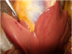
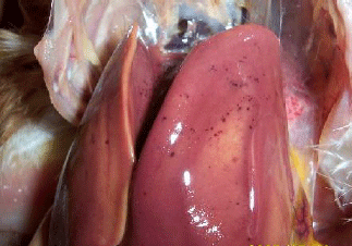
The most consistent findings in this condition were encountered in the liver. On gross examination, the appearance of the livers was very similar in all outbreaks. The liver is the primary organ affected in these birds which is enlarged, pale yellow with multiple petechial haemorrhages (Fig1a and 1b). The parenchyma of the liver was soft in consistency.
Fig 1a: liver from infected chicken (hypertrophy and discoloration)
Fig 1b: petechial hemorrhages in liver
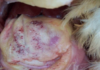
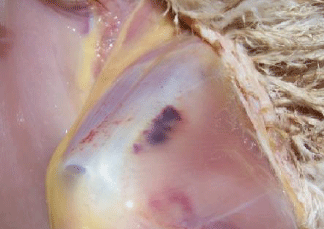
In some cases, kidneys appeared swollen, small and pale due to deposition of urates. Skin and body fat were yellow in color. The bursa, spleen and thymus were considerably smaller than those in non affected chickens. Petechias were observed also in the abdominal fat and on members (Fig 2, 3).
The post mortem lesions observed during the present investigation were similar to those reported by (Howell et al.,1970; Grimes et al.,1978; Fitzgerald, 2000; Hess, 2011).
Fig 2: Petechiae on the abdominal grease
Fig 3: Petechiae on the members
Liver histopathology
Hehmboldt and Frazier's term "hepatic catastrophe" to describe the severity of the changes observed in the liver. In the majority of cases, liver destruction was almost complete (Helmboldt and Frazier, 1963). Schonewille et al., 2008; found that fowl adenovirus (FAdV) can also cause depletion of B and T cells in lymphoid organs.
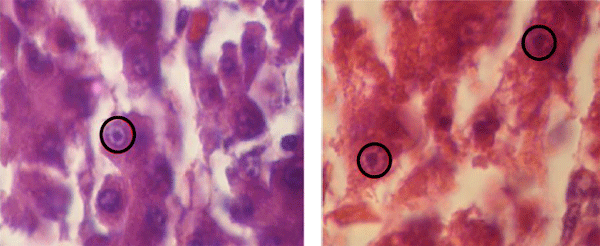
Varying degrees of pyknosis, karyorrhexis and karyolysis were observed in the majority of the hepatic cells (Fig 4). Multiple subcapsular hemorrhages, multifocal groups of hepatocytes and lipid degeneration were also present.
Fig 4: Hepatic cells with karyorrhexis and karyolysis (H&E X1000)
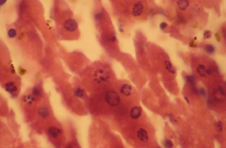
In some hepatic cells, swelling of the nuclei and margination of the chromatin were apparent. Large Cowdry Type A intranuclear inclusion bodies were noticed in many hepatocytes. They were surrounded by a clear halo and were irregular in outline. In some areas of the liver, inclusion bodies were very numerous (Fig 5).
Fig 5: Hepatocytes with large basophilic intranuclear inclusion bodies (H&E X1000).
CONCLUSIONS
The use of histopathological technique is an easy way to confirm the diagnosis of IBH. In spite of the limited diffusion of the IBH in comparison with other avian pathologies (Gumboro Disease, Marek Disease and Newcastle Disease) but this disease has a high economic importance because the high mortality rate, contagiousness, the least use of the vaccines and the absence of an effective treatment against this disease. The only way to control this disease remains the prophylactic vaccination of broiler-breeder flocks and implementation of a strong biosecurity program on farms to prevent contamination of the broiler farms environment and poultries.
REFERENCE
Campbell, T.W. 1995. Avian hematology and cytology. Iowa State University Press, Ames, 2 Eds. p 104.
Choi, K.S., S.J. Kye, J.Y. Kim, W.J. Jeon, E.K. Lee, K.Y. Park and H.W. Sung. 2012. Epidemiological investigation of outbreaks of fowl adenovirus infection in commercial chickens in Korea. Poult. Sci. 91: 2502-2506.
Dar, A., S. Gomis, I. Shirley, G. Mutwiri, R. Brownlie, A. Poter, V. Gerdts and S.K. Tikoo 2012. Pathotypic and molecular characterization of a fowl adenovirus associated with inclusion body hepatitis in Saskatchewan chickens. Avian Dis. 56: 73-81
Di Franco, E., G. Lussier, L. Berthiaume, S. Cloutier and P. Marois. 1974. Hépatite à corps d'inclusion chez le poulet de gril isolement d'un agent viral. Can. Vet. J. 15(5):144-147.
Fitzgerald, S.V., and A. Mc Connell. 2008. Group I adenovirus infections. In: Saif Y.M. Diseases of Poultry. 12 th Eds. Iowa, Wiley- Blackwell,pp: 252-266.
Gomis, S., A.R. Goodhope, A.D. Ojkis and P. Wilson. 2006. Inclusion body hepatitis as a primary disease in broilers in Saskatchewan, Canada. Avian Dis. 50, 550-555
Grimes, T.M., O.J. Fletcher and J.F. Munnell. 1978. Comparative study of experiment al inclusion body hepatitis of chickens caused by two serotypes of avian adenovirus. Vet. Pathol. 15: 249-263.
Helmboldt, C.F. and M.N. Frazier. 1963. Avian hepatic inclusion bodies of unknown significance. Avian Dis. 7: 446–450.
Hess, M. 2000. Detection and differentiation of avian adenoviruses: a review. Avian pathol. 29: 195-206
Hess, M. 2001. Fowl adenovirus infections in chickens: current status and control approaches. In: Proceeding of the XVII World Veterinary Poultry Congress, Cancun, Mexico, pp: 41-48
Howell, J., D.W. Mac Donald and R.G. Christian. 1970. Inclusion body hepatitis in chickens. Can. Vet. J. 11(5): 99-101
Luna, L.G. 1968. Manual of Histologic Staining Methods of the AFIP 3rd Eds., Mc Graw-Hill, NY, p: 131.
Mc Ferran, J.B and B.M. Adair. 2003. Group I adenovirus infections. In: Saif Y.M. Diseases of Poultry. 12 th Eds. Iowa, Wiley-Blackwell, pp: 214-227.
Schonewille, E., A.Singh, T.W. Gobet, W. Gemer, A. Saalmuller, and M. Hess. 2008. Fowl adenovirus (FAdV) serotype 4 causes depletion of B and T cells in lymphoid organs in specific pathogen-free chickens following experimental infection. Vet. Immunopathol. 121:130-139.
Villate, D. 2001. Maladie des volailles. Eds. France. Agricole, p: 282
Zadravec, M., B. Slavec, U. Krapež, G.L. Kaján, J. Racnik, P. Juntes, R. Juršic Cizerl, M. Benko and O.Z. Rojs. 2011. Inclusion body hepatitis associated with fowl adenovirus type 8b in broiler flock in Slovenia - A case report. Slov. Vet. Res. 48 (3/4): 107-113.
Citation
Laanani, I., N. Alloui, O. Bennoune, W. Laabaci, A. Ayachi and M.S. Benterki. 2015. Clinical and Histopathological Investigations on Inclusion Body Hepatitis in Chickens in the Ain Touta Area (Algeria). Global Journal of Animal Scientific Research. 3(1):72-76.
Received: 22 September 2014, Revised: 16 October 2014, Accepted: 18 October 2014