Home
Search
Explore
Publish
Explore

Advertise on Engormix
Home
Publish
Search

Methionine and Alpha-tocopherol supplementation improve liver health of aged laying hens

Published: October 22, 2026
Source : H. ZHANG 1, T. MAHMOOD 2, Y. MERCIER 2, A. HABTAMU 1, G. MA 1, J. WANG 1, J. LIN 1, S. WU 1, K. QIU 1, B. GUO 2 and G. QI 1 / 1 Feed Research Institute, Chinese Academy of Agricultural Sciences, Beijing 100081, China; 2 Adisseo France S.A.S.
Summary

Aged laying hens are exposed to oxidative stress which can contribute to liver damage. This study aimed to investigate the effects of methionine (Met) sources (DL-Met and DL-2-hydroxy-4- (methylthio)-butanoic acid (OH-Met)), Met levels (100, 110, and 120% of breeding company recommendation) and alpha-tocopherol (VE) levels (20 and 40 IU) supplementation on liver functioning in 70 wk aged laying hens. Met source and level interaction had significant effect on liver index (P < 0.01) along with significant interaction effect of Met source and VE level (P < 0.01). Further, OH-Met reduced liver fat percentage compared with DL-Met (P < 0.01). Met sources had no effect on liver Triglycerides (TG) and total cholesterol (TC) but Met level significantly affected TG (P < 0.05) with Met source and level interaction (P < 0.01), however, lower VE level reduced TC in liver (P < 0.05). Met source and Met level influenced superoxide dismutase (SOD), while catalase activities (CAT) was influenced by Met level only (P < 0.05). It was found that OH-Met at 110% level significantly increased SOD compared with other Met levels. DL-Met at 100% level and OH-Met at 110% exhibited higher CAT activities (P < 0.05). DL-Met at 110% level and OH-Met at 100% showed significant reduction of malonaldehyde (MDA; P < 0.05). Further, OH-Met significantly increased mRNA gene expression of glutathione synthase (GSS) and nuclear factor erythroid 2-related factor (Nrf2) compared with the DL-Met group (P<0.05). A higher number of normal livers were found at 100 % OH-Met with 20 IU VE supplementation. Taken together, this research demonstrates that the liver health of aged laying hens was slightly improved by OH-Met through enhanced hepatic antioxidative functions.

I. INTRODUCTION

Aged hens with their regressive antioxidant capacity are susceptible to reactive oxygen species (ROS) and reactive nitrogen species (RNS), thereby various liver metabolic disorders may occur (Gu et al., 2021; Ponnampalam et al., 2022). Though supplementation of Met and VE are involved in antioxidant defense system (Agostini et al., 2016; Mazur-Kusnirek et al., 2019), previous studies highlighted oxidative stress may increase the Met and VE demands beyond standard requirement of chickens (Lugata et al., 2022). Therefore, we hypothesized that current industry standards may be insufficient to fulfill the Met and VE dietary requirement of aged laying hens for antioxidation functions. Thus, this study aimed to investigate the effects of Met source, Met levels and VE levels on oxidative potential in aged laying hens.

II. METHODS

A total of 864, Hy-line brown laying hens (70 wk age) were randomly divided into 12 treatment groups with 6 repeats (12 birds in each repeat). The experimental birds were subjected to a factorial array of treatments using two Met sources (DL-Met and OH-Met) with three Met levels (100, 110, and 120% of breeding company recommendation), and two VE levels (20, 40 IU) The experimental birds were fed a corn-soybean meal diet. At the end of 12 wk of experimental period, one bird close to the average body weight of the replicate was selected per each replicate and euthanized by cervical dislocation. The body cavity was opened to collect and weigh the livers. The liver index was calculated by using the equation: Liver index (%) = [liver weight (g) / live weight (g)] × 100%.
The visible evaluation of the livers from different treatments was performed for lesions. Tissue samples from the liver were collected in RNA-free centrifuge tube, snap frozen in liquid nitrogen and stored at -80°C for mRNA analysis. Total RNA was extracted from liver tissue. The total RNA was reverse transcribed into cDNA. Quantitative real- time PCR for CDO, GSS, Nrf2, and GST was performed according to instructions of the manufacturer. Gene expression were analyzed using the relative PCR amplification analysis method (2−ΔΔCT). The content of fat in liver was detected by ether extraction method. The liver triglycerides (TG), total cholesterol (TC), SOD, MDA, reduced glutathione (GSH), oxidized glutathione (GSSG), CAT, were determined using a commercial kit.
The statistical differences were determined by three-way ANOVA in a 2 × 3 × 2 factorial arrangement with Duncan’s test for multiple comparisons. Meanwhile, one-way ANOVA and Duncan’s multiple comparisons were used when a significant interaction was observed. A value of P < 0.05 was considered significant. 0.05 < P < 0.10 was viewed as a trend.

III. RESULTS

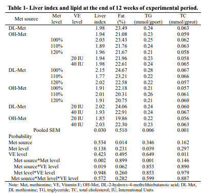
Met sources and Met levels interaction effect were observed on liver index, DL-Met at 100% level and DL-Met with 20 IU VE level exhibited higher liver index value (P < 0.05), as described in Table 1. Furthermore, OH-Met group had lower fat percentage than DL-Met (P < 0.05). We observed significant effect of Met level on TG wherein OH-Met had lower TG at 100% compared to DL-Met at the same level (P < 0.05). Total cholesterol in liver was significantly decreased by VE at 20 IU (P < 0.05). 
Table 1- Liver index and lipid at the end of 12 weeks of experimental period.
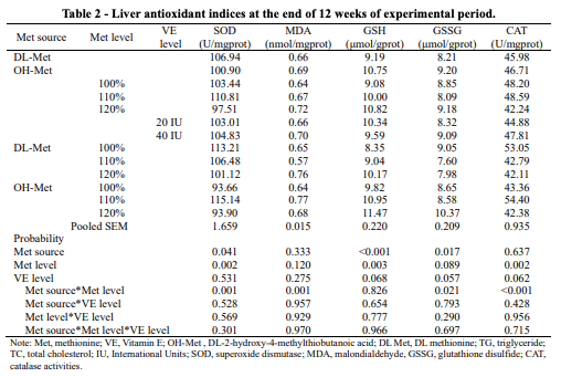
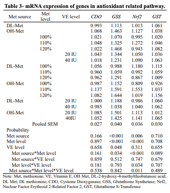
The supplementation of OH-Met increased both GSH and GSSG content compared with the DL-Met, while concomitant decrease in SOD activity in this group (P < 0.05) (Table 2). SOD activity decreased linearly with increasing level of DL-Met but OH-Met at 110% level had the highest SOD activity compared with other Met levels. In addition, Met at 110% level showed significantly higher SOD and CAT activities (P < 0.05). But GSH content linearly increased with the increase of Met level (P < 0.05). DL-Met at 110% level and OH-MET at 100% showed significant reduction of MDA content. DL-Met at 100% level and OH-Met at 110% level exhibited higher CAT activities compared to other supplementation level of the groups (P < 0.05). As shown in Table 3, GSS and Nrf2 expression in OH-Met was significantly higher than DL-Met (P < 0.05). With the increase of Met level, the GSS expression increased significantly, and the Nrf2 expression increased initially and then decreased significantly (P < 0.05). The GSS expression at 20 IU of VE was higher than at 40 IU (P < 0.05). For GSS and Nrf2 expression, a marked interaction between Met source and level was observed (P< 0.05). Additionally, distinct interaction between Met level and VE level on Nrf2 was found (P < 0.05). The highest Nrf2 expression was observed at 110% and 20 IU of Met level and VE level, respectively. 
Table 2 - Liver antioxidant indices at the end of 12 weeks of experimental period.
Table 3- mRNA expression of genes in antioxidant related pathway.
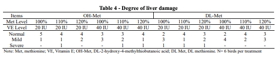
We found that OH-Met at 100% level had a higher number of normal livers than the DL-Met group. Further, OH-Met at 100% level with 20 IU VE supplementation had the most normal livers among all the groups, as described in Table 4. In conjunction with VE at 20 IU, OH-Met diets led to more normal livers and fewer livers with severe lesion. 
Table 4 - Degree of liver damage

IV. DISCUSSION

Liver indexes and hepatic lipid accumulation in aged layer hens can be an indication of liver health and metabolic function (Madeira et al., 2018). Antioxidant enzymes such as SOD, CAT, and GSH-Px are vital components of the antioxidant defense system (Cadenas and Davies, 2000). Previous research showed that antioxidant status or concentration can be manipulated through nutritional regulation, and the Met source (Wang et al., 2019). Consistently, our data confirmed that Met sources influenced antioxidant enzymes concentration. In the past, OH-Met was reported to increase GSH compared to DL-Met in broilers regardless of the tissue and level (Jankowski et al., 2017; Zhang et al., 2018). In line with our results, Ruan et al., (2018) also showed that GSH content increased linearly with the increase of Met. Thus, the increase GSH contents due to OH-Met supplementation indicates its antioxidant potential with scavenging free radical mechanism (Surai et al., 2019). The current study confirmed that Nrf2 expression in OH-Met group was significantly higher compared with DLMet. In this regard, the Nrf2-Keap1 system signal transduction pathways is one of the quickest responding systems to the changing environment, and it can upregulate the antioxidant defense networks (Pomatto and Davies, 2018). Additionally, OH-Met at 100% level with 20 IU VE had the most normal livers among all the groups. It can be argued that OH-Met supports antioxidant system better, resulting in normal liver functions. In conclusion, the study indicated that the inclusion of OHMet enhanced liver antioxidant capacity, lowered fat content, and promoted lipid metabolism through the up-regulation of key genes in aged laying hens.
ACKNOWLEDGEMENTS: We would like to thank National Key R&D Program of China (2021YFD1300204), Agricultural Science and Technology Innovation Program (ASTIP) of the Chinese Academy of Agricultural Sciences and Adisseo France S.A.S. for sponsoring this study.
   
Presented at the 35th Annual Australian Poultry Science Symposium 2024. For information on the latest and future editions, click here.

Agostini, P. S., Dalibard, P., Mercier, Y., van der Aar, P., & van der Klis, J. D. (2016). Poultry Science, 95, 560–569.

Cadenas, E., & Davies, K. J. A. (2000). Free Radical Biology and Medicine, 29, 222–230.

Gu, Y. F., Chen, Y. P., Jin, R., Wang, C., Wen, C., & Zhou, Y. M. (2021). Poultry Science, 100, 100949.

Jankowski, J., Ognik, K., Kubinska, M., Czech, A., Juskiewicz, J., & Zdunczyk, Z. (2017). Poultry Science, 96, 3229–3238.

Li, Y., Mei, H., Liu, Y., Li, Z., Qamar, H., Yu, M., & Ma, X. (2023). Foods, 12, 2302.

Lugata, J. K., Ortega, S. V., & Csaba, S. (2022). Agriculture, 12, 1701.

Madeira, M. S., Palmeira, M. M., Costa, F. G., Gomes, P. C., Ferreira, M. A., Lopes, P. A. A., Santos, C. E., Alfaia, M. M., & Prates, A. M. (2018). Poultry Science, 97, 2591–2599.

Mazur-Kusnirek, M., Antoszkiewicz, Z., Lipinski, K., Kaliniewicz, J., & Kotlarczyk, S. (2019). Archives of Animal Breeding, 62, 287–296.

Pomatto, L. C., & Davies, K. J. A. (2018). Free Radical Biology and Medicine, 124, 420–430.

Ponnampalam, E. N., Kiani, A., Santhiravel, S., Holman, W. B., Lauridsen, C., & Dunshea, F. R. (2022). Animals, 12, 3279.

Ruan, D., Fouad, A. M., Fan, Q., Xia, W., Wang, S., Chen, W., Lin, C., Wang, Y., Yang, L., & Zheng, C. (2018). British Journal of Nutrition, 119, 121–130.

Surai, P. F., Kochish, I. I., Fisinin, V. I., & Kidd, M. T. (2019). Antioxidants, 8, 235.

Wang, J., Yang, G., Zhang, K., Ding, X., Bai, S., & Zeng, Q. (2019). Poultry Science, 98, 341–349.

Zhang, S., Gilbert, E. R., Saremi, B., & Wong, E. A. (2018). Journal of Animal Physiology and Animal Nutrition, 102, 1274–1283.

Related topics:
Authors:
Tahir Mahmood
Yves Mercier
Recommend
Comment
Share
Profile picture
Would you like to discuss another topic? Create a new post to engage with experts in the community.
Featured users in Poultry Industry
Dr. Algis Martínez
Dr. Algis Martínez
DVM, Diplomado ACPV - Poultry Veterinarian North America Cargill
United States
Shivaram Rao
Shivaram Rao
PhD Director Principal de Nutrición y Servicios Técnicos de Pilgrim’s Pride Corporation
United States
Carolina Hall
Carolina Hall
United States