Infectious Laryngotracheitis
Forum: is this Infectious Laryngotracheitis (ILT)?
Is this Infectious Laryngotracheitis (ILT)? Please look at the nuclear area...
Please check the photo album


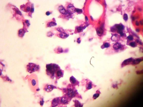
I'm Dr. Laila Tantawy, Avian Pathologist at Animal Health Research Institute. The intranuclear inclusion of the ILT may be appeared in this photo as round esinophilic body inside the nucleus. But in fact to be sure you must find syncytium, composed of different nuclei with marginalized chromatine. However, sometimes the virus destruct the cells after its replication, then in this case you cannot see the inclusion although the bird in infected with ILT.
See this picture: ILT,intranuclear inclusion bodies


I cannot see any typical intranuclear inclusions with clear margination of the chromatin which are diagnostic for ILT. The appearance of multinucleate syncytia would also be helpful but I am not convinced that they are present here. An important feature of...

Two nuclei under the two blood vessels in the right top corner of section appear suspicious. However, this H&E section does not contain adequate information for conclusive opinion.

Reference to topic on ILT
Characteristic inclusions are not always be seen, particularly as the lesions become old. It may be seen in initial stages only. Under field conditions we always get the typical cases for diagnosis after period of 3-4days of initial mortality by which time the mucosal lining would be completely degenerated and desquamated. In such cases it will be difficult to identify the typical inclusions. 2 or 3 tissues collected out of 15 - 20 birds may show the inclusions provided the tissues are properly proceesed and stained.
As noted from this section the inclusions are not so distinct. However, the grossn lesions with other serological studies might give conclusive diagnosis.
Dr A.G. Rao
Retired Professor Pathology, Orissa Veterinary College, Odisha
.jpg&w=3840&q=75)

Candidate Genes Associated with Survival Following Highly Pathogenic Avian Influenza Infection in Chickens


United States

Jonathan Cade comments on his new role as Chairman of the U.S. Poultry & Egg Association
