In vitro evaluation of CD40-targeting aptamer-based DNA vaccine adjuvants using HD11 cell culture
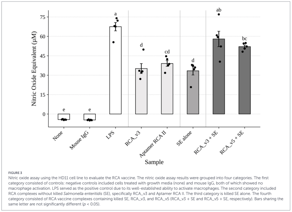
Salmonella is a major foodborne pathogen that colonizes the gastrointestinal tract of poultry, compromising bird health and productivity while posing significant public health threats. Consumption of contaminated poultry products is the leading cause of Salmonella-related illnesses in humans. Although numerous vaccination strategies exist, weak immune responses often limit the efficacy. One promising approach to address this limitation is targeting the cluster of Differentiation 40 (CD40), a costimulatory receptor expressed on antigenpresenting cells that plays a crucial role in initiating and regulating immune response. In this study, we developed two rolling circle amplification (RCA) vaccine complexes (RCA_v3 and RCA_v5) against Salmonella, each comprising a CD40-targeting component to enhance immune activation (Aptamer RCA II) and a Salmonella-binding component for antigen delivery (Anti-Salmonella RCA). We evaluated the two RCA vaccine complex designs for their ability to activate macrophages in the HD11 chicken cell line by quantifying nitric oxide production via the Griess assay. The results showed that the two RCA vaccine designs (RCA_v3 and RCA_v5) combined with formalin-killed Salmonella Enteritidis (SE) significantly (58.00 μM and 52.08 μM, respectively) enhanced macrophage activation (p < 0.05) compared to SE alone (33.37 μM). Also, the activation levels of RCA_v3 vaccine complex were significantly higher with SE as compared with the respective counterparts without SE, RCA_v3 alone, and Aptamer RCA II (35.13 μM and 39.02 μM, respectively) (p < 0.05). These findings offer valuable insights into developing effective vaccination strategies to control Salmonella colonization in poultry, presenting a promising approach to improving food safety and reducing public health risks associated with Salmonella infections in chickens.
KEYWORDS CD40-targeting, chicken, DNA aptamer, rolling circle amplification, Salmonella, vaccine adjuvant
Introduction
Materials and methods
Selection of Salmonella aptamers

RCA template and primer design
Production of rolling circle amplification (RCA) products
Assembly of the vaccine complex
Quantification of Salmonella binding to RCA vaccine construct
HD11 assay to quantify macrophage activation by the RCA vaccine complexes
Statistical analysis
Results and discussion
Rationale for the design of the RCA vaccine complexes
Characterization of the binding efficiency of selected Salmonella-specific aptamers
Verification of the optimized high-yield RCA products
Quantification of Salmonella binding to RCA constructs for vaccine dose selection
Macrophage activation by RCA-based vaccine complexes
Conclusion
- Shaji S, Selvaraj RK, Shanmugasundaram R. Salmonella infection in poultry: a review on the pathogen and control strategies. Microorganisms. (2023) 11:2814. doi: 10.3390/microorganisms11112814
- Kogut MH, Santin E. "Advances in vaccines for controlling foodborne Salmonella spp. in poultry". In: Venkitanarayanan K, Thakur S, Ricke S, editors. Food Safety in Poultry Meat Production. Cham, Switzerland: Spring Nature. (2019). p. 161–89.
- Desin TS, Köster W, Potter AA. Salmonella vaccines in poultry: past, present and future. Expert Rev Vaccines. (2013) 12:87–96. doi: 10.1586/erv.12.138
- Jajere SM. A review of Salmonella enterica with particular focus on the pathogenicity and virulence factors, host specificity and antimicrobial resistance including multidrug resistance. Vet World. (2019) 12:504–21. doi: 10.14202/vetworld.2019.504-521
- Sarrami Z, Sedghi M, Mohammadi I, Bedford M, Miranzadeh H, Ghasemi R. Effects of bacteriophage on Salmonella Enteritidis infection in broilers. Sci Rep. (2023) 13:12198. doi: 10.1038/s41598-023-38791-6
- Van Immerseel F, Methner U, Rychlik I, Nagy B, Velge P, Martin G, et al. Vaccination and early protection against non-host-specific Salmonella serotypes in poultry: exploitation of innate immunity and microbial activity. Epidemiol Infect. (2005) 133:959–78. doi: 10.1017/S0950268805004711
- Berry JD, Licea A, Popkov M, Cortez X, Fuller R, Elia M, et al. Rapid monoclonal antibody generation via dendritic cell targeting in vivo. Hybrid Hybridomics. (2003) 22:23–31. doi: 10.1089/153685903321538053
- Chen C-H, Abi-Ghanem D, Waghela SD, Chou W-K, Farnell MB, Mwangi W, et al. Immunization of chickens with an agonistic monoclonal anti-chicken CD40 antibody–hapten complex: rapid and robust IgG response induced by a single subcutaneous injection. J Immunol Methods. (2012) 378:116–20. doi: 10.1016/j.jim.2012.02.006
- Hatzifoti C, Heath AW. CD40-mediated enhancement of immune responses against three forms of influenza vaccine. Immunology. (2007) 122:98–106. doi: 10.1111/j.1365-2567.2007.02617.x
- van Kooten C, Banchereau J. CD40–CD40 ligand. J Leukoc Biol. (2000) 67:2–17. doi: 10.1002/jlb.67.1.2
- Al-Ogaili AS, Liyanage R, Lay JO, Jiang T, Vuong CN, Agrawal S, et al. DNA aptamer-based rolling circle amplification product as a novel immunological adjuvant. Sci Rep. (2020) 10:22282. doi: 10.1038/s41598-020-79420-w
- Banchereau J, Bazan F, Blanchard D, Briere F, Galizzi JP, Kooten C, et al. The CD40 antigen and its ligand. Annu Rev Immunol. (1994) 12:881–926. doi: 10.1146/annurev.iy.12.040194.004313
- Bullock TN. CD40 stimulation as a molecular adjuvant for cancer vaccines and other immunotherapies. Cell Mol Immunol. (2022) 19:14–22. doi: 10.1038/s41423-021-00734-4
- Chen C-H, Abi-Ghanem D, Njongmeta L, Bray J, Mwangi W, Waghela SD, et al. Production and characterization of agonistic monoclonal antibodies against chicken CD40. Devel Comp Immunol. (2010) 34:1139–43. doi: 10.1016/j.dci.2010.06.014
- Armitage RJ, Fanslow WC, Strockbine L, Sato TA, Clifford KN, Macduff BM, et al. Molecular and biological characterization of a murine ligand for CD40. Nature. (1992) 357:80–2. doi: 10.1038/357080a0
- Elgueta R, Benson MJ, De Vries VC, Wasiuk A, Guo Y, Noelle RJ. Molecular mechanism and function of CD40/CD40L engagement in the immune system. Immunol Rev. (2009) 229:152–72. doi: 10.1111/j.1600-065X.2009.00782.x
- Vonderheide RH. Prospect of targeting the CD40 pathway for cancer therapy. Clin Cancer Res. (2007) 13:1083–8. doi: 10.1158/1078-0432.CCR-06-1893
- Chou W-K, Chen C-H, Vuong CN, Abi-Ghanem D, Waghela SD, Mwangi W, et al. Significant mucosal sIgA production after a single oral or parenteral administration using in vivo CD40 targeting in the chicken. Res Vet Sci. (2016) 108:112–5. doi: 10.1016/j.rvsc.2016.08.013
- Omolewu A. Development of Aptamer Rolling Circle Amplification Platform as a Vaccine Adjuvant to Control Salmonella in Broiler Chickens. Fayetteville, Arkansas, U.S.: University of Arkansas (2021).
- Vuong CN, Chou W-K, Briggs W, Faulkner O, Wolfenden A, Jonas M, et al. Crude inactivated influenza a virus adjuvated with a bispecific antibody complex targeting chicken CD40 and AIV M2e confers protection against lethal HPAI challenge in chickens. Monoclon Antib Immunodiagn Immunother. (2018) 37:245–51. doi: 10.1089/mab.2018.0040
- Demidov VV. "Introduction: 20+ years of rolling the DNA minicircles—state of the art in the RCA-based nucleic acid diagnostics and therapeutics". In: Demidov VV, editor. Rolling Circle Amplification (RCA) Toward New Clinical Diagnostics and Therapeutics. Cham Switzerland: Springer International Publishing AG. (2016). p. 1–7.
- Goo N-I, Kim D-E. Rolling circle amplification as isothermal gene amplification in molecular diagnostics. Biochip J. (2016) 10:262–71. doi: 10.1007/s13206-016-0402-6
- Ali MM, Li F, Zhang Z, Zhang K, Kang D-K, Ankrum JA, et al. Rolling circle amplification: a versatile tool for chemical biology, materials science and medicine. Chem Soc Rev. (2014) 43:3324–41. doi: 10.1039/c3cs60439j
- Berr A, Schubert I. Direct labelling of BAC-DNA by rolling-circle amplification. Plant J. (2006) 45:857–62. doi: 10.1111/j.1365-313X.2005.02637.x
- Demidov VV. Rolling-circle amplification in DNA diagnostics: the power of simplicity. Expert Rev Mol Diagn. (2002) 2:542–8. doi: 10.1586/14737159.2.6.542
- Zhang Z, Ali MM, Eckert MA, Kang DK, Chen YY, Sender LS, et al. A polyvalent aptamer system for targeted drug delivery. Biomaterials. (2013) 34:9728–35. doi: 10.1016/j.biomaterials.2013.08.079
- Joshi R, Janagama H, Dwivedi HP, Senthil Kumar TMA, Jaykus L-A, Schefers J, et al. Selection, characterization, and application of DNA aptamers for the capture and detection of Salmonella enterica serovars. Mol Cell Probes. (2009) 23:20–8. doi: 10.1016/j.mcp.2008.10.006
- Dwivedi HP, Smiley RD, Jaykus L-A. Selection of DNA aptamers for capture and detection of Salmonella Typhimurium using a whole-cell SELEX approach in conjunction with cell sorting. Appl Microbiol Biotechnol. (2013) 97:3677–86. doi: 10.1007/s00253-013-4766-4
- Moon J, Kim G, Lee S, Park S. Identification of Salmonella Typhimurium-specific DNA aptamers developed using whole-cell SELEX and FACS analysis. J Microbiol Methods. (2013) 95:162–6. doi: 10.1016/j.mimet.2013.08.005
- Duan N, Wu S, Chen X, Huang Y, Xia Y, Ma X, et al. Selection and characterization of aptamers against Salmonella Typhimurium using whole-bacterium systemic evolution of ligands by exponential enrichment (SELEX). J Agric Food Chem. (2013) 61:3229–34. doi: 10.1021/jf400767d
- Park H-C, Baig IA, Lee S-C, Moon J-Y, Yoon M-Y. Development of ssDNA Aptamers for the sensitive detection of Salmonella typhimurium and Salmonella enteritidis. Appl Biochem Biotechnol. (2014) 174:793–802. doi: 10.1007/s12010-014-1103-z
- Lavu PSR, Mondal B, Ramlal S, Murali HS, Batra HV. Selection and characterization of aptamers using a modified whole cell bacterium SELEX for the detection of Salmonella enterica Serovar Typhimurium. ACS Comb Sci. (2016) 18:292–301. doi: 10.1021/acscombsci.5b00123
- Berman AJ, Kamtekar S, Goodman JL, Lazaro JM, de Vega M, Blanco L, et al. Structures of phi29 DNA polymerase complexed with substrate: the mechanism of translocation in B-family polymerases. EMBO J. (2007) 26:3494–505. doi: 10.1038/sj.emboj.7601780
- Lin C, Xie M, Chen J, Liu Y, Yan H. Rolling-circle amplification of a DNA nanojunction. Angew Chem Int Ed. (2006) 45:7537–9. doi: 10.1002/anie.200602113
- Rodríguez I, Lázaro JM, Blanco L, Kamtekar S, Berman AJ, Wang J, et al. A specific subdomain in φ29 DNA polymerase confers both processivity and strand-displacement capacity. Proc Natl Acad Sci. (2005) 102:6407–12. doi: 10.1073/pnas.0500597102
- Zhao W, Cui CH, Bose S, Guo D, Shen C, Wong WP, et al. Bioinspired multivalent DNA network for capture and release of cells. Proc Natl Acad Sci USA. (2012) 109:19626–31. doi: 10.1073/pnas.1211234109
- Green LC, Wagner DA, Glogowski J, Skipper PL, Wishnok JS, Tannenbaum SR. Analysis of nitrate, nitrite, and [15N] nitrate in biological fluids. Anal Biochem. (1982) 126:131–8. doi: 10.1016/0003-2697(82)90118-X
- Crippen TL, Sheffield CL, He H, Lowry VK, Kogut MH. Differential nitric oxide production by chicken immune cells. Dev Comp Immunol. (2003) 27:603–10. doi: 10.1016/S0145-305X(03)00035-1
- Balzer AH, Whitehurst CB. An analysis of the biotin–(strept) avidin system in immunoassays: interference and mitigation strategies. Curr Issues Mol Biol. (2023) 45:8733–54. doi: 10.3390/cimb45110549
- Lu Z, Zhang Y, Wang Y, Tan G-H, Huang F-Y, Cao R, et al. A biotin-avidin-system-based virus-mimicking nanovaccine for tumor immunotherapy. J Control Release. (2021) 332:245–59. doi: 10.1016/j.jconrel.2021.02.029
- Purow B, Staveley-O'Carroll K. Targeting of vaccinia virus using biotin-avidin viral coating and biotinylated antibodies. J Surg Res. (2005) 123:49–54. doi: 10.1016/j.jss.2004.04.022
- Lin K-Y, Matteucci MD. A cytosine analogue capable of clamp-like binding to a guanine in helical nucleic acids. J Am Chem Soc. (1998) 120:8531–2. doi: 10.1021/ja981286z
- Lou C, Dallmann A, Marafini P, Gao R, Brown T. Enhanced H-bonding and π-stacking in DNA: a potent duplex-stabilizing and mismatch sensing nucleobase analogue. Chem Sci. (2014) 5:3836–44. doi: 10.1039/C4SC00948G
- Wilds CJ, Maier MA, Tereshko V, Manoharan M, Egli M. Direct observation of a cytosine analogue that forms five hydrogen bonds to guanosine: guanidino G-clamp. Angew Chem Int Ed. (2002) 41:115–7. doi: 10.1002/1521-3773(20020104)41:1<115::AID-ANIE115>3.0.CO;2-R
- Lee Y, Han S, Maeng J, Cho Y, Lee S. In vitro selection of Escherichia coli O157:H7-specific RNA aptamer. Biochem Biophys Res Commun. (2012) 417:414–20. doi: 10.1016/j.bbrc.2011.11.130
- Shangguan D, Li Y, Tang Z, Cao ZC, Chen HW, Mallikaratchy P, et al. Aptamers evolved from live cells as effective molecular probes for cancer study. Proc Natl Acad Sci. (2006) 103:11838–43. doi: 10.1073/pnas.0602615103
- Wang K, Zeng Y, Yang X, Li W, Lan X. Utility of aptamer-fluorescence in situ hybridization for rapid detection of Pseudomonas aeruginosa. Eur J Clin Microbiol Infect Dis. (2011) 30:273–8. doi: 10.1007/s10096-010-1074-0
- Dwivedi H, Smiley R, Jaykus L. Selection and characterization of DNA aptamers with binding selectivity to Campylobacter jejuni using whole-cell SELEX. Appl Microbiol Biotechnol. (2010) 87:2323–34. doi: 10.1007/s00253-010-2728-7
- Liu G, Yu X, Xue F, Chen W, Ye Y, Yang X, et al. Screening and preliminary application of a DNA aptamer for rapid detection of Salmonella O8. Microchim Acta. (2012) 178:237–44. doi: 10.1007/s00604-012-0825-2
- Lee M, Shin S, Kim S, Park N. Recent advances in biological applications of aptamer-based fluorescent biosensors. Molecules. (2023) 28:7327. doi: 10.3390/molecules28217327
- Weaver SD, Whelan RJ. Characterization of DNA aptamer–protein binding using fluorescence anisotropy assays in low-volume, high-efficiency plates. Anal Methods. (2021) 13:1302–7. doi: 10.1039/D0AY02256J
- Ali MM, Li Y. Colorimetric sensing by using allosteric-DNAzyme-coupled rolling circle amplification and a peptide nucleic acid–organic dye probe. Angew Chem. (2009) 121:3564–7. doi: 10.1002/ange.200805966
- Ali MM, Su S, Filipe CD, Pelton R, Li Y. Enzymatic manipulations of DNA oligonucleotides on microgel: towards development of DNA–microgel bioassays. Chem Commun. (2007) 43:4459–61. doi: 10.1039/b709817k
- Qi X, Bakht S, Devos KM, Gale MD, Osbourn A. L-RCA (ligation-rolling circle amplification): a general method for genotyping of single nucleotide polymorphisms (SNPs). Nucleic Acids Res. (2001) 29:e116. doi: 10.1093/nar/29.22.e116
- Zhang T, Qing T, Xiao-Jun B, Qian C, Juan Y. Rapid visualized detection of Escherichia coli O157: H7 by DNA hydrogel based on rolling circle amplification. Chin J Anal Chem. (2021) 49:377–86. doi: 10.1016/S1872-2040(21)60085-3
- Zhao W, Gao Y, Kandadai SA, Brook MA, Li Y. DNA polymerization on gold nanoparticles through rolling circle amplification: towards novel scaffolds for three-dimensional periodic nanoassemblies. Angew Chem. (2006) 118:2469–73. doi: 10.1002/ange.200600061
- Cho EJ, Yang L, Levy M, Ellington AD. Using a deoxyribozyme ligase and rolling circle amplification to detect a non-nucleic acid analyte, ATP. J Am Chem Soc. (2005) 127:2022–3. doi: 10.1021/ja043490u
- Göransson J, Ke R, Nong RY, Howell WM, Karman A, Grawe J, et al. Rapid identification of bio-molecules applied for detection of biosecurity agents using rolling circle amplification. PLoS One. (2012) 7:e31068. doi: 10.1371/journal.pone.0031068
- Linck L, Reiß E, Bier F, Resch-Genger U. Direct labeling rolling circle amplification as a straightforward signal amplification technique for biodetection formats. Anal Methods. (2012) 4:1215–20. doi: 10.1039/c2ay05760c
- Sato K, Ishii R, Sasaki N, Sato K, Nilsson M. Bead-based padlock rolling circle amplification for single DNA molecule counting. Anal Biochem. (2013) 437:43–5. doi: 10.1016/j.ab.2013.02.016
- Ali MM, Kanda P, Aguirre SD, Li Y. Modulation of DNA-modified gold-nanoparticle stability in salt with concatemeric single-stranded DNAs for colorimetric bioassay development. Chem Eur J. (2011) 17:2052–6. doi: 10.1002/chem.201002677
- Xu W, Xie X, Li D, Yang Z, Li T, Liu X. Ultrasensitive colorimetric DNA detection using a combination of rolling circle amplification and nicking endonuclease-assisted nanoparticle amplification (NEANA). Small. (2012) 8:1846–50. doi: 10.1002/smll.201200263
- St John J, Quinn TW. Rapid capture of DNA targets. BioTechniques. (2008) 44:259–64. doi: 10.2144/000112633
- Barrow PA, Huggins MB, Lovell MA. Host specificity of Salmonella infection in chickens and mice is expressed in vivo primarily at the level of the reticuloendothelial system. Infect Immun. (1994) 62:4602–10. doi: 10.1128/iai.62.10.4602-4610.1994
- Gantois I, Ducatelle R, Pasmans F, Haesebrouck F, Gast R, Humphrey TJ, et al. Mechanisms of egg contamination by Salmonella Enteritidis. FEMS Microbiol Rev. (2009) 33:718–38. doi: 10.1111/j.1574-6976.2008.00161.x
- Hassan JO, Curtiss R. Development and evaluation of an experimental vaccination program using a live avirulent Salmonella typhimurium strain to protect immunized chickens against challenge with homologous and heterologous Salmonella serotypes. Infect Immun. (1994) 62:5519–27. doi: 10.1128/iai.62.12.5519-5527.1994
- Lin AW, Chang CC, McCormick CC. Molecular cloning and expression of an avian macrophage nitric-oxide synthase cDNA and the analysis of the genomic 5′-flanking region (*). J Biol Chem. (1996) 271:11911–9. doi: 10.1074/jbc.271.20.11911
- Sung Y-J, Hotchkiss JH, Austic RE, Dietert RR. L-arginine-dependent production of a reactive nitrogen intermediate by macrophages of a uricotelic species. J Leukoc Biol. (1991) 50:49–56. doi: 10.1002/jlb.50.1.49
- Orecchioni M, Ghosheh Y, Pramod A, Ley K. Macrophage polarization: different gene signatures in M1(LPS+) vs. classically and M2(LPS−) vs. alternatively activated macrophages. Front Immunol. (2019) 10:1084. doi: 10.3389/fimmu.2019.01084
- Sharif O, Bolshakov V, Raines S, Newham P, Perkins N. Transcriptional profiling of the LPS induced NF-κB response in macrophages. BMC Immunol. (2007) 8:1. doi: 10.1186/1471-2172-8-1
- Cha RS, Thilly WG. Specificity, efficiency, and fidelity of PCR. Genome Res. (1993) 3:S18–29. doi: 10.1101/gr.3.3.S18
- Budiarto B, Mustopa A, Ningrum R, Amilia N, Saepudin E. Gold nanoparticles (AuNP)-based aptasensor for enteropathogenic Escherichia coli detection. Mol Biol Rep. (2022) 49:9355–63.




















DICOSAN+

Hy-Line International


United States