Explore
Communities in English
Advertise on Engormix

Incidence of Avian Nephritis and Infectious Bronchitis Viruses in Broilers in Egypt

Published: July 17, 2018
By: Hatem S. Abd El-Hamid 1, Hany F. Ellakany 1, Elkholia S.Z. 2, Ahmed R. Elbestawy 1, Eman R. Diab 3. / 1 Department of Poultry and Fish Diseases; 2 Department of Animal Husbandry and Wealth Development, Faculty of Veterinary Medicine, Damanhour University, Egypt; 3 Veterinarian.
Summary

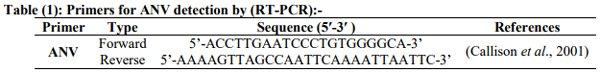
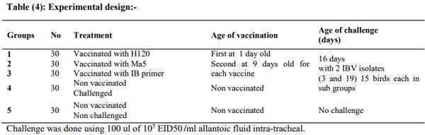
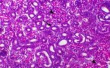
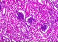
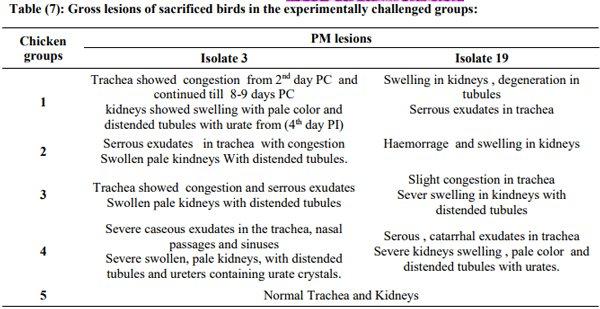
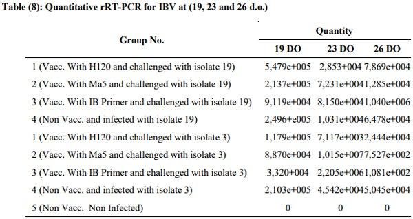
Avian nephritis virus is a new circulating virus in Europe, Australia, and Japan, this virus lead to growth retardation of young chickens by causing interstitial nephritis. Investigation was done in 5 Egyptian governorates (North Sinai, El-Minoufiya, ElGharbia and El-Behera and Kafr-Elsheikh) on 28 broiler chicken flocks and all the flocks were negative for Avian nephritis virus. In addition, we continued the detection of IBV in the same 28 broiler chicken flocks, resulting that 11 flocks out of 28 flocks, were positive for IBV. Regarding spike glycoprotein (S1) gene, sequencing was done to 5 isolates, which showed that they belonged to Eg/12120s/2012 spike glycoprotein (SP1) gene variant 2. Protectotyping was done using 2 isolates, and 3 types of IB live attenuated vaccines (H120, Ma5, IB primer). In conclusion, this study recorded the extensive circulation of variant 2 IBV in Egyptian broiler chickens. Regarding the pathogenicity of the tested variant and cross protection with available live IB vaccines, it was clear that there no complete cross-protection against IBV isolates although of using live attenuated IB vaccines so there a need for further studies to define a more effective program for IB control.

Key words: avian nephritis, infectious bronchitis viruses in broilers in Egypt

1. INTRODUCTION
Avian nephritis virus (ANV) is an etiological agent of growth retardation of young chickens by causing interstitial nephritis (Imada et al., 1979). Antibodies against ANV have been found in chicken and turkey flocks in the UK and Japan, suggesting a broad distribution (Nicholas et al., 1988; Takase et al., 2000).
It is originally considered to be a picornavirus, was later characterized as an avian astrovirus based on its nucleotide sequence (Imada et al., 2000). Different degrees of pathogenicity in chickens are exhibited by field strains of ANV, producing results ranging from subclinical infection to death, and there are at least two serotypes (Frazier et al., 1990 and Shirai et al., 1991).
While, Infectious bronchitis virus (IBV) is a ubiquitous, highly contagious respiratory pathogen of chickens that causes serious economic losses to the commercial poultry industry worldwide. IBV is a member of the order Nidovirales, family Coronaviridae, subfamily Coronavirinae, genus Gammacoronavirus (Carstens, 2010).
The Egyptian poultry industry in recent years has observed an increasing incidence of respiratory and nephritis pathologies related to infection with infectious bronchitis virus (IBV) in vaccinated and non-vaccinated flocks that caused severe economic losses (El-Mahdy et al., 2010).
IBV is typically associated with respiratory disease and/or renal damage, and increased mortality at the end of fattening cycle in broilers. Morbidity of IBV is usually 100%, but mortality varied between 0% and 82%, depending on the age and the immune status of the birds, the virus strain, and secondary bacterial or viral infections (Jackwood and De Wit, 2013). Co-infections of avian respiratory viruses including IBV may induce similar clinical signs or lesions and thus complicate diagnostic decisions, as well as complicating its control (Nguyen et al., 2013)
The genome of IBV is an approximately 27.6 kb single-stranded positive sense RNA molecule encoding 4 structural proteins, spike (S), membrane (M), envelope (E), and nucleoprotein (N) (Cavanagh ,2007). The S protein comprises two or three copies of each of its two subunits S1 and S2. However, S1 has been reported to be the most important protein in terms of coronavirus variability (Jackwood, et al., 2012). The S1 subunit protein is involved in infectivity and contains virus-neutralizing epitopes, serotype-specific sequences, and haemagglutinin activity (Carstens, 2010).
Several IBV serotypes or antigenic variant strains have been reported in many countries due to high rates of S1 gene mutation (Nguyen et al., 2013 and Lim et al., 2013). The different serotypes, subtypes, and variants of IBV are thought to be generated by nucleotide point mutations, insertions, deletion and recombination (Carstens, 2010 and Mahmood et al., 2011), which are responsible for outbreaks in vaccinated chicken flocks. In addition, one or more serotypes can be endemic in regions with intensive poultry production (Capua, et al., 1999).
Recently new IBV genotypes emerged (AbdelMoneim et al., 2012 and Zhou et al., 2014).
In general, different serotypes do not confer cross-protection against each other so the classification of IBV strains according to their ‘‘protectotype’’ had been proposed (Dhinakar and Jones, 1996). The cross protection tends to diminish as the degree of amino acid identity between the S1 proteins of 2 IBV strains decreases (Gelb et al., 2005 and Roh et al., 2013) . Ladman and coworkers (2006) found that partial S1 sequences identity is more strongly correlated with protective relatedness values than antigenic relatedness values.
IBV infection associated with renal lesions and mortality up to 30% was approved by the isolation of IBV from kidneys of naturally infected birds (Winterfield and Albassam, 1984 and Butcher et al., 1990).
In the past few years, kidney affections combined with respiratory manifestation in poultry had increased in Egypt (El-Mahdy et al., 2012).
Therefore, the aim of this study was to investigate the incidence of both Avian nephritis virus and Infectious bronchitis virus in broiler flocks in 5 governorates in Egypt (North Sinai, El-Minoufiya, El-Gharbia and El-Behera and Kafr Elsheikh).
2. MATERIAL AND METHODS
Kidney samples (5 samples/flock) were collected aseptically from samples from 28 broiler chicken flocks in 5 governorates (North Sinai, El-Minoufiya, El-Gharbia, El-Behera and Kafr-Elsheikh) in the period from (2013- 2014).
Tested flocks were suffering from respiratory signs including difficult breathing, tracheal rales, sneezing with or without nasal discharge and wet eyes with whitish or greenish whitish diarrhea. In addition, the examined flocks suffered from variable mortalities with PM lesions sometimes suggestive of viral infections. A generalized weakness accompanied by depression with feed consumption and markedly reduced body weight was markedly observed in broiler.
2.1. Virus inoculation in embryonated chicken eggs
Kidneys pooled samples homogenates were clarified and treated with antibiotics before inoculation into (9-11) day-old commercial embryonated chicken egg, 5 eggs/sample. Samples were passed for (3-5) blind serial passages and the –ve HA allantoic fluids were used for the detection of ANV and IBV using RT-PCR (Gelb and Jackwood, 1998).
2.2. Molecular detection of ANV and IBV:-
RNA was extracted from a pool of tracheas and a pool of kidneys from each flock using a QIAamp Viral RNA Mini Kit (Qiagen GmbH, Hilden, Germany) according to the manufacturer’s instructions. The RT-PCR for ANV and IBV detection was performed using one-step RT-PCR master mix. Sets for these primers are in tables 1 &2.
Incidence of Avian Nephritis and Infectious Bronchitis Viruses in Broilers in Egypt - Image 1
 
Incidence of Avian Nephritis and Infectious Bronchitis Viruses in Broilers in Egypt - Image 2
 
Incidence of Avian Nephritis and Infectious Bronchitis Viruses in Broilers in Egypt - Image 3
 
Incidence of Avian Nephritis and Infectious Bronchitis Viruses in Broilers in Egypt - Image 4
2.3. Sequencing of the amplified part of IBV S1gene
The sequencing data were initially checked by NCBI BLAST search, assembled, and edited using EditSeq (DNASTAR Inc., Madison, Wisconsin). Additional IBV sequences data available in GenBank were downloaded for comparative phylogenetic analysis. Selected sequences were subjected to Clustal W multiple sequence alignment. Residue analyses were done using the BioEdit 7.1.5 program. The amino acid sequences identity matrix was calculated to determine the homology between the isolates and other selected reference isolates.
2.4. Detection of other viruses
Real-time PCR (RRT-PCR) for the detection of NDV (F protein) and AI (M protein) (Table 3) was conducted to exclude these viruses from positive IBV samples. For NDV the cycling conditions for Fusion protein gene amplification primers were performed as follows: initial denaturation step at 95 °C for 15 min, 50 cycles of 94 °C for 10 s, 58 °C for 5 s, and 72 °C for 10 s. While for AI the cycling conditions for Matrix protein gene amplification primers as initial denaturation step at 95 °C for 15 min, 45 cycles of 94°C for 5 s, 60 °C for 20 s.
2.5. Experimental Trial
A total of about 150 one-day-old commercial broiler chicks were used for experimental infection and protection study.
2.5. a. IBV vaccines used in protection study:-
Three commercial IB live vaccines (H120 – IB primer – Ma5) were used. They were administrated at the manufactured recommended dose by intraocular and intra-Nasal route, at 1 day old and at 9 days old.
2.5.b. IBV challenge strains:-
The two isolates number (3 and 19) used for challenge test were identical to strain (Eg/12120s/2012-SP1).
The 2 viruses were titrated in SPF ECE as described by Villegas and Purchase, (1990), having 107 EID50 /ml according to the method of Reed and Muench, (1938).
2.5. c. Quantitative rRT-PCR for IBV at (19, 23 and 26 DO)
Five tracheal swabs were collected and pooled for RRT-PCR at 19, 23 and 26 days old DO (3, 7 and 10 Days Post Challenge - DPC). and The RRT-PCR was performed using One-Step RRT-PCR kit (thermo Scientific, Germany) with S1 for the detection of IBV (Cavanagh et al., 1999) and Sets of primers and taqman probe mentioned in table (5) used for conducting RRT-PCR using Quantitect probe RRT-PCR kit
Incidence of Avian Nephritis and Infectious Bronchitis Viruses in Broilers in Egypt - Image 5
2.6. Enzyme linked immunosorbent assay (ELISA):-
Five serum samples were collected at 5, 16 and 26 days old were separated and checked by ELISA (Biochek, Netherland) for detection of specific IBV antibodies.
2.7. Kidney function test
Three collected serum samples 5 days post challenge (DPC) at 21 DOES were used to measure the percent of uric acid and creatinine.
2.8. Histopathological examination:-
Samples for histopathological examination from trachea & kidney of 2 birds killed at 21 DO were kept in 10 % buffered formalin, routinely processed embedded in paraffin, sectioned and stained with haematoxylin and eosin (HE) (Bancroft and Steven, 1977).
3. RESULTS:
3.1. History of the examined flocks:
A total of 28 broiler flocks were investigated during the period between 2013 and 2014, the clinical signs of the investigated chicken flocks showed depression, illness, ruffled feathers and reduced weight gain due to decrease in feed consumption.
Respiratory signs ranged from mild to severe, including tracheal rales, gasping (mouth breathing), sneezing and nasal discharge with watery whitish or greenish whitish diarrhea.
Postmortem, lesions showed Kidneys were swollen, pale, enlarged with urates (Fig. 1) with occasionally swollen sinuses and tracheal catarrhal to caseous exudate and pneumonia.
3.2. RT-PCR for ANV and IBV:-
All flocks were negative for ANV even the RT-PCR was repeated 2 times, so the investigated parts from Egypt were free from ANV (Fig, 2).
After three blind passages, the inoculated embryos showed curling and dwarfing (fig.3) with subcutaneous hemorrhage. For IBV, 11 samples out of 28 total examined broiler flocks (39.3%), flocks number (2, 3, 7, 10, 19, 13, 21, 25, 26, 29 and 30) were positive using multiplex RT-PCR (Fig. 4) and the history of these samples is in table (6)
Incidence of Avian Nephritis and Infectious Bronchitis Viruses in Broilers in Egypt - Image 6
 
Fig. (1): Nephritis Nephrosis Syndrome (Nephropathogenic form of IBV) in the positive examined field samples.
Incidence of Avian Nephritis and Infectious Bronchitis Viruses in Broilers in Egypt - Image 7
 
Fig. (2): Negative RT-PCR results of ANV
Incidence of Avian Nephritis and Infectious Bronchitis Viruses in Broilers in Egypt - Image 8
 
Fig. (3): The effect of IBV (variant 2 identical) on the embryo as dwarfing and curling compared to the control.
Incidence of Avian Nephritis and Infectious Bronchitis Viruses in Broilers in Egypt - Image 9
 
Fig (4): Multiplex RT-PCR detection to distinguish IBV of Massachusetts, D274 and 793/B types; the relative location of the Massachusetts like strains is (298), D274 (220) and 793/B (154)
Incidence of Avian Nephritis and Infectious Bronchitis Viruses in Broilers in Egypt - Image 10
All positive IBV 11 isolates were –ve for NDV and AI using RRT-PCR
RESULTS OF GENETIC ANALYSIS:-
The allantoic fluid of positive IBV samples, 5 isolates from different localities (2, 3, 7, 10 and 19) was selected for further sequencing and genetic analysis.
Depending on spike glycoprotein (SP1) gene, phylogenetic analysis sequences of the 5 selected Egyptian IBV field isolates in this study was in the same group with Eg/12120s/2012-SP1 which known as variant 2 IBV.
Four isolates (2, 3, 10 and 19) were 98% related to variant 2 like Eg/12120s/2012, while isolate no. 7 was 97% identical to both infectious bronchitis virus isolate Eg/12120s/2012 and IBV/CK/MenofiaEgypt/USC-8/2013.
Experimental challenge results:
Clinical signs and PM lesions (Table, 7)
Challenged groups suffered from respiratory signs (gasping, tracheal rales, coughing and sneezing) with diarrhea and marked weight loss. Chickens in groups which challenged with isolate 3 IBV showed more severe signs than which challenged with isolate 19 IBV. This denotes that isolate 3 is more virulent than isolate 19 IBV.
Quantitative rRT-PCR (Table, 8)
IB primer vaccine in group 3 which challenged with isolate 3 was the best facing viral challenge while in isolate 19 it was the highest amount of viral shedding at 3 DPC. After 7th DPC, highest amount of virus was in group 1 (vaccinated with H120) challenged with isolate 3 while least amount was in group 2 (vaccinated with Ma5). In 10 DPC, the least amount of viral shedding for both isolates challenge (3 & 19) were the chickens of group 3 (vaccinated with IB primer).
Histopathological findings
Tracheas of normal birds showed normal mucosal lining cartilaginous ring and the Kidneys of normal birds showed normal renal glomeruli and tubules.
Infected birds with IB virus isolate 3-showed epithelial degeneration, congestion of the blood vessels of the mucosa and leukocytic infiltration. The chickens of this group also, showed mainly features of tubulo-interstitial nephritis (Fig. 5), which characterized mainly by degeneration of renal tubules associated with marked interstitial fibrosis as well as infiltration of inflammatory cells and acute tubular degeneration represented by degeneration of renal tubular epithelium could be detected in some cases. There was also some regenerative tubules which demonstrating marked tubular basophilia.
Infected birds with IBV isolate 19 revealed normal mucosa unless hyperplasia was detected, and demonstrated glomerulonephritis which characterized by hypercellularity of glomeruli due to increase in mesangial cells, with secondary tubular degeneration (Fig. 6). In addition to features of chronic interstitial nephritis was also noticed. In some cases glomeruli were atrophied due to sclerosis.
Vaccinated birds with H120, and infected with IBV isolates 3 and 19 revealed normal tracheal epithelium. While birds infected with isolate 3 revealed normal renal tubules (Fig. 7), and the birds infected with IB isolate 19 showed normal renal tubules with presence of focal regenerative tubules.
Birds vaccinated with Ma5 and infected with IB virus isolate 3 showed intact mucosal lining, with occasional focal epithelial degeneration and leukocytic infiltration (Fig. 8) and also, it caused multi-focal regenerative tubules in the kidneys. While birds infected with isolate 19 of IB virus demonstrated normal trachea and multi-focal regenerative tubules and interstitial hemorrhages in the kidneys (Fig. 9).
In vaccinated birds with IB primer, tracheas and kidneys of birds infected with IB virus isolate 3 (fig. 10) and/or isolate 19 were within the normal limits. Unless the isolate 19 showed mild glomerular nephropathy represented by mild sclerotic changes in glomeruli with dilated Bowman’s space (Fig. 11).
(Fig.5) Kidney of chicken infected with IBV isolate 3 revealed feature of chronic interstitial nephritis represented by tubular degeneration (arrowhead), interstitial fibrosis and regeneration of some renal tubules (arrow), H&E, X200
Incidence of Avian Nephritis and Infectious Bronchitis Viruses in Broilers in Egypt - Image 11
 
(Fig. 6) Kidney of chicken infected with IBV isolate 19 revealed feature of chronic glomerulo nephritis which showed hypercellularity of glomeruli (arrowhead) and tubular degeneration (arrow), H&E, X200
Incidence of Avian Nephritis and Infectious Bronchitis Viruses in Broilers in Egypt - Image 12
 
(Fig. 7) Kidney of chicken infected with IBV isolate 3 and vaccinated with H120 was within the normal limits, H&E, X200
Incidence of Avian Nephritis and Infectious Bronchitis Viruses in Broilers in Egypt - Image 13
 
(Fig. 8) Trachea of chicken infected with IB virus isolate 3 and vaccinated with MA5showed congestion of the mucosal blood capillaries and leukocytic infiltration (arrowhead), H&E, X200
Incidence of Avian Nephritis and Infectious Bronchitis Viruses in Broilers in Egypt - Image 14
 
(Fig. 9) Kidney of chicken infected with IBV isolate 19 and vaccinated with MA5 showed haemorrhage (arrow) and multifocal tubular regeneration areas (arrowhead), H&E, X200
Incidence of Avian Nephritis and Infectious Bronchitis Viruses in Broilers in Egypt - Image 15
 
(Fig. 10) Kidney of chicken infected with IB virus isolate 3 and vaccinated with IB primer was within the normal limits, H&E, X200
Incidence of Avian Nephritis and Infectious Bronchitis Viruses in Broilers in Egypt - Image 16
 
(Fig. 11) Kidney of chicken infected with IB virus isolate 19 and vaccinated with IB primer was within the normal limits, unless mild degree of glomerular sclerosis with dilated filtration spaces of the glomeruli H&E, X200
Incidence of Avian Nephritis and Infectious Bronchitis Viruses in Broilers in Egypt - Image 17
 
Incidence of Avian Nephritis and Infectious Bronchitis Viruses in Broilers in Egypt - Image 18
 
Incidence of Avian Nephritis and Infectious Bronchitis Viruses in Broilers in Egypt - Image 19
Serological examination (ELISA test):-
ELISA test for the collected serum samples from broiler chickens during the experimental challenge.
Incidence of Avian Nephritis and Infectious Bronchitis Viruses in Broilers in Egypt - Image 20
Biochemical identification using kidney function test:
Incidence of Avian Nephritis and Infectious Bronchitis Viruses in Broilers in Egypt - Image 21
3. DISCUSSION
Regarding avian nephritis surveys all samples proved negative results with PCR specific primers which used according to (Callison et al., 2001).
The investigation the IBV in 28 broiler flocks suffering from signs of depression, reduction in weight gain, respiratory manifestation and diarrhea with moderate to high mortalities and nephropathogenic effect was recorded and the clinical signs and gross lesions was suggestive of infectious bronchitis and were accorded to those described by many authors (Parsons et al., 1992, Capua et al ., 1999, Abd-almoniem et al .,2002 and Sedeik, 2005)
The most frequently published IBV sequences in GeneBank are localized at the S1 gene, which is a part of the IBV genome with high variability. Therefore, it provided obvious possibilities for the construction of strain-specific oligonucleotides. Our study was designed to identify the most genotypes of IBV circulating in Egypt. We used oligonucleotide primer designed by Adzhar et al., (1997), which was capable to detect the different genotypes of IBV (Classic and Variant).
Out of the 28 broiler flocks examined, 11 samples were positive (39.3%), and after three blind passages the inoculated embryos showed curling and dwarfing with subcutaneous hemorrhage in the embryos.
The 5 selected IBV isolates from 2 different governorates (Elbehira and Elgharbia) were analyzed by sequencing of the HVR 3 in S1 gene and 4 isolates (2, 3, 10 and 19) were 98% related to variant 2 like Eg/12120s/2012, while isolate no. 7 was 97% identical to both infectious bronchitis virus isolate Eg/12120s/2012 and IBV/CK/Menofia-Egypt/USC- 8/2013, (Arafa et al,. 2013).
Regarding egg inoculation, the 2 IBV isolates (3, and 19) were able to induce 50%, 30% mortality respectively in the inoculated embryonated eggs after passage 5.
The protectotype concept can explain the efficacy of the vaccination through boosting with heterologous vaccine strain that can protect the chicken from IBV challenge in a similar way to the homologous vaccine strain (Cook et al.,1999 , Terregino et al ., 2008 , Mahgoub et al., 2010 , De Wit et al., 2011 ,Lim et al ., 2012). This concept has been suggested to be a valuable one to consider in terms of developing strategies to control IBV infections (Lohr, 1988).
In Egypt, the situation of IBV necessitates the use effective vaccines to control the disease and to confirm broad protection against more than one serotype. However, it is not expected to have 100% protection but the maximum protection conferred by specific program be used as a guide for field application, in addition to the biosecurity procedures to reduce the losses by such infection.
So for protection estimation against these field isolates, we selected 3 commercial vaccines containing classical or variant strains as H120, Ma5 and IB primer vaccine (live IBV vaccine containing strains H120 and D274).
The Massachusetts serotype represented by H120 and Ma5 vaccines was chosen because it is the most commonly used IBV classical vaccines in Egypt and worldwide (Adzhar et al., 1996).
While use of IB primer which is unique combination (classical H120 +variant D274 strains) in a bivalent vaccine will make a synergism that will give a wide range of protection against various serotypes of IBV found worldwide (Cook et al., 1996).
After vaccination 2 times, first at 1 day old and second at 9 day old in each group, as the protection provided by a single vaccination may not be enough to cover the entire broiler production period. Application of a second IB vaccination may be beneficial in such situations, not only to prolong the duration of the protection obtained but also to broaden the spectrum of such protection (Malo et al., 1998).
The weight gain of chickens of group 1 vaccinated with H120 vaccine was less than group 2 vaccinated with Ma5 vaccine and the best weight gain was in the chicken group 3, which vaccinated with IB primer.
While in group number 4 which challenged at 16 day old & non vaccinated there was sever weight loss with severe respiratory signs & diarrhea compared to chicken group 5 which is negative control (non vaccinated, non challenged) in which the weight gain was in its normal rate.
Chickens challenged with isolate 19 IBV in all groups showed clinical respiratory signs that persist for (14 DPC) but it's severity milder than chicken groups that challenged with isolate 3 which had also some mortality rates rather than isolate 19 challenged groups.
Also on PM examination both IBV isolates (3 and 19) were able to induce renal lesions in all chicken groups. This was also indicated through the viral shedding in the tracheal swabs in which the virus in all groups was shed till 10 DPC but the least virus titer was in group 3 vaccinated with IB primer followed by Ma5 the H120 These findings agreed with Cook et al. 2001 and El-Mahdy et al. (2012).
The most severe respiratory signs were in chicken group challenged without IB vaccination accompanied with severe weight loss but there no mortalities. While in chicken groups vaccinated with Ma5 vaccine and challenged with both isolates, the clinical respiratory signs were higher than which vaccinated with H120 vaccine and IB primer vaccine. These findings agreed with Ignjatovic and Sapats (2000), who reported that strains of IBV differed in their virulence and pathogenicity for the respiratory tract and kidney.
Kidney function test was used to evaluate the efficacy of kidney and it's vitality under the effect of IBV infection with and without vaccination to estimate the ability of vaccines to protect renal system against IBV and effect of IBV on functions of kidneys. Uric acid and creatinine % were highest in group 4 which is non vaccinated and challenged with both IBV isolates number 3 and 19.
Chickens of group 1 which was challenged with isolate 3 IB and vaccinated with IB primer gave the least in uric acid and creatinine % which indicated that IB primer vaccine give best protection in comparison to Ma5 and H120 IB vaccines, matching with the results obtained by Cook et al., (1996).
Regarding the pathological finding, the kidneys of infected chickens with IB virus isolate 3 revealed feature of chronic interstitial nephritis characterized by marked interstitial inflammatory mononuclear cell infiltration. Kidney of chicken infected with IB virus isolate 19 revealed feature of chronic glomerulonephritis which showed hyper-cellularity of glomeruli and tubular degeneration. These finding agreed with (Cowen et al., 1987; Purcell et al., 1976; Albassam et al., 1986 and El-Mahdy et al., 2012).
Trachea of chicken infected with IB isolate 3 showed moderate tracheal lesions which represented by epithelial degeneration and mucosal leukocytic infiltration, and mild lesions as hyperplasia of the lining epithelial layer of trachea in chickens infected with isolate 19. These findings appeared similar to those previously recorded (Cavanagh and Naqi, 2003).
The widespread vaccination by using the Massachusetts type (H120) commercial vaccines did not protect against the IB Variant 2 isolates (EvenChen et al., 2013). While in IB primer vaccine when repeated is the best for protection against renal lesions of IBV variant 2 and this according to the results obtained by Ali, (2014) followed by H120 then Ma5 vaccine.
ELISA detected high levels of antibody against IBV on 10 day PI in chicken groups vaccinated with Ma5 and IB primer, respectively compared with the very low titers in H120 vaccinated birds, Also Ghadakchi et al. (2005) showed that ELISA titers could be reliable, repeatable, and sensitive for monitoring vaccination schedules and the rapid detection of the early rise of antibody against IBV.
CONCLUSION
The current study revealed that there was no ANV in the investigated governorates in Egypt.
Eleven flocks out of 28 were positive for IBV (39,3%) and after sequencing the circulating serotype was Eg/12120s/2012-SP1, which known as variant 2 IBV.
According to the results obtained from clinical signs, PM lesions, rRT-PCR, ELISA and Histopathological findings and biochemical Kidney function tests (Uric acid and Creatinine concentration tests) of experimentally challenged birds, isolate 3 IBV was more virulent than isolate 19 IBV and IB primer vaccine give better protection followed by Ma5 then H120 vaccines.
Protectotyping using IB primer vaccine 2 times may decrease the clinical signs, pathological lesions, renal infection and shedding of renal IBV strains.
Continuous isolation, identification and sequencing of Egyptian IBV isolates and performing protection studies against different commercially available vaccines must be done for better controlling IBV under the Egyptian situation
This article was originally published in Alexandria Journal of Veterinary Sciences, Vol. 51(2):36-47. November 2016 DOI: 10.5455/ajvs.233327. This is an Open Access article licensed under a Creative Commons Attribution-NonCommercial-ShareAlike 4.0 International License.

Abdel-Moneim A S, Madbouly H M, Gelb J Jr, and Landman S (2002): Isolation and identification of Egypt/Beni-Suef/01 a novel infectious bronchitis-virus genotype. Vet. Med. J. Giza, 50(4): 1065-1078.

Abdel-Moneim AS, Afifi MA, El-Kady MF (2012) Emergence of a novel genotype of avian infectious bronchitis virus in Egypt. Arch Virol 157: 2453-2457.

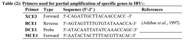
Adzhar A, Shaw K, Britton P, and Cavanagh D (1996):Universal digonucleotides for the detection of infectious bronchitits virus by the polymerase chain reaction. Avian Pathol. 25: 817-836.

Adzhar A, Gough R E, Haydon D, Shaw K, Brition P, and Cavanagh D (1997): Molecular analysis of the 793/B Seratype of infectious bronchitis virus in Great Britain. Avian Path, 26: 625-640.

Albassam MA, Winterfield BW, and Thacker HL (1986): Comparison of the nephropathogencity of four strains of infectious bronchitis virus. Avian Dis, 30, 468-476.

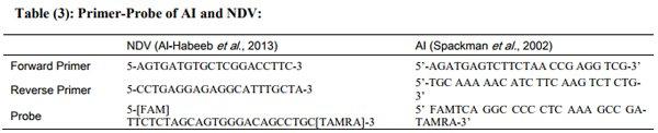
Al-Habeeb, A.M; Mohamed, M. H. A. and Sharawi, S. (2013): Detection and characterization of Newcastle disease virus in clinical samples using real time RTPCR and melting curve analysis based on matrix and fusion genes amplification. Vet. World 6(5):239-243.

Ali MMZ (2014): Epidemiological and Molecular studies on the status of Infectious Bronchitis disease in Egypt. Ph.D., Thesis, Poultry Dis.Fac.Vet.Med.Beni-suef Univ.Egypt.

Arafa, A., Samir, M., Hasan, M., Selim, A.,Khaliel, S. (2013): Molecular characterization of infectious bronchitis virus in Egypt, National Laboratory for Veterinary Quality Control on Poultry Production, Animal Health Research Institute, 7 Nadi ElsaidStreet, Dokki, Giza12618, Egypt

Bancroft, J.D. and Stevens, A. (1977): Theory and practices of histologic techniques 2nd Eds. Churchill, Living Stone Edingburgh, London Melborne and New York.

Butcher G D, Winterfield R W, and Shapira D P (1990): Pathogenesis of H13 nephropathogenic infectious bronchitis virus. Avian Dis. 34: 916-921.

Callison S A, M W Jackwood, and D A Hilt, (2001): virus isolates foreign to the United States and associated with nephritis nephrosis syndrome in Molecular characterization of infectious bronchitis chicken. J. Egypt. Vet. Med. Ass., 37(2): 71-79.

Capua I, Minta Z, Karpinska E, Mawditt, Britton P, Cavanagh D, and Gough R E (1999):Co-circulation of four types of infectious bronchitis virus (793/B, 624/I, B1648 and Massachusetts). Avian Pathol. 28: 587-592.

Carstens EB (2010) Ratification vote on taxonomic proposals to the International Committee on Taxonomy of Viruses (2009). Arch Virol 155: 133-146. Cavanagh D (2007) Coronavirus avian infectious bronchitis virus. Vet Res 38: 281-297. 5.

Jackwood MW, Hall D, Handel A (2012) Molecular evolution and emergence of avian gammacoronaviruses. Infect Genet Evol 12: 1305-1311.

Cavanagh D, and Naqi S A(2003): Infectious Bronchitis In: Diseases of Poultry,11thed,Y.M.Saif,H.J.Barsones,J.R.Glisson,A.M.Fa dly,L.R.McDougald and D.E.Swwayne,eds.Iowa State University press ,Amess,Iowa:pp. 101-119.

Cavanagh D, Mawditt K, Britton P, Naylor CJ (1999) Longitudinal field studies of infectious bronchitis virus and avian pneumovirus in broilers using type-specific polymerase chain reactions. Avian Pathology 28: 593– 605.

Cook J.K., Orbell S.J., Woods M.A. and Huggins M.B. (1996): A survey of the presence of a new infectious bronchitis virus designated 4/91 (793B). Vet Rec, 138, 178-180.

Cook, J.K.A., Orbell, S.J., Woods, M.A. & Huggins, M.B. (1999): Breadth of protection of respiratory tract provided by different liveattenuated infectious bronchitis vaccines against challenge with infectious bronchitis viruses of heterologous serotypes. Avian Pathology, 28 , 477-485.

Cowen B S, Widemen R F, Braune M O, and Owen R L (1987): An infectious bronchitis virus isolated from chickens experiencing a urolethiasis outbreak. I. In vitro characterization studies. Avian Dis. 31: 878-883.

De Wit J J, J Nieuwen Huiesen-Van Wilgen, A Hoogkamar, H Van de Sande , G J Zuidam, and T H Fabri (2011):Induction of cystic oviducts and protection against early challenge with infectious bronchitis virus serotype D388(genotype QX)by maternally derived antibodies and by early vaccination .Avian Pathol.40:463-471.

Dhinakar Raj G, Jones RC (1996) Protectotypic di ? ?erentiation of avian infectious bronchitis viruses using an in vitro challenge model. Veterinary Microbiology 53: 239–252.

El-Mahdy, S.S.; El-Hady, M.M. & Soliman,Y. A. (2010): Isolation and characterization of Nephropathogenic strain of infectious Bronchitis virus in Egypt; Journal of American Science 6 (9)669-674.

El-Mahdy SS, Salama E, and Ahmed A (2012): Efficacy of some living classical and variant infectious bronchitis vaccines against local variant isolated from Egypt. Nat Sci; 10(12):292-299.

Even-Chen T, Heifetz S, Perelman B, Kin E, Finger, Avner, Ashash U, And Banet-Noach C (2013): Development of New Infectious Bronchitis Variant 2 Vaccines from a Local Israeli Isolate as a Solution for Regional Virulent Field Strains.World Veterinary Poultry Association French branch (GF-AMVA) 19th - 23rd August 2013.

Frazier JA, Howes K, Reece RL, Kidd AW, Cavanagh D (1990): Isolation of non-cytopathic viruses implicated in the aetiology of nephritis and baby chick nephropathy and serologically related to avian nephritis virus. Avian Pathol. 19: 139-160.

Gelb J Jr, and Jackwood M K (1998):Infectious bronchitis. In: A laboratory manual for the Isolation and Identification of Avian Pathogens. 4th Ed. AAAP: 169- 174.

Gelb Jr J, Weisman Y, Ladman BS, Meir R (2005) S1 gene characteristics and efficacy of vaccination against infectious bronchitis virus field isolates from the United States and Israel (1996 to 2000). Avian Pathol 34: 194- 203.

Ghadakchi H, Pourbakhsh SA, and Hosseini SMH(2005): standardization of an Enzyme –LinkedImmunosorbent Assay for detection of Infectious Bronchitis Virus Antibody ,''Archives of Razi Institute ,59,pp57-83.

Ignjatovic J, and Sapats S (2000): Avian infectious bronchitis virus. Rev. Sci. Off. Int. Epiz. 19: 493-508.

Imada T., Yamaguchi S. and Kawamura H. (1979): Pathogenicity for baby chicks of the G-4260 strain of the picornavirus ‘avian nephritis virus’. Avian Diseases, 23, 582– 588.

Imada T., Yamaguchi, S., Mase M., Tsukamoto K., Kubo, M. and Morooka A (2000): Avian nephritis virus (ANV) as a new member of the family Astroviridae and construction of infectious ANV cDNA. Journal of Virology, 74, 8487_8493.

Jackwood MW, De-wit S (2013) Infectious bronchitis in Diseases of poultry. Swayne DE, Glisson JR, McDougald LR, Nolan LK, Suarez DL et al. John Wiley and Sons Inc Ames Iowa 139–159.

Jackwood MW, Hall D, Handel A (2012) Molecular evolution and emergence of avian gammacoronaviruses. Infect Genet Evol 12: 1305-1311.

Ladman BS, Loupos AB, Gelb JJr (2006) Infectious bronchitis virus S1 gene sequence comparison is a better predictor of challenge of immunity in chickens than serotyping by virus neutralization. Avian Pathology 35: 127–133.

Li M, Mo ML, Huang BC, Fan WS, Wei TC, Li KR, Wei P (2013) Continuous evolution of avian infectious bronchitis virus resulting in different variants cocirculating in southern China. Archives of Virology 158: 1783–1786.

Lim T H, Kim J H, Lee D H, Park J K, Youn H N, Lee J B, Park S Y, Chai I S, and Song C S (2012):live attenuated nephropathogenic infectious bronchitis virus vaccine provide broad protection against new variant strains .Poul Sci.91(1):89-94.

Lohr JE (1988):Differentiation of infectious bronchitis virus strains.In E.F. Kaleta & U. Heffels-Redmann (Eds.), Proceedings of the Ist International Symposium on Infectious Bronchitis Rauischholzhausen, Germany.199-208

Mahgoub K M,Bassiouni A A, Aly M A,and Rabie N S(2010): The prevalence of Infectious Bronchitis (IB)Outbreaks in some chicken farms 3.cross protection of vaccinated chickens versus field IB virus. Journal of American science 6(9):94-108.

Mahmood ZH, Sleman RR, Uthman AU (2011) Isolation and molecular characterization of Sul/01/09 avian infectious bronchitis virus, indicates the emergence of a new genotype in the Middle East. Veterinary Microbiology 150: 21–27.

Malo A, Orbell S, Huggins M, Woods M, and Cook J (1998): Cross protection studies after the use of live attenuated IBV vaccines Nobilis® IB 4-91 and Nobilis® IB Ma5 (Massachusetts type). Intervet, VSD Newsletter: 17:1- 6.

Meir, R.; Maharat, O.; Farnushi, Y. and Simanov, L. (2010): Development of a real-time TaqMan® RT-PCR assay for the detection of infectious bronchitis virus in chickens, and comparison of RT-PCR and virus isolation. J Virol. Methods 163:190–194.

Nguyen TT, Kwonb HJ, Kima H, Hong SM, Seong WJ, Jang JW, Kim JH (2013) Multiplex nested RT-PCR for detecting avian influenza virus, infectious bronchitis virus and Newcastle disease virus. Journal of Virological Methods 188: 41-46.

Nicholas R.A., Goddard R.D., Luff P.R. (1988): Prevalence of avain nephritis virus in England. Veterinary Record, 123, 398.

Parsons D, Ellis M M, Cavanagh D, and Cook J K, (1992): Characterization of an infectious bronchitis virus isolated from IB-vaccinated broiler breeder flocks. Vet. Rec., 131: 408-411.

Purcell DA, Tham VL, and Surman P G (1976): The histopathology of infectious bronchitis in fowls infected with a nephrotropic T strain of virus. Australian Vet. J. 52:85-91.

Reed L J and Muench H (1938):A simple method for estimating fifty percent endpoint. Am. J. Hyg., 27: 493- 496.

Roh HJ, Hilt DA, Williams SM, Jackwooda MW (2013) Evaluation of infectious bronchitis virus Arkansas-type vaccine failure in commercial broilers. Avian Dis 57: 248-259.

Sediek M M (2005): Studies on infectious bronchitis in chickens. M.V.Sc, thesis, poultry Dis, Fac. Vet. Med. Alex Univ. Egypt.

Shirai J, Nakamura K, Shinohara K, Kawamura H (1991) : Pathogenicity and antigenicity of avian nephritis isolates. Avian Dis. 35: 49–54.

Spackman, E; Dennis A. Senne; T. J. Myers; Leslie L. Bulaga; Lindsey P. Garber; Michael L. Perdue; Kenton Lohman; Luke T. Daum and David L. Suarez (2002): Development of a Real-Time Reverse Transcriptase PCR Assay for Type A Influenza Virus and the Avian H5 and H7 Hemagglutinin Subtypes. Journal of Clinical Microbiology, p. 3256–3260

Takase K., Murakawa Y., Ariyoshi R., Eriguchi S., Sugimura, T. and Fujikawa H. (2000): Serological monitoring on layer farms with specific pathogen-free chickens. Journal of Veterinary Medical Science, 62, 1327–1329.

Terregino C, Toffan A, and Serena Beato M (2008): “Pathogenicity of a QX strain of infectious bronchitis virus in specific pathogen free and commercial broiler chickens, and evaluation of protection induced by a vaccination programme based on the Ma5 and 4/91 serotypes,” Avian Pathol. vol. 37, no. 5, pp. 487–493.

Villegas P and Purchase G H (1990): Preparation of chicken embryo kidney cell cultures (CEKC). In: Laboratory manual for the isolation and identification of avian pathogens. AAAP. Ames, Iowa, USA. pp: 3-4.

Winterfield R W and Albassam M A (1984): Nephropathogenicity of infectious bronchitis. Poultry Sc, 63: 2358-2363.

Zhou S, Tang M, Jiang Y, Chen X, Shen X (2014) complete genome sequence of a novel infectious bronchitis virus strain circulating in China with a distinct S gene. Virus Genes 49: 152-15.

Related topics:
Authors:
Hany Ellakany
Damanhour University, Egypt
Ahmed Elbestawy
Recommend
Comment
Share
tamem
30 de julio de 2018

Does the h9n2 play role to increase the lose of I.b and how to early controle to early lnfection of h9n2?

Recommend
Reply
Mangesh Shirke
26 de julio de 2018

Infectious Bronchitis has been always a big challenge in prevention and control the disease. The intensive farming tech. has added and increased the bio burden to a level that almost every flock gets exposed to it. The early exposure to immunosuppressive agents adds to increase the severity of the disease. The problem has also been observed all over the world including US, which has more severity than rest. The common techniques used over there is use of auto vaccines, has also been of little help. The disease needs to be addressed judiciously with all care, taking utmost care at biosecurity, control of immunosuppressive agents, serological (antibody) monitoring and vaccination and subsequent monitoring of same.

Recommend
Reply
Profile picture
Would you like to discuss another topic? Create a new post to engage with experts in the community.
Join Engormix and be part of the largest agribusiness social network in the world.