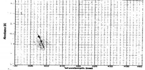
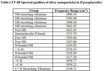
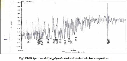
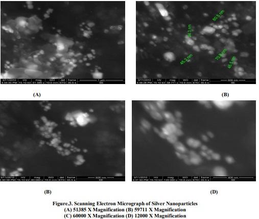
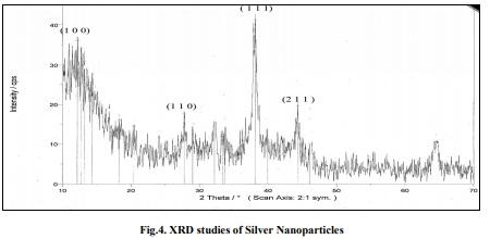
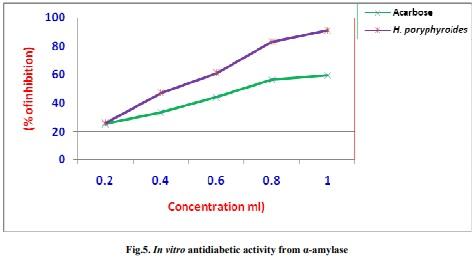
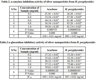
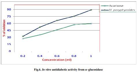
Biogenic silver nanoparticles by Halymenia poryphyroides and its in vitro anti-diabetic efficacy
1] CB Field; M Behrenfeld; JT Randerson; and P Falkowski. Science., 1998, 281, 237-240.
[2] CI Febles; A Arias et al. In vitro study of antimicrobial activity in algae (Chlorophyta, Phaeophyta and
Rhodophyta) collected from the coast of Tenerife (in Spanish). Anuario Del Estudios Canarios., 1995, 34, 181-192.
[3] N Bottini; T Vang; F Cucca; T Mustelin. Seminars in Immunology., 2006, 18, 207- 213.
[4] EW Roderick. Diabetes Research and Clinical Practice., 2004, 65, 53-58.
[5] H Gin and V Rigalleau. Diabetes and Metabolism., 2000, 26, 265-272.
[6] DV Goia; ENJ Matijevic. Chem., 1998, 22, 1203.
[7] C Taleb; M Petit; P Pileni. Chem. Mater., 1997, 9, 950.
[8] K Esumi; T Tano; K Torigoe; K Meguro. Chem.Mater., 1990, 2, 564.
[9] A Henglein. Langmuir., 2001, 17, 2329.
[10] L Rodriguez-Sanchez; MC Blanco; MA Lopez – Quintela. J. Phys. Chem., B 2004, 104, 9683.
[11] JJ Zhu; SW Liu; O Palchik; Y Koltypin; A Gedanken. Langmuir., 2000, 16, 6396.
[12] Pastoriza- Santos; LM Liz – Marzan. Langmuir., 2002, 18, 2888.
[13] NA Begum; S Mondal; S Basu; RA Laskar; D Mandal. Colloids and Surfaces B: Biointerferaces., 2009, 71 (1), 113-118
[14] H Bar; DK Bhui; GP Sahoo; P Sarkar; SP De; A Misra. Colloids and Surfaces A: Physichemical and
Engineering Aspects., 2009, 339, 134-139.
[15] JY Song; BS Kim. Bioprocess Biosyst. Eng., 2009, 32, 79-84.
[16] CP Malik and MB Singh. Plant Enzymology and Histoenzymology,Kalyani publishers, New Delhi, 1980, 278.
[17] S Krishnaveni; B Theymoli and S Sadasivam. Food Chem., 1984, 15, 229.
[18] M Sastry; V Patil; SR Sainkar. J Phys Chem B., 1998, 102, 1404.
[19] P Mulvaney. Langmuir., 1996, 12, 788.
[20] P Mukherjee; S Senapati; D Mandal; A Ahmad; MI Khan; R Kumar & M Sastry. Chem. Bio. Chem., 2002, 3,
461.
[21] J Gonzalo; R Serna; J Sol; D Babonneau & CN Afonso. J. Phys : Condens. Matter., 2003, 15, 3001.
[22] I Sondi and B Salopek- Sondi. J. Colloid Interface Sci., 2004, 275, 177.
[23] C Nabanita; T Ghosh; S Sinha; C Kausik; P Karmakar and Bimalendu Ray. Food Chemist., 2010, 118(3), 823-
829.
[24] S Murugesan; M Elumalai and R Dhamotharan. Biosci.Biotech. Res. Comm., 2011, 4(1), 105-110.
[25] Inbakandan; Venkatesan and Ajmal Khan. Colloids and Surfaces B: Biointerface., 2010, 81(2), 634–639.
[26] H Xu; M Kall. J.Nanosci.Nanotechnol., 2002, 4, 254.
[27] H Kurihara; J Ando; M Hatano; J Kawabata. Bioorg.Med. Chem Lett., 1995, 5, 1241-1244.
[28] L Xiancui; N Rough; F Xiao; H Lijun; Z Lixin. Chin. J. Oceanol. Limmol., 2005, 23, 354-356.
[29] B Harekrishna; KB Dipak; PS Gobinda; S Priyanka; P Santanu; M Ajay. Colloids and Surfaces A:
Physicochem. Eng. Aspects., 2009, 348, 212-216.
[30] N Xu; X Fan; X Yan; X Li; R Niu and CK Tseng. Phytochemistry., 2003, 62, 1221–1224.
[31] H Kurihara; T Mitani; J Kawabata and K Takahashi. Fish Sci., 1999a, 65, 300–303.
[32] H Kurihara; T Mitani; J Kawabata and K Takahashi. J Nat Prod., 1999b, 62, 882–884.
[33] L Xiancui; L Rough; N Xiao; H Lijun; Z Lixin. Chin. J. Oceanol. Limmol., 2005, 23, 354-356.


United States


