Explore
Communities in English
Advertise on Engormix

Supranutrition of microalgal docosahexaenoic acid and calcidiol improved growth performance, tissue lipid profles, and tibia characteristics of broiler chickens
Published: April 3, 2023
By: Sahil Kalia 1, Andrew D. Magnuson 1, Tao Sun 1, Guanchen Liu 2, Woo Kyun Kim 2, Zackary Johnson 3 and Xin Gen Lei 1 / 1 Department of Animal Science, Cornell University, 252 Morrison Hall, Ithaca, NY 14850, USA; 2 Department of Poultry Science, University of Georgia, Athens, GA, USA; 3 Nicholas School of the Environment, Duke University, Beaufort, NC, USA.
Background
Biofortifications of chicken with bioactive nutrients such as DHA and calcidiol [25(OH)D3] have been viewed as an effective strategy to produce health-promoting meat for human consumption [1, 2]. Relatively low to moderate inclusion levels of calcidiol (1600 to 2800 IU/kg diet) [3, 4] and DHA-rich microalgal biomass or oil (0.55 to 2.55  g DHA/kg diet) [5–7] in broiler diets caused no negative effects on growth performance, lipid profile of tissues, or bone strength. In contrast, a high inclusion of calcidiol (27,600 IU/kg diet) in broiler diet decreased body weight by 5% [8], and a high inclusion of DHA-rich microalgal biomass (6.8  g DHA/kg diet) decreased growth performance by 19% and breast muscle weight by 21% [9]. However, these past studies were focused on fortifying chicken with DHA [10] and calcidiol singularly [4]. Little research was attempted to enrich chicken simultaneously with these two nutrients or to look out for potential adverse effects of a combined high supplementation of these two nutrients on growth performance, lipid metabolism, and bone integrity of chickens.
To fill in the gap of knowledge, we conducted two experiments to examine those effects of supplementing high levels of these two bioactive nutrients in broiler chickens. In the first experiment, we supplemented 1% DHA-rich Aurantiochytrium sp. biomass (1.2 g DHA/kg) as the source of DHA in diets for the enrichment of DHA according to our previous findings [11]. In a subsequent experiment, we used a commercial source of DHA-rich microalgal oil (1.5 to 3.0  g DHA/kg) as the source of DHA, along with an EPA-rich Nannochloropsis sp. CO18 biomass (0.3 to 0.6  g EPA/kg). In both experiments, we supplemented a feed grade of synthetic calcidiol (6000 to 12,000 IU/kg) in vitamin D-adequate diets (300 IU/kg) as the source of bioactive cholecalciferol.
Materials and methods
Animal, diets, and management
Our animal protocols were approved by the Cornell University Institutional Animal Care and Use Committee. The DHA-rich microalgal biomass (Aurantiochytrium, 12% DHA in the biomass) and oil (45% DHA in the oil) were provided by Heliae (Gilbert, AZ, USA) and Archer Daniels Midland Company (ADM, Decatur, IL, USA), respectively. The EPA-rich microalgal biomass (Nannochloropsis sp. CO18, 1.6% EPA in the biomass) and calcidiol (Rovimix HyD Premix, 138 mg calcidiol/kg of premix) were provided by Duke University (Beaufort, NC, USA) and Royal DSM N.V. (DSM, Parsippany, NJ, USA), respectively. In Exp. 1, a total of 144 day-old Cornish male broiler chicks were purchased from Moyer’s Chicks (Quakertown, PA, USA) and housed in a temperature-controlled unit at Cornell University Poultry Research Farm. Chicks were allotted into 4 treatment diets (6 cages/treatment, 6 birds per cage). Birds were fed 1 of the 4 diets: a corn-soybean meal basal diet (BD), BD+10,000  IU calcidiol/kg of diet (BD+Cal), BD+1% DHA-rich microalgal biomass (Aurantiochytrium, 1.2  g DHA/kg diet; BD+DHA), and BD+Cal+DHA. The supplemental dietary level of DHA was based on the results of our previous study in which grade levels of the same microalgal biomass were used for enriching DHA in tissues of chickens [11]. The 10,000 IU of calcidiol/kg was chosen based on the safe range of supplementations reported in literature for the enrichments of calcidiol or testing of toxicity [12, 13]. In the BD and all treatment diets, cholecalciferol was supplemented at 1.5-fold (300 IU/kg) of the recommendation by National Research Council (NRC, 1994) [14].
In Exp. 2, a total of 180  day-old Cornish male broiler chicks were purchased from same supplier as in Exp. 1 and allotted into 5 treatment diets (6 cages/diet, 6 birds/ cage): BD, BD+DHA (0.33% and 0.66% of the DHArich microalgal oil to provide 1.5 and 3.0 g DHA/kg diet for 0–3 and 4–6  weeks, respectively), BD+DHA+EPA (1.9% and 3.8% of EPA-rich Nannochloropsis sp. CO18 to provide 0.3 and 0.6 g EPA/kg diet for 0–3 and 4–6 weeks, respectively); BD+DHA+Cal (6000 and 12,000  IU of calcidiol/kg diet for 0–3 and 4–6  weeks, respectively); and BD+DHA+EPA+Cal (a combination of all 3 supplements at the doses used in diets of 2 supplements). Different from the design of Exp. 1, we used the commercial source of microalgae DHA oil, with more concentrated DHA than the microalgal biomass, to provide higher supplementations of DHA for a better enrichment outcome. We also intended to determine if supplemental both DHA and EPA (at a ratio of 5:1) could enrich both in the chicken tissues. The design for doubling the supplementation of DHA, EPA, and calcidiol from the starter diet to the grow diet was for a high enrichment efficiency of these nutrients by avoiding a potential feedback or homeostatic regulation [15, 16]. Likewise, the BD and all treatment diets were supplemented with cholecalciferol at 1.5-fold (300 IU) of the NRC recommendation [14]. All other nutrients in all diets used in both experiments were formulated to meet the nutrient requirements for broilers by NRC [14]. Compositions of starter and finisher diets used in both experiments are presented in Additional file 1: Table S1–4. Both experiments lasted for 6 weeks, Birds had free access to feed and water and received a lighting schedule of 22 h light and 2 h dark throughout.
Growth performance and sample collections
During both experiments, body weights of individual birds were recorded at week 3 and week 6. Feed disappearance of cages were recorded weekly to calculate feed intakes. Chicken health and mortality were checked daily. At the end of week 3 and week 6, 2 birds per cage were euthanized via asphyxiation with carbon dioxide. Blood was drawn from heart puncture by using heparinized needles to prepare plasma samples that were stored at –20 °C until analysis. Liver, breast, thigh, and tibia samples were removed and stored at –20 °C for later analyses.
Laboratory analyses
Concentrations of non-esterified fatty acids (NEFAs), total cholesterol (TC), triglycerides (TGs), and phospholipids (PL) in plasma, liver, breast, and thigh samples were determined using commercially available kits (Wako Chemicals, Richmond, VA, USA) as described in previous studies [17, 18]. In Exp. 1, tibia bone (week 6) characteristics were determined following the protocol described previously [19]. Briefly, soft tissues were removed manually from the bone. The length, width, and depth were measured at the center of the shaft for both tibias and averaged for each bird. Bone breaking strength was measured on the right tibia with the use of an Instron 5965 (Instron Corporation, Norwood, MA, USA) equipped with a 5-kN load cell and a cross-head speed of 20 mm/min. Bluehill 3 Testing Software (Instron Corporation, Norwood, MA, USA) was used to perform flexure tests with a 38-mm supported length. Maximum slope, maximum load, and energy to maximum load were recorded for each tibia. In Exp. 2, characteristics of tibia (week 6) bone were determined using Micro-CT using the method described by Sharma et al. [20]. Briefly, tibia bones were thawed at 4 °C and cleaned of all soft tissues, and analyzed by Skyscan X-ray microtomography (Bruker MicroCT, Billerica, MA, USA). The X-ray source was set at 75 kV and 133 µA. The pixel size was fixed at 25 µm, the rotation angle of 0.4o was applied at each step, and 4 images per rotation were captured. A series of 2D images were captured, which were later used to reconstruct a 3D image using N-Recon (Brucker MicroCT, Billerica, MA, USA). Microtomography was performed on the distal epiphyses of the tibia, and a part of the distal supracondylar region was selected as a volume of interest wherein all bone sections (cortical bone and trabecular bone) were present. Percentage bone volume and bone mineral density (BMD) were measured from the whole total volume of interest, cortical bone, and trabecular bone sections. From trabecula bone, trabecular thickness, trabecular separation, and degree of anisotropy were also measured.
Statistical analysis
Data from Exp. 1 and 2 were analyzed by two-way (2 by 2 factorial arrangement of treatments) and one-way analysis of variance (ANOVA) using a completely randomized design, respectively. Data were presented as mean±SEM and P< 0.05 was assumed to be statistically significant. Means of different treatment groups were compared using Duncan’s multiple range test. Pen was served as an experimental unit (n=6).
Results
Growth performance
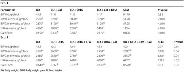
In Exp. 1, there was no difference in the body weight gain (BWG) or feed intake of chicks among the 4 treatment diets at week 3 (Additional file 1:  Table S5). Compared with those fed BD, birds fed BD+Cal had 11% higher (P< 0.05) BWG, and 7% higher (P< 0.05) gain:feed ratio at week 6 (Table 1). Although birds fed BD+DHA had 6% higher BWG and 8% higher feed intake (8%) than those fed BD, the differences were not statistically significant. Moreover, birds fed BD+Cal+DHA also showed nonsignificantly higher BWG (9%) and feed intake (12%) compared with birds fed BD at week 6 (Table 1).
In Exp. 2, birds fed BD+DHA+EPA had 17%–27% higher BWG and 6%–25% higher gain:feed ratio than those fed the other diets at week 3, but the differences were not statistically significant (Additional file 1: Table S5). Compared with birds fed the BD, birds fed BD+DHA+EPA had 10% higher (P< 0.05) BWG and 14% higher (P< 0.05) feed intake at week 6 (Table 1). Birds fed the BD+DHA diet had 3% higher BWG and gain:feed ratio, compared with birds fed BD, but the differences were not statistically significant (Table 1). Moreover, birds fed BD+DHA+EPA+Cal had 15% higher (P< 0.05) feed intake, compared with those fed BD. No difference in the BWG, feed intake, or gain:feed ratio was shown in birds fed BD+DHA+Cal, compared with those fed BD (Table 1).
Plasma and tissue lipid profiles
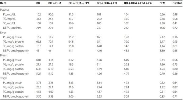
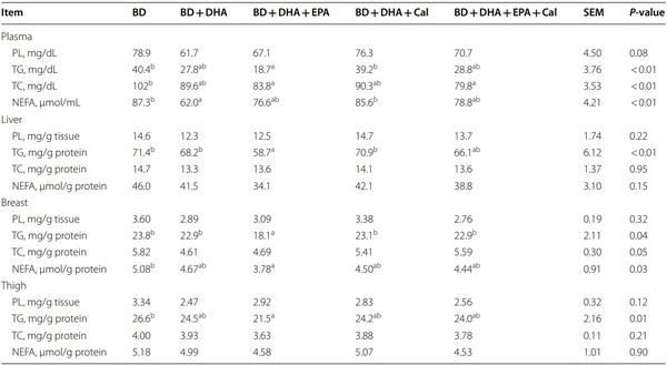
In Exp. 1, there was no difference in concentrations of plasma TGs, TC, and NEFAs among all 4 diets at week 3 (Table 2). At week 6, birds fed BD+Cal had 22% and 29% lower (P< 0.05) plasma TC concentrations compared with birds fed BD+DHA or BD alone, respectively. In Exp. 2, diets produced no significant effects on any of the lipid profiles in plasma or tissues at week 3 (Table 3). However, at week 6, birds fed BD+DHA+EPA had lower (P< 0.05) concentrations of TGs (plasma 54%; liver 18%; breast 24%; thigh 19%), TC (plasma 18%; liver 8%; breast 19%; thigh 9%), and NEFA (plasma 12%; liver 26%; breast 26%; thigh 13%), than birds fed BD, respectively (Table 4).
Table 1 Effects of supplementation of calcidiol, DHA-rich microalgal biomass or oil, and EPA-rich microalgal biomass on growth performance of broiler chickens in Exp. 1 and 2 (0–6 weeks)
Supranutrition of microalgal docosahexaenoic acid and calcidiol improved growth performance, tissue lipid profles, and tibia characteristics of broiler chickens - Image 1
BW Body weight, BWG Body weight gain, FI: Feed intake
Exp. 1; BD=Corn-soybean basal diet; BD+Cal=BD+10,000 IU calcidiol/kg of diet; BD+DHA=BD+1% DHA-rich microalgal biomass; BD+Cal+DHA=BD+Cal+1% DHA-rich microalgal biomass
Exp. 2; 0–3 weeks: BD=Corn-soybean basal diet; BD+DHA=BD+1.5 g of DHA oil/kg; BD+DHA+EPA=BD+DHA+0.3 g/kg Nannochloropsis sp. CO18; BD+DHA+Cal=BD+DHA+6000 IU calcidiol/kg; BD+DHA+EPA+Cal=BD+DHA+EPA (0.3 g/kg Nannochloropsis sp. CO18)+6000 IU calcidiol/kg
Exp. 2; 4–6 weeks: BD=Corn-soybean basal diet; BD+DHA=BD+3.0 g DHA oil/kg; BD+DHA+EPA=BD+DHA+0.6 g/kg Nannochloropsis sp. CO18; BD+DHA+Cal=BD+DHA+12,000 IU calcidiol/kg; BD+DHA+EPA+Cal=BD+DHA+EPA (0.6 g/kg Nannochloropsis sp. CO18)+12,000 IU calcidiol/kg
a,b Means bearing the different superscripts in a row differ (P< 0.05)
Data are expressed as means (n=6 cages and 6 birds/cage) and were analyzed by two-way and one-way ANOVA in Exp. 1 and 2, respectively
Table 2 effects of supplementation of calcidiol and DHA-rich microalgal biomass on plasma lipid profile of broiler chickens in Exp. 1
Supranutrition of microalgal docosahexaenoic acid and calcidiol improved growth performance, tissue lipid profles, and tibia characteristics of broiler chickens - Image 2
NEFA Non-esterified fatty acids, TC Total cholesterol, TG Triglycerides
BD=Corn-soybean basal diet; BD+Cal=BD+10,000 IU calcidiol/kg of diet; BD+DHA=BD+1% DHA-rich microalgal biomass; BD+Cal+DHA=BD+Cal+1% DHA-rich microalgal biomass 
a,b Means bearing the different superscripts in a row differ (P< 0.05)
Values are expressed as means of 6 birds/treatment and data were analyzed using two-way ANOVA
Tibia bone health
In Exp. 1, tibia from birds fed BD+Cal had 8%–24% greater (P< 0.05) breaking strength than that of birds fed the other diets at week 6 (Table 5). However, there was no difference in other measured variables among the 4 treatment diets. In Exp. 2, birds fed BD+DHA+Cal had 19% higher (P< 0.05) BMD than birds fed BD+DHA diet and 13% higher (P< 0.05) total bone volume compared with birds fed BD and BD+DHA diets at week 6 (Table 6). Diets produced no significant effects on other measured variables including cortical BMD, cortical percentage bone volume, trabecular BMD, trabecular percentage bone volume, trabecular thickness, trabecular separation, or degree of anisotropy.
Table 3 effects of supplementation of DHA-rich microalgal oil, EPA-rich microalgal biomass, and calcidiol on plasma and tissue lipid profiles of broiler chickens at week 3 in Exp. 2
Supranutrition of microalgal docosahexaenoic acid and calcidiol improved growth performance, tissue lipid profles, and tibia characteristics of broiler chickens - Image 3
BD=Corn-soybean basal diet; BD+DHA=BD+1.5 g of DHA oil/kg; BD+DHA+EPA=BD+DHA+0.3 g/kg Nannochloropsis sp. CO18; BD+DHA+Cal=BD+DHA+6000 IU calcidiol/kg; BD+DHA+EPA+Cal=BD+DHA+EPA (0.3 g/kg Nannochloropsis sp. CO18)+6000 IU calcidiol/kg
Values are expressed as means of 6 birds/treatment and data were analyzed using one-way ANOVA
Discussion
Two consecutive experiments were conducted in the present study to determine responses of growth performance, tissue lipid profiles, and bone characteristics of broiler chickens to supranutrition of DHA and calcidiol during the full starter and grower periods. The sources and concentrations of these two nutrients were chosen based on our previous findings [9, 11] and literature [12, 13] to enrich them in the chicken tissues singularly or together. Indeed, supplementing these two nutrients into diets at the doses used in the present study led to 4–19- fold increases in DHA and 44%–52% increase in calcidiol contents of the liver, breast, and thigh (Kalia et al., manuscript in preparation). The most relevant finding for the broiler industry is that the high dietary supplemental levels of DHA and calcidiol exerted no negative effects on growth performance [8, 21], tissue lipid profiles, or bone characteristics. In contrast, those high supplementations actually exhibited moderate benefits to several measures. To our best knowledge, this was the first attempt to evaluate such effects of the two bioactive nutrients supplemented simultaneously at higher doses, although prior trials determined the effects of calcidiol [3, 4] and DHArich microalgal biomass or product [6, 22] singularly.
The enhanced BW, BWG, and gain:feed ratio by feeding birds with BD+Cal over BD in Exp. 1 were consistent with the reported improvements in body weight and feed efficiency resulted from feeding high doses of calcidiol to broiler chickens [13, 23] and breeders [24]. Interesting, birds fed BD+DHA+Cal had an increased feed intake and numerical improvements in BW and BWG. In Exp. 2, supplementing EPA into the BD+DHA diet further improved BW, BWG, and FI of broilers to be significantly higher than those of birds fed BD. In contrast, supplementing EPA into the BD+DHA+Cal diet improved only FI. This implies a potential benefit of supplemental EPA and a unique interaction between EPA and DHA and calcidiol on growth performance of chickens [25–27]. Long et al. [5] and Ribeiro et al. [28] reported that high doses of n-3 fatty acids in broiler diets improved BWG and feed conversion ratio. However, supplementing calcidiol alone decreased feed intake of chickens in two studies [13, 29]. Meanwhile, a number of studies indicated that broiler chickens or breeders responded well to dietary supplementations of 1400 to 2800  IU of cholecalciferol/kg diet [3, 4, 30] and 0.55 to 2.55  g DHA/kg diet [5–7]. Notably, Yarger et al. [8] found no evidence of renal calcification caused by supplemental 27,600  IU of calcidiol/kg, suggesting that our supplementation doses of calcidiol (6000 to 12,000  IU/kg) were within the safe range. Intriguingly, feeding broilers with 5520  IU of calcidiol/kg increased breast meat yield [31].
Table 4 effects of supplementation of DHA-rich microalgal oil, EPA-rich microalgal biomass, and calcidiol on plasma and tissue lipid profiles of broiler chickens at week 6 in Exp. 2
Supranutrition of microalgal docosahexaenoic acid and calcidiol improved growth performance, tissue lipid profles, and tibia characteristics of broiler chickens - Image 4
0–3 weeks: BD=Corn-soybean basal diet; BD+DHA=BD+1.5 g of DHA oil/kg; BD+DHA+EPA=BD+DHA+0.3 g/kg Nannochloropsis sp. CO18; BD+DHA+Cal=BD+DHA+6000 IU calcidiol/kg; BD+DHA+EPA+Cal=BD+DHA+EPA (0.3 g/kg Nannochloropsis sp. CO18)+6000 IU calcidiol/kg
4–6 weeks: BD=Corn-soybean basal diet; BD+DHA=BD+3.0 g DHA oil/kg; BD+DHA+EPA=BD+DHA+0.6 g/kg Nannochloropsis sp. CO18; BD+DHA+Cal=BD+DHA+12,000 IU calcidiol/kg; BD+DHA+EPA+Cal=BD+DHA+EPA (0.6 g/kg Nannochloropsis sp. CO18)+12,000 IU calcidiol/kg
a,b Means bearing the different superscripts in a row differ (P< 0.05)
Values are expressed as means of 6 birds/treatment and data were analyzed using one-way ANOVA
Table 5 effects of supplementation of calcidiol and DHA-rich microalgal biomass on tibia bone properties of broiler chickens at week 6 in Exp. 1
Supranutrition of microalgal docosahexaenoic acid and calcidiol improved growth performance, tissue lipid profles, and tibia characteristics of broiler chickens - Image 5
BD = Corn-soybean basal diet, BD+Cal = BD+10,000 IU calcidiol/kg of diet, BD+DHA = BD+1% DHA-rich microalgal biomass, BD+Cal+DHA = BD+Cal+1% DHA-rich microalgal biomass
a,b Means bearing the different superscripts in a row differ (P< 0.05)
Values are expressed as means of 6 birds/treatment and data were analyzed using two-way ANOVA
Because DHA and calcidiol are fat-soluble, bioactive nutrients that play major roles in regulating lipid metabolism [32, 33], we determined how the supranutrition of them intended for their enrichments in the chicken affected lipid profiles of plasma and tissues of chickens.
Table 6 effects of supplementation of DHA-rich microalgal oil, EPA-rich microalgal biomass, and calcidiol on tibia bone properties of broiler chickens at week 6 in Exp. 2
Supranutrition of microalgal docosahexaenoic acid and calcidiol improved growth performance, tissue lipid profles, and tibia characteristics of broiler chickens - Image 6
BMD Bone mineral density
0–3 weeks: BD=Corn-soybean basal diet; BD+DHA=BD+1.5 g of DHA oil/kg; BD+DHA+EPA=BD+DHA+0.3 g/kg Nannochloropsis sp. CO18; BD+DHA+Cal=BD+DHA+6000 IU calcidiol/kg; BD+DHA+EPA+Cal=BD+DHA+EPA (0.3 g/kg Nannochloropsis sp. CO18)+6000 IU calcidiol/kg
4–6 Weeks: BD=Corn-soybean basal diet; BD+DHA=BD+3.0 g DHA oil/kg; BD+DHA+EPA=BD+DHA+0.6 g/kg Nannochloropsis sp. CO18; BD+DHA+Cal=BD+DHA+12,000 IU calcidiol/kg; BD+DHA+EPA+Cal=BD+DHA+EPA (0.6 g/kg Nannochloropsis sp. CO18)+12,000 IU calcidiol/kg
a,b Means bearing the different superscripts in a row differ (P< 0.05)
Values are expressed as means of 6 birds/treatment and data were analyzed using one-way ANOVA
Such effects will have both animal and human health implications. Whereas substantial changes in the plasma and tissue lipid profiles may reflect a metabolic shift to impair overall health status of broilers, the decreases in lipid contents of the tissues (muscles) may render chicken a more desired animal-sourced protein for human nutrition to reduce risks of chronic diseases. In Exp. 1, birds fed BD+Cal had lower plasma TC concentrations than those fed BD at week 6. This decrease agreed with the reported reduced plasma cholesterol levels in rodents fed high doses of calcidiol [34, 35]. Because 3-hydroxy3-methylglutaryl coenzyme A (HMG-CoA) reductase catalyzes the major rate-limiting step in cholesterol biosynthesis [34], these supplemental calcidiol-mediated cholesterol decreases might be due to an inhibition of the enzyme by calcidiol [34, 35]. In Exp. 2, supplementing EPA into the BD+DHA diet, compared with BD, caused consistent decreases of concentrations of TGs in the plasma, liver, and breast and thigh muscles, concentrations of TC in the plasma, and concentrations of NEFAs in the breast muscle at week 6. At the same time, supplementing DHA into BD decreased plasma concentrations of NEFAs and the combined supplementations of DHA, EPA, and calcidiol decreased plasma concentrations of TC compared with BD. Previous studies [5, 36] showed hypocholesterolemic and hypotriacylglycerolemic effects of EPA and DHA via inhibiting squalene epoxidase enzyme [36, 37] and reducing very low-density lipoprotein (VLDL) synthesis and secretion [5, 38, 39], respectively. These regulations may be used to explain the decreases of TGs and TC by supplemental DHA and EPA in our study. The lack of any treatment effects on plasma and tissue lipid profiles of chickens at week 3 suggests that a longer time than 3 weeks was required for the dietary supplementations of DHA, EPA, and calcidiol to alter lipid metabolism in the broilers.
It is practically relevant to show an elevated energy at maximum load of the tibia bone from chickens fed BD+Cal compared with those fed BD in Exp. 1. This elevation implies that adding extra 10,000  IU of calcidiol/kg into the BD contained 300  IU of vitamin D3 improved tibia bone strength. Improvements in tibia bone strength and bone volume were produced by supplementing calcidiol at 2760 IU/kg [40] and 12,000 IU/kg [13], respectively, in the poultry diet. It will be interesting to fnd out if these improvements are still detectable when 10,000 to 12,000  IU calcidiol/kg is added to commercial diets supplemented with > 2000 IU of vitamin D3/kg. The lack of supplemental DHA alone or with calcidiol effect on tibia bone characteristics in Exp. 1 are consistent with outcomes of DHA supplementation on bone structural integrity and strength in several earlier studies [41–43]. In Experiment 2, supplementing high levels of calcidiol into the BD+DHA diets appeared to improve total bone volume and total BMD, implying synergistic potential of DHA and calcidiol in improving bone mineral metabolism [44, 45] and reducing risk of bone fracture [46]. Because broiler chickens are among the most fast-growing animals and are susceptible to tibial dyschondroplasia (TD) that reduces the stability of leg bones and deteriorates the quality of meat from the legs [3], supranutrition of DHA and calcidiol may help not only enrichments of chicken with those nutrients but also prevention of TD and associated losses.
Conclusions
Feeding broiler chickens with supranutritional levels of DHA, either from microalgal biomass or oil, and synthetic calcidiol from day 1 to day 42 of age produced no adverse effects on growth performance, plasma and tissue lipid profiles, or tibia characteristics. Instead, some of these singular or combined supplementations led to moderate beneficial responses of the three types of measures. Supplementing low levels of EPA into the BD+DHA diet or the BD+DHA+Cal diet resulted in rather consistent improvements in a number of those measures. There was a synergistic potential between supranutritions of DHA and calcidiol in improving tibial traits. Overall, it seems to be not only safe but also metabolically beneficial to supplement high levels of dietary DHA and calcidiol, much higher than the nutrient requirements, for biofortifying chicken with these bioactive nutrients.
     
This article was originally published in Journal of Animal Science and Biotechnology (2023) 14:27. https://doi.org/10.1186/s40104-023-00842-3. This is an Open Access article is licensed under a Creative Commons Attribution 4.0 International License.

1. Patel A, Desai SS, Mane VK, Enman J, Rova U, Christakopoulos P, et al.
Futuristic food fortifcation with a balanced ratio of dietary ω-3/ω-6 omega fatty acids for the prevention of lifestyle diseases. Trends Food Sci
Technol. 2022;120:140–53. https://doi.org/10.1016/j.tifs.2022.01.006.
2. Neill HR, Gill CIR, McDonald EJ, McRoberts WC, Pourshahidi K. The future is bright: Biofortifcation of common foods can improve vitamin D status.
Crit Rev Food Sci Nutr. 2021. https://doi.org/10.1080/10408398.2021.1950609.
3. Ledwaba MF, Roberson KD. Efectiveness of twenty-fve-hydroxycholecalciferol in the prevention of tibial dyschondroplasia in ross cockerels depends on dietary calcium level. Poult Sci. 2003;82:1769–77. https://doi. org/10.1093/ps/82.11.1769.
4. Wideman RF, Blankenship J, Pevzner IY, Turner BJ. Efcacy of 25-OH vitamin
D3 prophylactic administration for reducing lameness in broilers grown on wire fooring. Poult Sci. 2015;94:1821–7. https://doi.org/10.3382/ps/pev160.
5. Long SF, Kang S, Wang QQ, Xu YT, Pan L, Hu JX, et al. Dietary supplementation with DHA-rich microalgae improves performance, serum composition, carcass trait, antioxidant status, and fatty acid profle of broilers.
Poult Sci. 2018;97:1881–90. https://doi.org/10.3382/ps/pey027.
6. Moran CA, Keegan JD, Vienola K, Apajalahti J. Broiler tissue enrichment with docosahexaenoic acid (DHA) through dietary supplementation with aurantiochytrium limacinum algae. Food Nut Sci. 2018;9;1160–73. https:// doi.org/10.4236/fns.2018.910084.
7. Khan IA, Parker NB, Lohr CV, Cherian G. Docosahexaenoic acid (22:6 n-3)-rich microalgae along with methionine supplementation in broiler chickens: efects on production performance, breast muscle quality attributes, lipid profle, and incidence of white striping and myopathy.
Poult Sci. 2021;11:865–74. https://doi.org/10.1016/j.psj.2020.10.069.
8. Yarger JG, Quarles CL, Hollis BW, Gray RW. Safety of 25-hydroxycholecalciferol as a source of cholecalciferol in poultry rations. Poult Sci.
1995;74:1437–46. https://doi.org/10.3382/ps.0741437.
9. Sun T, Tolba SA, Magnuson AD, Lei XG. Excessive Aurantiochytrium acetophilum docosahexaenoic acid supplementation decreases growth performance and breast muscle mass of broiler chickens. Algal Res.
2022;63:102648. https://doi.org/10.1016/j.algal.2022.102648.
10. Gatrell S, Lum K, Kim J, Lei XG. Potential of defatted microalgae from the biofuel industry as an ingredient to replace corn and soybean meal in swine and poultry diets. J Anim Sci. 2014;92:1306–14. https://doi.org/10.
2527/jas.2013-7250.
11. Tolba SA, Sun T, Magnuson AD, Liu GC, Abdel-Razik WM, Gamal MF, et al.
Supplemental docosahexaenoic-acid-enriched microalgae afected fatty acid and metabolic profles and related gene expression in several tissues of broiler chicks. J Agric Food Chem. 2019;67:6497–507. https://doi.org/
10.1021/acs.jafc.9b00629.
12. Adhikari R, White D, House JD, Kim WK. Efects of additional dosage of vitamin D3, vitamin D2, and 25-hydroxyvitamin D3 on calcium and phosphorus utilization, egg quality and bone mineralization in laying hens.
Poult Sci. 2021;99:364–73.
13. El-Safty SA, Galal A, El-Gendi GM, El-Azeem NAB, Ghazaly MA, Abdelhady
AYM. Efect of 25-hydroxyvitamin D supplementation, ultraviolet light and their interaction on productive performance, bone characteristics, and some behavioral aspects of broiler chicks. Ann Agric Sci. 2022;67:72–
8. https://doi.org/10.1016/j.aoas.2022.05.002.
14. National Research Council. Nutrient Requirements of Poultry. 9th rev. ed.
Washington: National Academy Press; 1994.
15. Cachaldora P, Garcia-Rebollar P, Alvarez C, Blas JCD, Mendez J. Efect of type and level of fsh oil supplementation on yolk fat composition and n-3 fatty acids retention efciency in laying hens. Br Poult Sci.
2006;47:43–9.
16. Feng J, Long S, Zhang HJ, Wu SG, Qi GH, Wang J. Comparative efects of dietary microalgae oil and fsh oil on fatty acid composition and sensory quality of table eggs. Poult Sci. 2020;99:1734–43.
17. Sun T, Yin R, Magnuson AD, Tolba SA, Liu G, Lei XG. Dose-dependent enrichments and improved redox status in tissues of broiler chicks under heat stress by dietary supplemental microalgal astaxanthin. J Agric Food
Chem. 2018;66:5521–30. https://doi.org/10.1021/acs.jafc.8b00860.
18. Magnuson AD, Liu G, Sun T, Tolba SA, Xi L, Whelan R, et al. Supplemental methionine and stocking density afect antioxidant status, fatty acid profles, and growth performance of broiler chickens. J Anim Sci.
2020;98:skaa092. https://doi.org/10.1093/jas/skaa092.
19. Manor ML, Derksen TJ, Magnuson AD, Raja F, Lei XG. Inclusion of dietary defatted microalgae dose-dependently enriches ω-3 fatty acids in egg yolk and tissues of laying hens. J Nutr. 2020;149:942–50. https://doi.org/
10.1093/jn/nxz032.
20. Sharma MK, White D, Chen C, Kim WK, Adhikari P. Efects of the housing environment and laying hen strain on tibia and femur bone properties of diferent laying phases of Hy-Line hens. Poult Sci. 2021;100:100933. https://doi.org/10.1016/j.psj.2020.12.030.
21. Chou SH, Chung TK, Yu B. Efects of supplemental 25-hydroxycholecalciferol on growth performance, small intestinal morphology, and immune response of broiler chickens. Poult Sci. 2009;88:2333–41. https://doi.org/
10.3382/ps.2009-00283.
22. Yan L, Kim IH. Efects of dietary ω-3 fatty acid-enriched microalgae supplementation on growth performance, blood profles, meat quality, and fatty acid composition of meat in broilers. J Appl Anim Res. 2013;4:392–7. https://doi.org/10.1080/09712119.2013.787361.
23. Fritts CA, Waldroup PW. Efect of source and level of vitamin D on live performance and bone development in growing broilers. J Appl Poult
Res. 2003;12:45–52.
24. Atencio A, Edwards HM, Pesti GM. Efect of the level of cholecalciferol supplementation of broiler breeder hen diets on the performance and bone abnormalities of the progeny fed diets containing various levels of calcium or 25-hydroxycholecalciferol. Poult Sci. 2005;84:1593–603. https://doi.org/10.1093/ps/84.10.1593.
25. Wei H-K, Zhou Y, Jiang S, Tao Y-X, Sun H, Peng J, et al. Feeding a DHAenriched diet increases skeletal muscle protein synthesis in growing pigs: association with increased skeletal muscle insulin action and local mRNA expression of insulin-like growth factor 1. Br J Nutr. 2015;110:671–80. https://doi.org/10.1017/S0007114512005740.
26. Betiku OC, Barrows FT, Ross C, Sealey WM. The efect of total replacement of fsh oil with DHA-Gold® and plant oils on growth and fllet quality of rainbow trout (Oncorhynchus mykiss) fed a plant-based diet. Aqua Nutr.
2016;22:158–69. https://doi.org/10.1111/anu.12234.
27. Liu B, Jiang J, Yu D, Lin G, Xiong YL. Efects of supplementation of microalgae (Aurantiochytrium sp.) to laying hen Diets on fatty acid content, health lipid Indices, oxidative stability, and quality attributes of meat.
Food. 2020; https://doi.org/10.5713/ajas.2012.12455.
28. Ribeiro T, Lordelo MM, Alves SP, Bessa RJB, Costa P, Lemos JPC, et al.
Direct supplementation of diet is the most efcient way of enriching broiler meat with n-3 long-chain polyunsaturated fatty acids. Br Poult Sci.
2013;54:753–65.
29. Garcia AFQM, Murakami AE, Duarte CRA, Rojas ICV, Picoli KP, Puzotti
MM. Use of vitamin D3 and its metabolites in broiler chicken feed on performance, bone parameters, and meat quality. Asian-Aust J Anim Sci.
2013;26:408–15. https://doi.org/10.5713/ajas.2012.12455.
30. Atencio A, Edwards HM Jr, Pesti GM, Ware GO. The vitamin D3 requirements of broiler breeders. Poult Sci. 2006;85:674–92. https://doi.org/10.
1093/ps/85.4.674.
31. Vignale K, Greene ES, Caldas JV, England JA, Boonsinchai N, Sodsee P, et al. 25-hydroxycholecalciferol enhances male broiler breast meat yield through the mTOR pathway. J Nutr. 2015;145:855–63. https://doi.org/10.
3945/jn.114.207936.
32. Mansoori A, Sotoudeh G, Djalali M, Eshraghian MR, Keramatipour M,
Nasli-Esfahani E, et al. Efect of DHA-rich fsh oil on PPARγ target genes related to lipid metabolism in type 2 diabetes: A randomized, doubleblind, placebo-controlled clinical trial. J Clin Lipidol. 2015;9:770–7. https://doi.org/10.1016/j.jacl.2015.08.007.
33. Surdu AM, Pinzariu O, Ciobanu DM, Negru AG, Cainap SS, Lazea C, et al.
Vitamin D and its role in the lipid metabolism and the development of atherosclerosis. Biomedicines. 2021;9:172. https://doi.org/10.3390/biome dicines9020172.
34. Gupta AK, Sexton RC, Rudney H. Efect of vitamin D3 derivatives on cholesterol synthesis and HMG-CoA reductase activity in cultured cells.
J Lipid Res. 1989;30:379–86.
35. Quach HP, Dzekic T, Bukuroshi P, Pang KS. Potencies of vitamin D analogs,
1α-hydroxyvitamin D3, 1α-hydroxyvitamin D2 and 25-hydroxyvitamin D3, in lowering cholesterol in hypercholesterolemic mice in vivo. Biopharm
Drug Dispos. 2018;39:196–204. https://doi.org/10.1002/bdd.2126.
36. Froyland L, Vaagenes H, Asiedu DK, Garras A, Lie O, Berge RK. Chronic administration of eicosapentaenoic acid and docosahexaenoic acid as ethyl esters reduced plasma cholesterol and changed the fatty acid composition in rat blood and organs. Lipids. 1996;31:169–78. https://doi. org/10.1007/BF02522617.
37. Bahety P, Nguyen THV, Hong Y, Zhang L, Chan ECY, Ee PLR. Understanding the cholesterol metabolism-perturbing efects of docosahexaenoic acid by gas chromatography-mass spectrometry targeted metabonomic profling. Eur J Nutr. 2017;56:29–43. https://doi.org/10.1007/s00394-015-1053-4.
38. Grimsgaard S, Bonaa H, Hansen J-B, Nordoy A. Highly purifed eicosapentaenoic acid and docosahexaenoic acid in humans have similar triacylglycerol-lowering efects but divergent efects on serum fatty acids.
Am J Clin Nutr. 1997;66:649–59. https://doi.org/10.1093/ajcn/66.3.649.
39. Bernstein AM, Ding EL, Willett WC, Rimm EB. A meta-analysis shows that docosahexaenoic acid from algal oil reduces serum triglycerides and increases HDL-Cholesterol and LDL-cholesterol in persons without coronary heart disease. J Nutr. 2011;142:99–104. https://doi.org/10.3945/jn.111.148973.
40. Chen C, Turner B, Applegate TJ, Litta G, Kim WK. Role of long-term supplementation of 25-hydroxyvitamin D3 on laying hen bone 3-dimensional structural development. Poult Sci. 2020;99:5771–82. https://doi.org/10.
1016/j.psj.2020.06.080.
41. Sirois I, Cheung AM, Ward WE. Biomechanical bone strength and bone mass in young male and female rats fed a fsh oil diet. Prostaglandins
Leukot Essent Fatty Acid. 2003;68:415–21. https://doi.org/10.1016/s0952-
3278(03)00066-8.
42. Damsgaard CT, Molgaard C, Matthiessen J, Gyldenlove SN, Lauritzen L.
The efects of n-3 long-chain polyunsaturated fatty acids on bone formation and growth factors in adolescent boys. Pediatric Res. 2012;71:713–9. https://doi.org/10.1038/pr.2012.28.
43. Anez-Bustillos L, Cowan E, Cubria MB, Villa-Camacho JC, Mohamadi A, Dao
DT, et al. Efects of dietary omega-3 fatty acids on bones of healthy mice.
Clin Nutr. 2019;38:2145–54. https://doi.org/10.1016/j.clnu.2018.08.036.
44. Zhao B, Nemere I. 1,25(OH)2D3-mediated phosphate up-take in isolated chick intestinal cells: efect of 24,25(OH)2D3, signal transduction activators, and age.
J Cell Biochem. 2002;86:497–508. https://doi.org/10.1002/jcb.10246.
45. Bar A. Calcium homeostasis and vitamin D metabolism and expression in strongly calcifying laying birds. Comp Biochem Physiol A Mol Integr
Physiol. 2008;151:477–90. https://doi.org/10.1016/j.cbpa.2008.07.006.
46. Ammann P, Rizzoli R. Bone strength and its determinants. Osteoporos Int.
2003;14:13–8. https://doi.org/10.1007/s00198-002-1345-4.

Related topics:
Authors:
Dr. Sahil Kalia, Ph.D.
Dr. Woo Kyun Kim
University of Georgia
Recommend
Comment
Share
Profile picture
Would you like to discuss another topic? Create a new post to engage with experts in the community.
Join Engormix and be part of the largest agribusiness social network in the world.