Introduction
Ileitis (proliferative enteropathy of pigs), caused by the obligate intracellular parasite, the bacterium Lawsonia intracellularis, is the most common intestinal infection worldwide, affecting piglets in the rearing and fattening pigs. Arnold et al. (2018) detected a whole group of pathological changes in the small intestine. The disease occurs in four different forms, namely: regional ileitis, intestinal adenomatosis, hemorrhagic enteropathy, necrotic enteritis. It is characterized by intestinal disorders and progressive weight loss in pigs (Lawson et al., 2000; McOrist et al., 2006; Karuppannan & Opriessnig, 2018; Leite et al., 2019). In general, Ileitis has a negative effect on pork’s production, by reducing the average daily weight gain, feeding efficiency and deteriorating of the intestines function of the animals (Kukushkin et al., 2012; Yermolenko, 2019; Jacobset al., 2019; Visscher et al., 2018).
Our studies, conducted on 20 pig farms during 2011-2015confirmed the circulation of the stimulator agent of proliferative enteropathy of pigs, including serologically in IFA among the pig’s population in Ukraine (Yermolenko, 2019). In pig farms, the infection tends to be spread among fattening piglets (28.9%). The source of infection was the main herd (male pigs - 90.0% of seropositive, replacement gilts - 65.9%, sows - 42.1%).According to this, 36.5% of serum had antibodies to Lawsonia intracellularis. In 2019, our scientists conducted a solid-phase blocking ELISA to detect specific antibodies to the pathogen Lawsonia intracellularis (Ayshpur et al., 2019).A total of 653 serum samples of different age pigs groups from 17 domestic farms were studied. 46.4% of seropositive animals were detected. Seropositive pigs were found in all the farms, which were researched, namely, among sows - 74.0%, replacement gilts - 79.6%, piglets for rearing 10.5% and pigs for fattening - 55.3%. These results verified the decease increasing in domestic pig farms, especially during the period of fattening, which caused significant economic losses (Ayshpur et al., 2019).
Ileitis diagnosis is based on clinical and epizootiological analysis, microscopic, serological methods, immunofluorescence method (IFA) and immunoperoxidase monolayer assay (IPMA) for the detection of the antibodies of Lawsonia intracellularis, detection of pathogen DNA by polymerase chain reaction, immunohistochemical analysis (IHC) and histopathological analysis (Bonaet al., 2003; Hardge et al., 2006; Ayshpur et al., 2019; Resendeet al., 2019; Visscheret al., 2018; Guedeset al., 2017; Pérez Gaudioet al., 2018; Yeh& Ga, 2018).
Material and methods
We evaluated the sick, forcibly killed and dead piglets of different ages in Ukrainian pig farms during 2018-2019 in animal fattening groups. Materials were taken from piglets aged 120-150 days in farms where the ileitis was diagnosed. To assess the complex of pathologgical and anatomical changes detected during autopsies of piglets, V.P. Shishkov’s methods were used (Shishkov et al., 1984).
Macro- and microscopic methods of morphological studies were used. Materials from tissues and organs were taken according to the generally accepted histological methods (Lawson et al., 2000). The fragments of the affected areas of intestines (5-7 cm lenght) were taken for the microscopic researches, namely ileum - 10 cm before the drainage of ileocecal valve (initial sample); cecum - 5 cm from the ileocecal branch (second sample); the upper maximum frequency of the colon - 10 cm from the ileocecal branch (third sample).
The detected samples were labeled and marked with the inventory number corresponded to animal and tissue area sampling. The samples were fixed in 10% water buffered formalin solution and filled with paraffinwithout damaging the surface of the mucous membrane. The sections of 3-5 μm were prepared from the created paraffin blocks and painted with hematoxylin and eosin.
Results and discussion
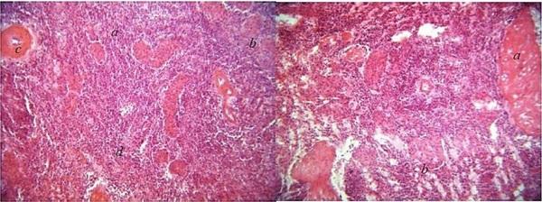
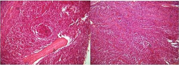
The histological examination of the spleen has shown a slight thickening of the connective tissue capsule and trabeculae extending from it. A deformation of lymph nodes was noted in some parts of the histopreparation. As a result, the barrier between white and red pulps of the spleen was smoothed. Minor activation of follicles were recorded indirectly. The red pulp of the spleen was infiltrated by lymphoid cells (Figures 1a, 1b, 2a, 2b).
All blood vessels, even the smallest capillaries, were moderately dilated, filled with blood cells.
Figure 1A. Capture of the spleen of a piglet aged 140 days; a - red pulp; b - trabeculae; c - a vessel; d - infiltration by lymphoid cells. Hematoxylin and eosin. X 200. B. Capture of the spleen of a piglet aged 150 days; a - red pulp; b - deformed follicles. Hematoxylin and eosin. X 200.
Figure 2A. Capture of the spleen of a piglet aged 14 5 days; a - red pulp; b - trabeculae; c - follicles. Hematoxylin and eosin. X 200. B. Capture of the spleen of a piglet aged 145 days; a – red pulp; b – lymphoid tissue. Hematoxylin and eosin. X 200.
In some areas, the typical to the lungs histostructure was destroyed due to thinning of the interalveolar septa, the lumen of the alveoli was unevenly expanded with thinned and sometimes torn parts, and the blood capillaries were compressed. Such alveoli occupied a significant part of the histopreparation. Often the alveoli became rounded with the irregular shapes. Some alveoli were torn, which caused cavities of different sizes in the lungs. This was typical for alveolar emphysema (Figure 3a). Around large vessels and bronchial tubes these changes were not seen very well. Intensive infiltration of the interstitium of lymphoid-histiocytic cells was registered for some of the cases. In such cases, a desquamation of the bronchial epithelium (desquamative interstitial pneumonia) and infiltration of peribronchial connective tissue by cells of the lymphoid-histiocytic series were detected (Figure 3b-3c).
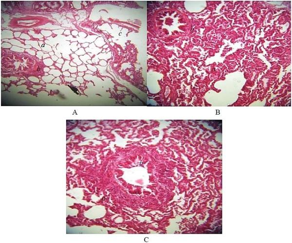
Figure 3A. Fragment of the lungs of piglets aged 140 days; a - alveoli; b - bronchus; c - alveolar emphysema. Hematoxylin and eosin. X 200. B. Fragment of the lungs of piglets aged 150 days; a - alveoli; b - bronchus; c - infiltration of the interstitium by lymphoid-histiocytic cells. Hematoxylin and eosin. X 200.. Fragment of the lungs of piglets aged 140 days; a - alveoli; b - bronchus; c - infiltration of the interstitium by lymphoid-histiocytic cells. Hematoxylin and eosin. X 200.
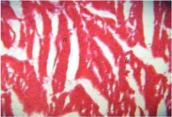
We recorded the most impressive heart changes in the myocardium. On histopreparations of myocardium of piglets, accoding to the elite, we registered some areas with uneven coloration of muscle fibers. Its striation was not expressed. The cardiomyocytes were in a state of granular dystrophy. Small oxyphilic granulars were noticeable in the enlightened protoplasm of such cells. The core of cardiomyocytes were enlarged, with a cigar-shaped, rounded, and a chromatin was sparse. In some places, the absence of the core was rarefied. The lumen of the vessels was moderately expanded and filled with a blood (Figure 4).
Figure 4. Fragment of the myocardium of piglets aged 120 days; a - myocardium; b - cardiomyocytes in a state of granular dystrophy. Hematoxylin and eosin. X 200.
A histological examination of the kidneys revealed the defection of the microscopic structure of the convoluted and part of the straight tubules, while the lumen of some individual tubules was narrowed. In the cortical substance of the kidneys, along with normal tubules with the same colored protoplasm of cells and empty lumens, tubules with turbid swelling and protein grains in the protoplasm of epithelial cells (granular dystrophy) were registered. The lumens of such tubules are mostly closed by swollen epithelium and they contained a fine-grained protein mass. There was a weakly transparent (turbid) mass or fine granularity in the protoplasm of the cells (Figure 5a).
In some tubules, the epithelium was separated from the basement membrane or exfoliated. The lumen of the straight and part of the twisted tubules contained eosinophilic protein mass that consisted of single epitheliocytes. In the cerebral substance, part of the epithelium of the straight tubules was in a state of fatty dystrophy of the infiltrative type. In the protoplasm, parts of epithelial cells of straight tubules were seen, instead of small transparent "vacuoles" - fat droplets. The lumen of such tubules was significantly reduced, and the epithelial cells were swollen (Figure 5b). The mesangium of vascular glomeruli was clearly expressed. The lumen of the Bowman’s capsule cavity was seen. The structure of the vascular glomerulus was kept overall. The vessels of both cortical and cerebral substances were moderately filled with blood cells.
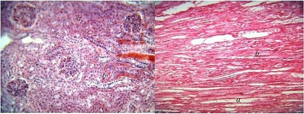
Figure 5A. Fragment of the kidneys of piglets aged 140 days; a - cortical substance; b - turbid swelling and granular dystrophy of tortuous tubules. Hematoxylin and eosin. X 200. B. Fragment of the kidneys of piglets aged 150 days; a - cortical substance; b - fatty infiltration of the epithelium of the straight tubules. Hematoxylin and eosin. X 200.
The histological examination of the liver has shown significant changes in all structural components of the body. The blood vessels of the stroma of the liver were dilated, overloaded with the blood cells (especially veins and venules). In the interparticle connective tissue, especially in the area of hepatic triads, lympho-histiocytic infiltration was observed. We detected the chains of lymphoid cells between the beams, along the course of blood vessels. Central veins and intraparticle capillaries were sharply dilated and filled with blood. The hepatic beams were compressed accordingly. We did not notice the diffuse infiltration of the liver tissue. We observed the protein granular dystrophy in liver cells. The hepatocytes were increased in size due to an increase in the volume of the cytoplasm, which was turbid and dull, and as well as contained different sizes and shapes of protein grains. Dystrophic changes in the cytoplasm of liver cells were accompanied by marked changes in their nuclei. In many cells, the nuclei had sparse chromatin, and acquired an irregular oval-elongated shape. On the periphery, the structure remained normal (Figure 6).
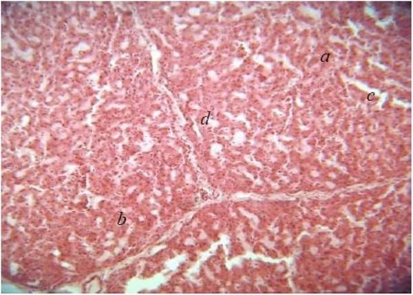
Figure 6. Fragment of the piglets livered aged 150 days; a - the central vein; b - hepatocytes with signs of granular dystrophy; c - dilated sinusoidal capillaries; d - chains of lymphoid substances. Hematoxylin and eosin. X 200.
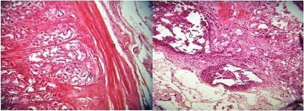
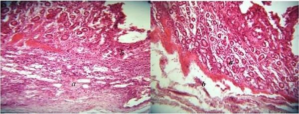
The histological examination of the large intestine revealed desquamation of enterocytes of the apical parts of the cilia and proliferation of enterocytes near the basement membrane and a decrease in the number of goblet cells. Slight thickening of the intestinal wall occurs due to the accumulation of cell proliferates in its own plate of the mucous membrane and intense infiltration of lymphoid cells. These processes occur against the background of hypoxia (vessels are empty, containing only single erythrocytes). Reduced activity of intestinal lymphoid follicles, their centers are enlightened, devastated (Figures 7a and 7b).
Figure 7A. Fragment of the walls of the ileum of a piglet aged 145 days; a - intestinal wall; b - crypts; c - goblet cells; enterocyte proliferation. Hematoxylin and eosin. X 200. B. Fragment of the wall of the colon near the ileocecal opening of piglets aged 125 days; a) - intestinal wall; b) lymphoid follicles; c) exudate. Hematoxylin and eosin. X 200.
Indirectly, along with the germinal layer, it is possible to detect crypts that contain dozens of goblet cells and the formation of single retention cysts in such areas. In such areas, the vessels of the submucosal layer are dilated and contain a moderate number of erythrocytes. In some areas, quite intense infiltration of the connective tissue base by lymphoid cells was registered, or the latter was impregnated with transudate (Figures 8a and 8b).
Figure 8A. Fragment of the wall of the colon near the ileocecal opening of the piglet aged 140 days; a - intestinal wall; b - infiltration of lymphoid cells of the own plate of the mucosa and submucosal layer. Hematoxylin and eosin. X 200. B. Fragment of the wall of the colon near the ileocecal opening of piglets aged 145 days; a - intestinal wall; b - transudation of the submucosal layer of the intestine.
Thus, we revealed a characteristic morphology of proliferative lesions. We believed that the histological examination could serve as an important diagnostic method and an alternative to direct isolation of Lawsonia intracellularis, which is very difficult to cultivate.
Conclusion
We determined that the histopathological changes in pig organs indicated a chronic effect caused by animal pathogen. We revealed that Lawsonia intracellularis caused the profound pathological changes in pig digestive tracts, which affected the functioning of all organs and tissues of animals and lead to severe disease and possible animal death. We suggested that the histological examination is an important step in diagnosing and detecting of profound pathological changes in the organs of sick pigs, which caused the decrease in their productivity and increase the mortality.