Detoxify Aflatoxic Diets of Tilapia Fish
Nutritious Attempts to Detoxify Aflatoxic Diets of Tilapia Fish
There were severe histological alterations in all tested organs (livers, kidneys, intestines and gills, except in the gonads of the treated fish). Dietary ginger inclusion alleviated aflatoxicosis symptoms by fish, since it improved all the above tested parameters of the aflatoxicated fish. Generally, the obtained results in the present study indicated that ginger was the best detoxifying agent of aflatoxin, followed by aspirin and chamomile flowers, res pectively.
Key words: Nile tilapia - Ginger - Aspirin - Chamomile flowers-Aflatoxin
INTRODUCTION
Mycotoxins are secondary metabolites produced by specific filamentous fungi that contaminate agricultural commodities. They are toxic to humans and animals, cause significant reductions in crop yield, and cause economic losses (Gourama and Bullerman, 1995; Gqaleni et al., 1996) and losses worldwide in condemned agricultural products and in animal and human health (CAST, 2003). Long term effects of diets containing aflatoxin are correlated with high incidence of liver disease in certain regions (Abbott, 2002).
The most acutely and chronically toxic member of the aflatoxin family is AFB1. It is the most frequent of all aflatoxins in contaminated food (Kennedy et al., 1998 and Hussein and Brasel, 2001). Aflatoxin B1 is a natural hepatotoxin produced by the ubiquitous fungi Aspergillus flavus and parasiticus. Aflatoxin B1 (AFB1) is known as the most toxic one-it exerts mutagenic, carcinogenic, teratogenic and cytotoxic action. The target organ of AFB1 is especially the liver (Denissenko et al., 1999). Mistry et al. (1996 & 1997) demonstrated that AFB1 stimulated the activity of the rat hepatic phosphatidylinositol kinase and the protein kinase C-the key enzymes in the cell signalling system. The stimulation of the phosphatydylinositol cycle might contribute to the activation of DNA synthesis and evoke the later toxic and carcinogenic effects of AFB1. AFB1 is probably the competitive inhibitor of the cyclic nucleotide phosphodiesterase (PDE). It is believed that changes of cellular cyclic nucleotide levels may be an important way of aflatoxin action (Bonsi et al., 1999).
Aquatic vertebrates of widely divergent taxes are known to suffer toxic effects of dietary AFB1. For example, dietary levels of AFB1 at or below 1 μg/kg have shown to cause liver tumors in rainbow trout (Lovell, 1989). Also it has been reported that sensitivity to these toxins depends on species development temperature. Warm water species are generally less sensitive to AFB1 than cold water species (Lovell, 1989).
Some scientific efforts were conducted to use dietary supplements which detoxify the drastic effects of aflatoxins on some animals such as, glucomannan (Karaman et al., 2005), yeast cell wall mannanoligosaccharide (MOS) (Devegowda et al., 1998), or Saccharomyces cerevisiae which were found to have beneficial effects during mycotoxicosis (Raju and Devegowda 2000), chamomile (Abdelhamid et al., 1985; Soliman and Badeaa 2002 and Ibrahim, 2004), and ginger (Vimala et al., 1999 and Abdelhamid et al., 2002d). Nile tilapia Oreochromis niloticus may represent a sensitive model for mycotoxicosis, since this fish extremely vulnerable to toxic insult from various chemicals and poisons including aflatoxin B1 (AFB1). Therefore, the present work aimed to study the drastic effects of AFB1 on the clinical lesions and postmortem examination, biochemical studies (total protein and esterase (EST) isozyme) and some histological alterations of the experimented fish O. niloticus. Also, this study was conducted to evaluate the ability of some dietary supplements, namely Bio-Buds-2x, chamomile flowers, aspirin and ginger (at a level of 0.5%) to detoxify the drastic effects of this dangerous toxin AFB1 on the Nile tilapia fish for 14 weeks.
MATERIALS AND METHODS
This study was conducted to evaluate the ability of some dietary supplements namely Bio-Buds-2x (T3), chamomile flowers (T4), aspirin (T5), and ginger (T6) (at a level of 0.5%), to detoxify the drastic effects of this dangerous toxin AFB1 on Nile tilapia fish for 14 weeks. A group of 180 mono-sex Nile tilapia O. niloticus fingerlings (obtained from the private fish farm at Tolombat 7, Kafr El-Sheikh), with an average initial body weights of 10g were used in this study. Fish were maintained in the aquaria for one month before the beginning of the experiment for acclimatization purpose. Twelve glass aquaria were used (60×35×40cm) each aquarium was continuously supplied with a compressed air from an electric compressor. Dechlorinated tap water was used to change one third of the water in each aquarium every day. The fish were distributed into the aquaria at stocking rate of 15 fish per aquarium. The experimental treatments were tested in two aquaria for each.
A basal diet (30.38% crude protein, 8.79% ether extract, 4.40% crude fiber, 6.24% ash, 478.4 Kcal/100g DM gross energy and 63.5mg CP/Kcal GE, P/E ratio) was formulated from the local commercial ingredients (fish meal 10%, soybean meal 38%, yellow corn 35.5%, sunflower oil 4%, wheat bran 12% and vit. & min.0.5%). The basal diet was considered as a negative control (T1) whereas the aflatoxic diet (T2) was considered as a positive control. These ingredients were pressed by manufacturing machine (pellets size 1mm), they were milled and AFB1 was added at a concentration of 100ppb to all diets (T2,T3,T4,T5,T6), except the negative control (T1). Each anti-toxin was added at a concentration of 0.5%.The ingredients and supplements were bought from the local market, aflatoxin B1 was produced through pellets fermentation using Aspergillus parasiticus NRRL 2999 according to the method described by Abdelhamid and Mahmoud (1996).
The experiment continued for 14 weeks. During the experimental period the fish were fed the experimental diets at a rate of 3% of the live body weight daily, six days a week. The diet was introduced twice daily, at 8 a.m. and 2 p.m.. The amount of food was adjusted bi-weekly based on the actual body weight changes. Light was controlled by a timer to provide a 14h light: 10h dark as a daily photoperiod.
From 1st week of the experiment and through all the intervals periods, the clinical lesions and postmortem examination of the aflatoxicated fish were recorded by photo camera. However, at the end of the experiment, samples from fish muscles and livers were extracted for biochemical examination according to El-Fadly et al. (1990). Muscles and livers were mixed from six fishes/treatment.
Two hundreds mg of fish muscles or 200 mg of fish liver were homogenized in one ml of sucrose 20% solution for total determination protein according to Laemmli (1970) and isozyme activity according to Ahmed (1994). Also, at the end of the experiment, all fish were sacrificed and the target organs (liver, kidneys, gills, intestine and gonads) were sampled. Samples were fixed in 10% neutralized formalin solution followed by washing with tab water, then dehydrated by different grades of alcohol (70, 85, 96 and 99%). Samples were cleared by xylene and embedded in paraffin wax. The wax blocks were sectioned to six micron. The sections were stained by hematoxyline and eosin and then subjected to a histological examination according to Pearse (1968).
RESULTS AND DISCUSSION
1-The clinical lesions and postmortem examination:
External symptoms and postmortem signs of aflatoxicated O. niloticus fish were recorded from the 1st week and continued at the different experimental intervals, which are presented in the following Figures (1-4).
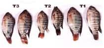
Fig. (1a):From left to right: Aflatoxicated O. niloticus with 100 ppb AFB1 with 0.5% Bio-Buds-2x (T3), aflatoxicated diet without additives (T2) and control group (T1) showing gradual decreases in sizes of aflatoxicated fish comparing with the negative control group.
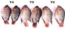
Fig. (1b):From left to right: Aflatoxicated O. niloticus with 100 ppb AFB1 and with 0.5% chamomile flowers (T4), aspirin (T5) and ginger (T6) showing gradual decreases in sizes of aflatoxicated fish between different treatments.
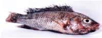
Fig. (2): O. niloticus fed diet containing 100 ppb AFB1 (T2), showing severe discarded scales, and dorsal and caudal fins erosion.
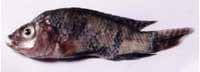
Fig. (3):O. niloticus fed diet containing 100 ppb AFB1 + 0.5% Bio-Buds-2x (T3), showing discarded scales, dark patches (discoloration), caudal fin erosion and abdominal shrinkage.
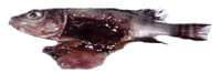
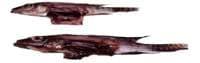
Fig. (4):Dietary aflatoxicated O. niloticus with 100 ppb AFB1 (T2) and with 0.5% chamomile flowers (T4), respectively. Pictures from left to right showing uncharacterized liver and all viscera of fish and the abdomen cavity was filled with heavy amounts of mucus.
These findings of the present work are in agreement with those mentioned by Hussein et al.(2000), Soliman et al. (2000) and Abdelhamid et al. (2002 b). Abdelhamid et al. (2002c) confirmed that dietary inclusion of 0, 500, 1000 and 1500 ppb AFB1 led to a gradual decrease in survival rate in the small fish size (2g weight) by increasing dietary level of AFB1. Also, the authors added that those fed the AFB1 showed (from the 5th week) cloudy eyes (which were pumped thereafter), yellow-greenish infiltrations near the gills and erosion of the caudal fins and the abdomen. Other symptoms appeared as slow-motion, lethargy, less feed acceptability and discarded scales of fish. While the large fish (30 g) reflected fins erosion and discarded scales. Also, Abdelhamid et al.
(2004d) recorded that the aflatoxin- B1 caused severe external clinical lesions (protrusive eyes, abdominal distension, hardening of the body, discarding viscera, fins erosion, discarded scales, hemorrhage, discoloration of skin, abdominal shrinkage, operculum erosion and cataract) and postmortem symptoms (enlarged gall bladder and stomach, distended yellowish liver, viscera covered by a thick layer of mucus and uncharacterized liver and viscera) in the internal organs of the aflatoxicated O. niloticus. These signs were recorded from the 1st week and continued throughout the experiment (8 weeks). Also, they added that all groups fed the diets containing 200 ppb AFB1 with and without the additives died at the end of 4th week of the experiment. In the same trend, Cagauan et al. (2004) reported that AF led to eye opacity, cataract, blindness, lesions on the body surface, fin and tail rot, yellowing of the body surface, abnormal swimming, feeble and stationary on one place, and reduced appetite of Nile tilapia fish.
2- Biochemical studies:
2.1-Muscles total protein:
Figure (5) and Table (1) presented the banding patterns and their molecular weight against standard protein (Ladder, SM0669, Fermentas Life Sciences, 10-200 KDa). As shown from the results obtained, the highest band numbers (14) were detected in the last treatment (T6, 100 ppb AFB1+ 0.5% ginger), the intensity of these bands was also differed. On the other hand, the lowest number (9) was detected in the fourth treatment (100 ppb AFB1 + 0.5% chamomile flowers). In addition, it was noticed that, no differences among the control treatment (T1) and all treatments for bands No. 6, 11, 17, 18 and 21.With regard to bands No. 14 and 19, the results showed that no differences were detected among the control treatment (T1) and all treatments, except in the fourth treatment, since these bands were absent. It was also noticed that the band No.7 was changed from faint to dark in the last two treatments, T5 and T6, while some bands were absent among different treatments, i.e. band No. 8, 9, 13, 15 and 16. Only one band with molecular size < 10 KDa (No. 20) was detected in the best treatment in this experiment, while it was absent in the control (T1) and the rest treatments. However, AFB1 is known as protein depressant (Sahoo and Mukherjee, 2001), since it inhibits RNA/DNA synthesis (Lovell, 1992; Abdelhamid et al., 1998, 2002 a,b&c and 2004 a,b,c&d and Abdelhamid 2000 and 2005). 2.2- Esterases isozyme:
Table (2) and Figure (6) represented the esterases (EST) electrophoretic banding patterns of muscle following the treatments with the tested toxin and toxin with 0.5% of either Bio-Buds-2x, chamomile flowers, aspirin and ginger. All treatments exhibited the same effects in the number of bands since they appeared nine bands, except the treatment T5, which appeared seven bands. In addition, the same effect on the bands intensity was appeared after the treatment with toxin and toxin with 0.5% of Bio-Buds-2x, except bands No.7 and 8 which were altered from dark to very dark. Also, it was noticed that the effect of T5 and T6 treatments on EST isozymes was identical as well as untreated treatment (control, T1), except bands No. 3 and 5 which were absent and bands No. 1 and 4 which were altered from faint and very dark to very faint. The lowest number of bands were detected in the five treatment, since they exhibited seven bands with different intensity. AFB1 was found to be genotoxic, since it affects microsomal cytochrome P-450 isozymes in fish (Carpenter et al., 1995). Different stressors also negatively affected lysozymes activity of different tissues of Nile tilapia (Abdelhamid et al., 2006 a).
Table (1):Effect of aflatoxin B1 on muscle protein of O. niloticus at the end of the experiment.
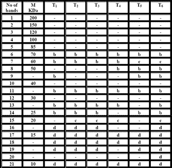
- =Absent a= Very faint b= Faint c= Dark d= Very dark
Table (2):Effect of aflatoxin B1 on muscle EST isozymes of O. niloticus after 14- weeks of treatments
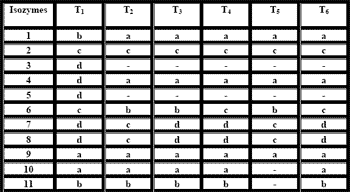
- =Absent a= Very faint b= Faint c= Dark d= Very dark
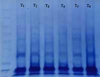
Fig. (5):Effect of aflatoxin B1 (AFB1) on muscle protein of O. niloticus at the end of the 14- weeks experimental feeding.
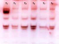
Fig. (6):Effect of aflatoxin B1 on muscle EST isozymes of O. niloticus at the end of the 14- weeks experimental feeding.
3-Histological effects:
3.1- Liver:
The present histopathological findings of AFB1 at 100 ppb and different sources of dietary additives in the different treatments are shown at the following Figures (7-12). The observed pathological effects of AFB1 were nearly similar to those reported by Abdelhamid et al. (2002 c) on big or small sized O. niloticus fish fed diets contaminated with 1500 and 1000 ppb AFB1, respectively. Also, Nguyen et al. (2002) found similar lesions in liver of Nile tilapia fish fed 100 mg AFB1/kg diet for 8 weeks. Hussein et al. (2000) and Soliman et al. (2000) reported similar pathological effects on Nile tilapia fish fed dietary AFB1.
Recently, also Abdelhamid et al. (2004 d) reported that 100 and 200 ppb AFB1 in the O. niloticus fish diets led to severe histological alterations in the liver. These alterations in the liver of the aflatoxicated fish included mild congestion, enlargement and dilatation of the blood vessels with microscopic focci of hemorrhage in the portal lobules, thickening of the lobular endings with basophilic cells, congestion of the bile ducts and widening the adjacent blood sinusoids and regularity within the hepatic lobules and some fibroblast cells were spread within abnormal blood sinusoids. The authors added that these alterations increased by increasing level of aflatoxin B1 (200 ppb AFB1).
It is of interest to note that the experimented additives to diets contaminated with aflatoxin for elimination of the toxic effects reduced the pathological signs in different degrees. Addition of 0.5% ginger (T6) showed the best results, followed by 0.5% aspirin (T5) and 0.5% chamomile flowers (T4). However, adding 0.5% Bio-Buds-2x (T3) failed to improve the toxic effects of aflatoxin on hepatic histogenesis. In this respect, Abdelhamid et al. (2004 d) suggested that using of adsorbent agents (namely 1% egg shell and 2% shrimp wastes) alleviated the adverse effects of AFB1on the histopathological changes in the internal organs of the aflatoxicated fish. The positive effects of these additives (ginger or aspirin) may be due to their chemical and physical properties and/or their positive effects on immune system of fish. Ginger stimulates digestion as it influences positively the terminal enzymes of digestive process (Platel and Srinivasan, 1996 & 2000 and Ahmed and Sharma, 1997). However, aspirin (acetylsalicylic acid) is known to inhibit the cyclooxygenases and enhancement of cellular immune response, or induction of apoptosis (Shiff and Rigas, 1999 and Subongkot et al., 2003).
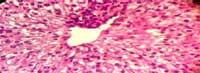
Fig. (7): Section in liver of the control O. niloticus (T1, zero ppb AFB1) showing normal hepatic lobules with normal hepatocytes arrangement around the central vein. (X 460, H&E stains)
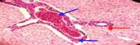
Fig. (8):Section in liver of O. niloticus fed the diet contaminated with 100 ppb AFB1 (T2) showing severe congestion, enlargement of the portal vein with monocytes, fibroblast infiltration (
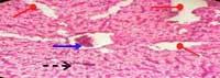
Fig. (9): Section in liver of O. niloticus fed the diet contaminated with 100 ppb AFB1+ 0.5% Bio-Buds-2x (T3) showing mild congestion and infiltration of monocytes within the central vein (
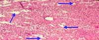
Fig. (10): Section in liver of O. niloticus fed the diet contaminated with 100 ppb AFB1+ 0.5% chamomile flowers (T4) showing higher incidence of small areas of degenerated hepatocytes within the hepatic lobules (
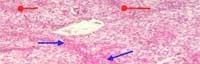
Fig. (11):Section in liver of O. niloticus fed on diet contaminated with 100 ppb AFB1+ 0.5% aspirin (T5) showing normal hepatic lobular architecture with slight necrosis in hepatocytes (
Fig. (12): Section in liver of O. niloticus fed the diet contaminated with 100 ppb AFB1+0.5% Ginger (T6) showing normal hepatic lobules with normal hepatocytes arrangement around the central vein. (X 51.2, H&E stains)
3.2- Kidney:
The histological examination of the kidney in the experimental fish at the different treatments is shown at the following Figures (13-18). The present pathological signs of 100 ppb AFB1 diets were observed too by Abdelhamid et al. (2002 b) on fish fed different levels of AFB1 (500-2000 ppb). Also, Hussein et al. (2000), Soliman et al. (2000) and Abdelhamid et al. (2002 c) found similar findings. Recently, Abdelhamid et al. (2004 d) reported that 100 and 200 ppb AFB1 in the O. niloticus fish diets led to severe histological alterations in the kidney. These alterations included neoplastic signs and periglomerular and peritubular cell infiltration, severe and mild congestion of the glomeruli within the renal cortex. Also, markedly degenerated and interstitial hemorrhage of the epithelium of renal tubules and chronic nephritis were seen. The authors added that these alterations increased by increasing level of aflatoxin B1 (200 ppb AFB1).
Interestingly to note that the changes in histological structure of the kidney were associated with those occurred in the liver of fish in all treated groups. Some histological improvements in the kidney structure were attributed to the dietary additives especially to ginger, which are due to its chemical and physical properties and/or their positive effects on immune system (Platel and Srinivasan, 1996 & 2000 and Ahmed and Sharma, 1997). Also, Abdelhamid et al. (2004 d) mentioned that using of adsorbent agents (namely 1% egg shell and 2% shrimp wastes) alleviated the adverse effects of AFB1 on the histopathological changes in the internal organs of the aflatoxicated fish.
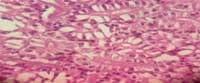
Fig. (13): Cross-section in kidney of the control O. niloticus (T1) showing normal kidney tissue and normal proximal and distal convoluted tubules of the renal cortex. (X 640, H&E stains)
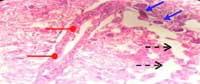
Fig. (14):Cross-section in kidney of O. niloticus fed the diet contaminated with 100 ppb AFB1 (T2) showing renal cortex with normal melpigain corpuscles, degeneration between glomeruli (
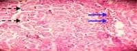
Fig. (15):Cross-section in kidney of O. niloticus fed the diet contaminated with 100 ppb AFB1 + 0.5% Bio-Buds-2x (T3) showing mild congestion of the epithelium lining the adjacent renal tubules (
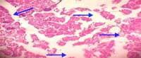
Fig. (16): Cross-section in kidney of O. niloticus fed the diet contaminated with 100 ppb AFB1 + 0.5% chamomile flowers (T4) showing abnormal architecture of the renal tissue and severe degeneration of the epithelium lining the adjacent renal tubules (
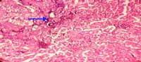
Fig. (17): Cross-section in kidney of O. niloticus fed the diet contaminated with 100 ppb AFB1 + 0.5% aspirin (T5) showing nearly normal architecture of the renal tissue and glomeruli with slight congestion (
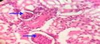
Fig. (18):Cross-section in kidney of O. niloticus fed the diet contaminated with 100 ppb AFB1 + 0.5% ginger (T6) showing normal intact kidney tissue, tubules and intact glomeruli (
3.3- Intestine:
The histological examination in the small intestine in the experimental fish at the different treatments is shown at the following Figures (19-25). The examined lesions in the intestine of fish as affected by AFB1 are nearly similar to those obtained by Kandil et al. (1991) on broiler chicks fed the aflatoxicated diet 100 ppb. However, Abdelhamid et al. (2002 b) found an increase of number of goblet cells and marked inflammatory cellular infiltration with edema in intestine of Nile tilapia fish fed 500-2000 ppb AFB1.The same authors found similar lesions without edema in fish fed aflatoxicated diets with 2 or 4% Biogen®. The recent observation of Abdelhamid et al. (2004 d) on O. niloticus fish fed diets containing 100 or 200 ppb AFB1 included severe histological alterations in intestine. These alterations included abnormal intestinal architecture of mucosa and thickening musculosa layer, absence of mucosa layer and wider and shorter intestinal villi compared with the control. The authors added that these alterations were increased by increasing level of aflatoxin B1 (200 ppb). Moreover, they reported that using adsorbent agents (namely 1% egg shell and 2% shrimp wastes) alleviated the adverse effects of AFB1on the histopathological changes in the intestine of the aflatoxicated fish. The present histopathological findings of 100 ppb AFB1 on the intestine may affect nutrients observation within the intestine, which was associated with marked reduction in growth performance and significant changes in blood parameters of fishes fed AFB1 diets as compared with the control group (Abdelhamid et al., 2006 b). Some histological improvement in the intestine were attributed to the dietary additives, especially to ginger which may be, due to its chemical and physical properties and/or their positive effects on immune system (Platel and Srinivasan, 1996 & 2000 and Ahmed and Sharma, 1997).
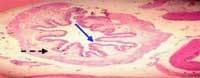
Fig. (19):Cross-section in intestine of O. niloticus fed the control diet (T1) showing intact intestinal layers (
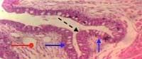
Fig. (20):Magnification of the previous figure (Fig. 19) showing intact and normal lamina epithelialis mucosa (
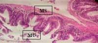
Fig. (21):Cross-section in intestine of O. niloticus fed the contaminated diet with 100 ppb AFB1 (T2) showing abnormal intestinal architecture of tunica mucosa (MU), mild breakdown on the free surfaces of the epithelium cells and thicker of tunica musculosa (MS). (X 160, H&E stains)
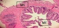
Fig. (22): Cross-section in intestine of O. niloticus fed the contaminated diet with 100 ppb AFB1 + 0.5% Bio-Buds-2x (T3) showing slight abnormality in intestinal architecture of mucosa (MU), with normal structure of the intestinal villi and thicker of tunica musculosa (MS). (X 160, H&E stains)
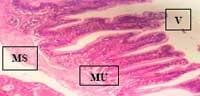
Fig. (23): Cross-section in intestine of O. niloticus fed the contaminated diet with 100 ppb AFB1 + 0.5% chamomile flowers (T4) showing undeveloped intestinal villi (V) with normal tunica musculosa (MS), mucosa (MU) and thicker of tunica submucosa. (X 160, H&E stains)
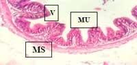
Fig. (24): Cross-section in intestine of O. niloticus fed the contaminated diet with 100 ppb AFB1 + 0.5% aspirin (T5) showing intact mucosa (MU) and musculosa (MS) architecture, and wide and short intestinal villi (V). (X 160, H&E stains)
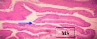
Fig. (25): Cross-section in intestine of O. niloticus fed the contaminated diet with 100 ppb AFB1 + 0.5% ginger (T6) showing developed structure of the intestinal villi (
3.4- Gills:
The histological examination of gills in the experimental fish at the different treatments is shown at the following Figures (26-31). The obtained pathological effects of 100 ppb AFB1 are in agreement with those reported by Abdelhamid et al. (2002 b) on Nile tilapia fed aflatoxicated diets with 500-2000 ppb AFB1. Also, Roberts (1978) and Hussein et al. (2000) found similar results. Effects of addition of Bio-Buds-2x are similar to those obtained by adding 2 or 4 g/kg diet from Biogen® to AFB1 diets (Abdelhamid et al. 2002 b). In the same respect, Abdelhamid et al. (2004 d) reported that 100 and 200 ppb AFB1 in the O. niloticus fish diets led to severe histological alterations in gills. These alterations included congested lamellae and hyperplasia of the lining epithelial layer of the secondary lamellae, severe lesions in term of pronounced degeneration of the secondary lamellae, mild congestion and marked lesions in the epithelial layer lining the lamellae, slight inflammentation within filament interstitium and slight congestion hemorrhage of the epithelial layer. The authors added that these alterations were increased by increasing the level of aflatoxin B1 (200 ppb AFB1) but using adsorbent agents (namely 1% egg shell and 2% shrimp wastes) alleviated the adverse effects of AFB1on the histopathological changes in affected gills of the aflatoxicated fish. Based on these findings, it is of interest to note that the toxic effects of 100 ppb AFB1 on gills of fish decreased by adding 0.5% ginger (T6) and appearently were eliminated by adding 0.5% aspirin (T5) to aflatoxicated diets. The positive effects of these additives may be due to their chemical and physical properties and/or their positive effects on immune system (Platel and Srinivasan, 1996 & 2000 and Ahmed and Sharma, 1997).
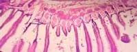
Fig. (26):Cross-section in gills of the control O. niloticus (T1) showing intact architecture of the lamellae. (X 160, H&E stains)
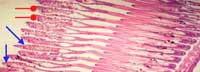
Fig. (27):Cross-section in gills of O. niloticus fed the contaminated diet with 100 ppb AFB1 (T2) showing severe lesions in term of pronounced degeneration of the primary lamellae (
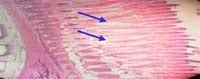
Fig. (28):Cross-section in gills of O. niloticus fed the contaminated diet with 100 ppb AFB1 + 0.5% Bio-Buds-2x (T3) showing mild congestion and marked lesions in the epithelial layer lining the lamellae (
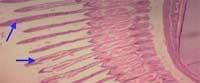
Fig. (29):Cross-section in gills of O. niloticus fed the contaminated diet with 100 ppb AFB1 + 0.5% chamomile flowers (T4) showing desquamation of the epithelial layer, congestion of blood vessels in the primary lamellae (
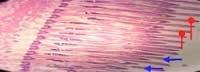
Fig. (30):Cross-section in gills of O. niloticus fed the contaminated diet with 100 ppb AFB1 + 0.5% aspirin (T5) showing normal structure of the primary lamellae (
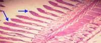
Fig. (31):Cross-section in gills of O. niloticus fed the contaminated diet with 100 ppb AFB1 + 0.5% ginger (T6) showing intact lamella architecture with slight congestion hemorrhage of the epithelial layer of the primary lamellae (
3.5- Gonads:
The histological examination of O. niloticus fish in this experiments is shown at the following Figures (32-34). All examined testes were not affected mainly by the treatment of AFB1 but the major effects on gonads were found to be from the hormonal treatment during producing the mono-sex tilapia fish with 17α- methyl testosterone in fry stage in the commercial fish hatchery. Also, it could be confirmed that from the obtained results it was observed that aflatoxin B1 (AFB1) led to histological changes in all tested organs of fish, except gonads. Generally, addition of aspirin and/or ginger at level 0.5% showed milder lesions on all organs, except the gonads of fish.
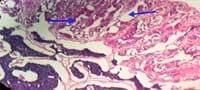
Fig. (32):Section in testis of O. niloticus fed the control diet (T1) showing abnormal structure of gonad which tended to be testis with irregular seminiferous tubules (
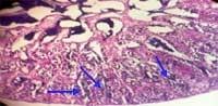
Fig. (33):Section in testis of O. niloticus fed the contaminated diet with 100 ppb AFB1 (T2) showing abnormal architecture of testis and degeneration of seminiferous tubules with depressing some cells intraluminally (
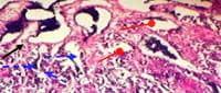
Fig. (34):Section in testis of O. niloticus fed the contaminated diet with 100 ppb AFB1 (T2) showing interrupted and breakdown of basement membrane of the seminiferous tubules (
CONCLUSIONS
From the foregoing results it could be concluded that aflatoxin contamination of fish diets caused many drastic effects on all the tested parameters. Also, AFB1 is very dangerous from the view point of fish production and public health. It could be recommended for the beneficial using of 0.5% ginger and/or 0.5% aspirin is dietary additives to alleviate the toxic effects of AFB1 contaminated fish diets. Also, it is a must to conduct a lot of scientific efforts in this trend to use the medical herbs and other natural agents to detoxify the aflatoxic diets of fish. But, the wisdom still right, that prophylaxis, from toxic effects of mycotoxins especially AFB1, is more useful than treatments.
REFERENCES
Abbott, S.P. (2002). Mycotoxins and Indoor Molds. Indoor Environment CONNECTIONS. 3: 14-24.
Abdelhamid, A. M. (2000). Fungi and Mycotoxins, 1st Ed. Dar Anashr for Universities, Cairo, Deposition No. 13738/97, ISBN: 977/5526180/9, 539p.
Abdelhamid, A. M. (2005). Carcinogens. Dar Alnashr for Universities, Cairo. Deposit No.: 1949/2005, ISBN: 977-316-149-8.
Abdelhamid, A.M. and Mahmoud, K.I. (1996). Elimination or adsorption of aflatoxin from poultry feedstuffs. Proc. Conf. Foodborne contamination & Egyptian’s Health, Mansoura Univ., 26-27 Nov., pp. 61-69.
Abdelhamid, A.M., Abdel- Khalek, A.E., Mehrim, A.I. and Khalil, F.F. (2004 c). An attempt to alleviate aflatoxicosis on Nile tilapia fish by dietary supplementations with chicken-hatchery by-products (egg shells) and shrimp processing wastes (shrimp shells). 1- On fish performance and feed and nutrients utilization. J. Agric. Sci. Mansoura Univ., 29 : 6157 -6173.
Abdelhamid, A.M., Abdel- Khalek, A.E., Mehrim, A.I. and Khalil, F.F. (2004 d). An attempt to alleviate aflatoxicosis on Nile tilapia fish by dietary supplementations with chicken-hatchery by-products (egg shells) and shrimp processing wastes (shrimp shells). 2- On clinical, blood and histological parameters. J. Agric. Sci. Mansoura Univ.,29 : 6175 - 6196.
Abdelhamid, A.M., Ahmed, A.A. and El- Meleigy, Kh.M. (2004 a). An attempt to alleviate the histological alterations of some internal organs of rats fed on aflatoxin contaminated diets. J. Agric. Sci. Mansoura Univ., 29: 2355-2370.
Abdelhamid, A.M., Ahmed A.M. and El-Meleigy Kh.M. (2002 a). Detoxification of aflatoxins – contaminated diet by some physical and chemical means. J. Agric. Sci. Mansoura Univ., 27: 8213 – 8224.
Abdelhamid, A. M., Khalil, F. F. M., El-Barbary, M. I., Zaki, V. H. and Hussein, H. S. (2002 b). Feeding Nile tilpaia on Biogen® to detoxify aflatoxic diets. Proc.1st Conf. Animal & Fish Prod., Mansoura, 24&25, Sept., pp:207-230.
Abdelhamid, A. M., Khalil, F. F. and Ragab, M. A. (1998). Problem of mycotoxins in fish production. Egypt. J. Nutr. and Feeds, 1: 63-71.
Abdelhamid, A. M., Magouz F.I., Salem M.F.E., Mohamed A.A. and Mohsen M.K. (2002 c). Effect of graded levels of aflatoxin B1 on growth performance and biochemical, chromosomal and histological behaviour of Nile tilapia Oreochromis niloticus. Proc.1st Conf. Animal & Fish Prod., Mansoura, 24&25, Sept., pp: 231-250.
Abdelhamid, A. M., Mehrim, A. I. and Khalil, F. F. (2004 b). Detoxification of aflatoxin–contaminated diet of tilapia fish using dietary supplementation with egg shell, Betafin, clay or silica. J. Agric. Sci. Mansoura Univ., 29: 3163-3174.
Abdelhamid, A. M., Nemetallah, B. R., Abd Allah, M. A. and Mousa, T. A. E. (2006 a). Hemalytic activity in blood serum of Oreochromis niloticus under different types of stress. The 3rd Int. Conf. for Develop. and the Env. in the Arab World, March 21-23, Assuit Univ., pp: 153-169.
Abdelhamid, A.M., Salem, M.F.I., Mehrim, A. I. and El-Shaarawy, M.A.M.(2006 b). Nutritious attempts to detoxify aflatoxic diets of tilapia fish: 1-Fish performance, feed and nutrients utilization, organs indices, residues and blood parameters. (Under publication).
Abdelhamid, A.M., Sadik, E.A. and Fayzalla, E.A. (1985). Preserving power of some additives against fungal invasion and mycotoxin production in stored-crushed-corn containing different levels of moisture. Acta Phytopathologica Academiae Scientiarum Hungaricea, 20: 309-320.
Abdelhamid, A. M., Sallam, A. E., Abd Allah, G. A. and El-Samra, S. H. (2002 d). Effect of feeding male rats on aflatoxic diets without or with medicinal herbs (thyme, safflower, ginger, black cumin and/or garlic). Proc. 2nd Conf. Foodborme Contamination and Egyptian’s Health, 23-24 April, El-Mansoura, pp: 99-121.
Ahmed, E. S. E. (1994). The parallel response of enyzme loc in Drosophlia and yeast to the environmental stresses of pollutants. M.Sc. Thesis. Fac. of Agric. Ain shams Univ.
Ahmed, R.S. and Sharma, S.B. (1997). Biochemical studies on combined effects of garlic (Allium sativum Linn) and ginger (Zingiber officinal Rose) in albino rats. Indian J. Exp. Biol., 35: 841.
Bonsi, P., Augusti-Tocco, G., Palmery, M. and Giorgi, M. (1999). Aflatoxin B1 is an inhibitor of cyclic nucleotide phosphodiesterase activity. General Pharmacology, 32: 615–619.
Cagauan, A. G., Tayaban, R. H., Sanga, J. R. and Bartolome, R. M. (2004). Effect of aflatoxin- contaminated feeds in Nile tilapia (Oreochromis niloticus L.). Proc. 6th Inter. Symp. On Tilapia in Aquacultre, Manila, Philippines, Sept. 12-16, P: 172.
Carpenter, H. M., Zhang, Q., El- Zahar, C., Selivonchick, D. P., Brock, D. E. and Curtis, L. R. (1995). In vitro and in vivo temperature modulation of hepatic metabolism and DNA adduction of aflatoxin B1 in rainbow trout. J. Biochm. Toxicol., 10: 1-10.
CAST (Council for Agricultural Science and Technology) (2003). Mycotoxins: risks in plant, animal, and human systems. Vol. Task Force Report 138, Ames, IA, USA, 199 pp.
Denissenko, M.F., Cahill, J., Koudriakova, T.B., Gerber, N. and Pfeifer, G.P., (1999). Quantitation and mapping of aflatoxin B1-induced DNA damage in genomic DNA using aflatoxin B1-8,9-epoxide and microsomal activation systems. Mutation Research, 425: 205–211.
Devegowda, G., Raju, M. V. L. N., Afzali, N., and Swamy, H. V. L. N. (1998). Mycotoxins picture worldwide: Novel solutions for their counteraction. In: T.P. Lyons and K.A. Jacques (Eds.) Biotechnology in the Feed Industry, pp.241-255. Proc. of All Tech's 14th Annual Symposium, Nottingham, U.K.
El-Fadly, G., Sidaros, S. and Dif, A. A. (1990). Effect of boistin on gene expression and yield components of faba bean Vicia faba infected with broad bean strain virus. Proc. 3th Conf. Agric. Dev. Res. Fac. Agric., Ain shams Univ., Cairo, Egypt.
Gourama, N. and Bullerman, L.B., (1995). Aspergillus flavus and Aspergillus parasiticus: Aflatoxigenic fungi of concern in foods and feeds: A review. J. Food Protect., 58: 1395–1404.
Gqaleni, N., Smith, J.E. and Lacey, J. (1996). Co-production of aflatoxins and cyclopiazonic acid in isolates of Aspergillus flavus. Food Additives and Contaminants, 13: 677– 685.
Hussein, H.S. and Brasel, J.M. (2001). Toxicity, metabolism, and impact of mycotoxins on humans and animals. Toxicology, 167: 101–134.
Hussein, S.Y., Mekkawy, I.A.A., Moktar, Z.Z. and Mubarak, M. (2000). Protective effect of Nigella sativa seed against aflatoxicosis in Oreochromis niloticus. Proc. Conf. Mycotoxins and Dioxins and the Environment, Bydgoszcz, 25 – 27 Sept., pp: 109 – 130.
Ibrahim, D.H.E. (2004). Biochemical studies on fungi toxins of feedstuffs in Dakahlia and Damietta governorates. M. Sc. Thesis, Fac. of Agric. Mansoura Univ.
Kandil, W.M., Sirag, S.M., Abdelhamid, A.M. and Dorra, T.M. (1991). Histopathological studies on mycotoxicoses in broiler chicks Mansoura Medical Journal, 21: 193 – 204.
Karaman, M., Basmacioglu, H., Ortatatli, M. and Oguz, H.(2005). Evaluation of the detoxifying effect of yeast glucomannan on aflatoxicosis in broilers as assessed by gross examination and histopathology. Br. Poult. Sci., 46: 394-400.
Kennedy, D., Delaney, K.A. and Koren, G. (1998). Mutagens, carcinogens, and teratogens. In: Goldfrank’s Toxicologic Emergencies, sixth ed. Appleton & Lange, Stamford, CT, pp. 262–273.
Laemmli, U. K. (1970). Clevage of structure protein during assembly of head bacteriophage T4. Nature, 227: 680-685.
Lovell, T. (1989). Nonnutrient diet components. In: Lovell, T. (Ed.), Nutrition and Feedings of Fish. Van Nostrand Reinhold, New York, N.Y., pp. 93– 105.
Lovell, T. (1992). Mycotoxins: Hazardous to farmed fish. Feed International, March, pp: 24 – 28.
Mistry, K.J., Krishna, M. and Bhattacharya, R.K. (1997). Modulation of aflatoxin B1 activated protein kinase C by phenolic compounds. Cancer Letters, 121: 99–104.
Mistry, K.J., Krishna, M., Pasupathy, K., Murthy, V. and Bhattacharya, R.K. (1996). Signal transduction mechanism in response to aflatoxin B1 exposure: protein kinase C activity. Chemico-Biological Interactions, 100: 177–185.
Nguyen, A. T., Grizzle, J. M., Lovell, R. T., Manning, B. B. and Rottinghaus, E. G. (2002). Growth and hepatic lesions of Nile tilapia Oreochromis niloticus fed diets containing aflatoxin B1. Aquaculture, 212: 311-319.
Pearse, G. W. (1968). Histological effects and diagnostic problems of mycotoxins in poultry. Proc. 25th West States Poult. Dis. Conf., pp.76-79.
Platel, K. and Srinivasan, K. (1996). Influence of dietary spices or their active principles on pancreatic digestive enzymes of small intestinal mucosa in rats. Int. J. Food Sci. Nutr., 47: 55.
Platel, K. and Srinivasan, K. (2000). Influence of dietary spices and their active principles on pancreatic digestive enzymes in albino rats. Nahrung, 44: 42.
Raju, M.V. and Devegowda, G. (2000). Influence of esterified- glucomannan on performance and organ morphology, serum biochemistry and haematology in broilers exposed to individual and combined mycotoxicosis (aflatoxin, ochratoxin and T-2 toxin). British Poultry Science, 41: 640-650.
Roberts, R. J. (1978). The pathophysiology and systemic pathology of teleosts. In: Fish Pathology. Ronald J. Roberts (Ed.): 55-91, Balliere Tindall, London.
Sahoo, P.K. and Mukherjee, S. C. (2001). Immunosuppressive effects of aflatoxin B1 in Indian major carp (Labeo rohita). Comparative Immun., Microbi., Infectious Diseases, 24: 143 – 149.
Shiff, S. J. and Rigas, B. (1999). The role of cyclooxygenase inhibition in the antineoplastic effects of nonsteroidal antiinflammatory drugs (NSAIDs). J. Exp. Med., 190: 445–450.
Soliman, K.M. and Badeaa, R.I. (2002). Effect of oil extracted from some medicinal plants on different mycotoxigenic fungi. Food and Chemical Toxicology, 40: 1669-1675.
Soliman, M.K., Khalil, R.H., Youssef, S.A. and Mahfouz, N.M. (2000). Afleatoxicosis among cultured freshwater fish. Abstracts AQUA-2000, Nice-France, May 2 – 6, p: 801.
Subongkot, S., Frame, D., Leslie, W. and Drajer, D. (2003). Selective cyclooxygenase-2 inhibition: A target in cancer prevention and treatment. Pharmacotherapy, 23: 9–28.
Vimala, S., Northanom, A. W. and Yadav, M. (1999). Anti-tumor promoter activity in Malaysian ginger rhizobia used in traditional medicine. Br. J. Cancer, 80: 110.
Authors: Mehrim, A. I.*; A. M. Abdelhamid*, A. A. M. Abo Shosha***, M.F.I. Salem**, M.A.M.M. El-Sharawy*
* Department of Animal Production, Faculty of Agriculture, Mansoura University, Egypt. **Central Laboratory for Aquaculture Research, Kafr El-Shiekh Aquaculture Research Unit, Egypt.
*** Department of Genetics, Faculty of Agriculture, Kafr El-Shiekh University, Egypt.


just want to know if aflotoxins contamination is this common (percentage) and it impact is this important