Case study: pathology and histopathology as a tool for diagnosis of reproductive disorders in pigs related to mycotoxins
Published: March 15, 2022
By: Margarita Trujano
Clinical case
Two commercial farms in Mexico with reproductive disorders were studied. They have a total of 2.000 sows, one farm has 800 and the other 1.200. Abortions at different stages of gestation were observed, both gilts and sows were affected (Fig.1). Different types of antibiotics were used to diminish the clinical signs or abortions but they were not effective, the abortions continued. The abortions did not include mummies but the fetuses and embryos were degenerated, macerated or missed organs. The vaginal discharges present on the animals in the farm were minimal. A great variety of clinical signs and symptoms were noticed in sows (Fig. 2) such as abdominal breathing, epiphora, skin erythema, cyanosis in the abdominal region, several tortuous blood vessels in the abdominal region and in fore- and hindlegs. Another symptom observed in both pregnancy and maternity animals was low or no feed consumption. Some even showed anorexia.
Wet gangrene in the abdominal area was observed in some aborted animals. 70% of sows had either dysgalactia or agalactia. In the farrowing areas piglets were weak, small and almost 50% in each litter showed necrosis of tail in newborn animals (Figure 3). The location of the necrosis varied, some were observed at the top, others at the bottom. The color was dark brown to black depending on the severity. The necrotic tails eventually dried and fell off. Figure 4 shows the grain (sorghum) used in the farm.
These farms had never experienced this kind of pathological event. Blood samples were taken and sent for serological anal-yses. They were negative for PRRS, Parvovirus and Influenza.
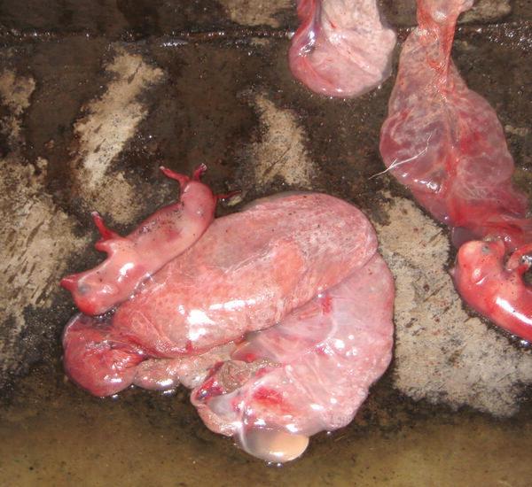
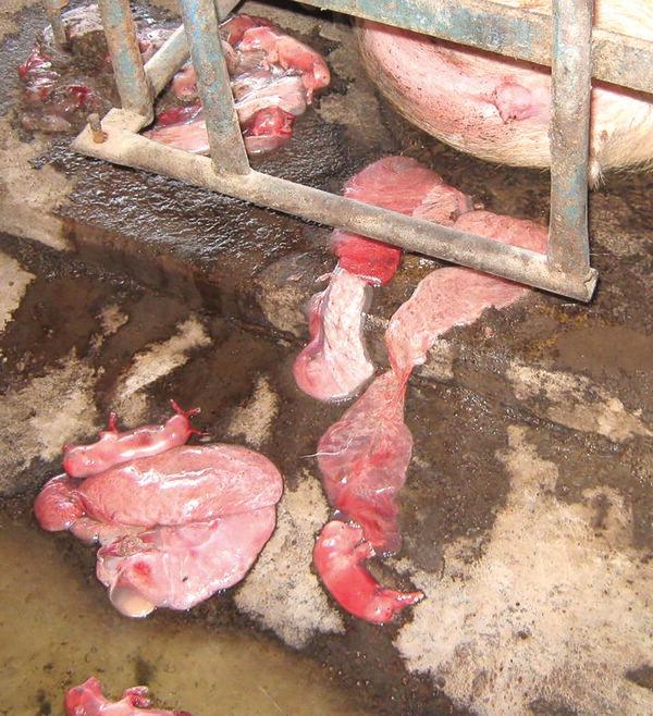
Fig. 1 Characteristics of aborted fetuses.
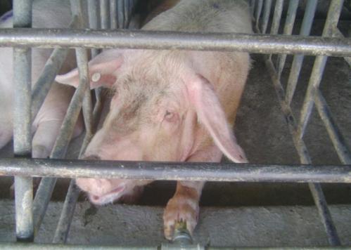
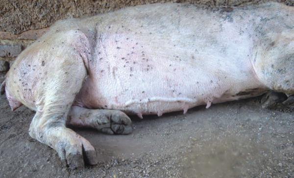
Fig. 2 Sows affected, anorexia and convoluted blood vessels
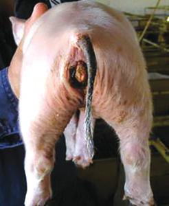
Fig. 3 Tail necrosis observed in newborn piglets
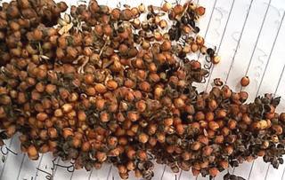
Fig. 4 Sorghum used in the farm
Material & methods
Mycotoxin analysis: determination by enzyme immunoassay (ELISA)
Mycotoxins: aflatoxin B1 (AFLA), ochratoxin A (OTA), deoxynivalenol (DON), T-2 Toxin (T2), fumonisin B1 (FUM), citrinin and zearalenone (ZEA) in gestation feeds
Qualitative determination of ergotoxines (Van Urk technique)
Mycotoxins: aflatoxin B1 (AFLA), ochratoxin A (OTA), deoxynivalenol (DON), T-2 Toxin (T2), fumonisin B1 (FUM), citrinin and zearalenone (ZEA) in gestation feeds
Qualitative determination of ergotoxines (Van Urk technique)
Results
Post-mortem findings in fetuses and sows
Petechial, ecchymosis and bleeding in organs like kidney, heart, liver, spleen, skin, lung, subcutaneous tissue and placenta were observed. Some other lesions included hemorrhagic ascites, subcutaneous edema and reddish amniotic fluid in embryos (Fig. 5).
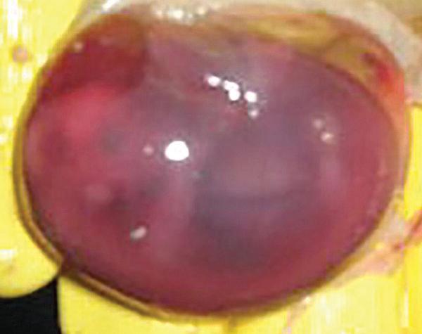
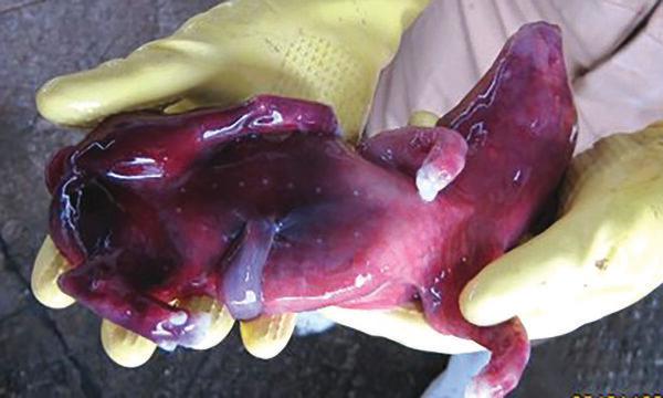
Fig. 5 Lesions observed in aborted fetuses.
Histopathology findings
The lesion most commonly observed in the organs was the presence of thrombi in blood vessels (Fig. 6). Hemorrhagic lesions were observed in all the organs.
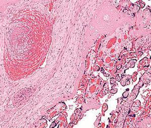
Fig. 6 Thrombi in fetal vessels
Mycotoxins
Tables 1 and 2 show the presence of ergotoxines, ZEA and DON in the feed samples analyzed.
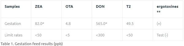
*Methods ELISA (R-Biopharm), above limit
** Van Urk positive reaction to p-dimetil aminobenzaldehido sulfuric from the extracts with ammoniacal ether
** Van Urk positive reaction to p-dimetil aminobenzaldehido sulfuric from the extracts with ammoniacal ether
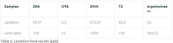
*Methods ELISA (R-Biopharm), above limit
** Van Urk positive reaction to p-dimetil aminobenzaldehido sulfuric from the extracts with ammoniacal ether
** Van Urk positive reaction to p-dimetil aminobenzaldehido sulfuric from the extracts with ammoniacal ether
Discussion
One of the common mycotoxins in our environment but little studied is ergotoxines. In this study both macroscopic and microscopic lesions, as well as the analyses of mycotoxins, verified the presence of ergotoxines. This mycotoxin is important because they are powerful initiators of the contraction of the smooth muscles present in the uterus and in the muscular layer in arteries. These alkaloids simulate in addition the action of dopamine in the central nervous system and inhibit the liberation of prolactin which prevents the development of the mammary gland and disables the lacteal secretion. The cause of abortions can be explained as a result of the vasoconstriction in the arteries which produces ischemia, lack of irrigation and therefore nutrients to the embryos or fetuses. It is interesting that in both feed samples analyzed concentrations of different mycotoxins were found. Considering that mycotoxins are accumulated, the animals are in danger of continuously showing this type of outbreaks since the majority of mycotoxins are immunosuppressive and pass from sows to piglets through maternal milk. Other mycotoxins involved were DON (feed intake refusal) and ZEA. We can assume that at the beginning the animals ate feed with lower levels of these mycotoxins. The feed samples were taken when the problem was at its peak. Ergot related lesions were the ones that had the biggest impact since they culminated in abortions. The feed was removed and a mycotoxin adsorbent was added to to the new feeds in order to prevent challenges in the future.
Conclusion
Mycotoxins should be considered when no response to any kind of treatment is observed during reproductive problems in pigs.
Related topics:
Recommend
Comment
Share


8 de abril de 2022
AFB1 was also quantified but not discussed..Anyways, this exemplifies the need of preventive approach to improve feed quality...
Recommend
Reply

Would you like to discuss another topic? Create a new post to engage with experts in the community.


Featured users in Pig Industry


Francis Simard
Trouw Nutrition
Agr., M. Sc. / Nutrition and Development Director at Trouw Nutrition Canada
United States
United States
