Explore
Communities in English
Advertise on Engormix

Mycoplasma bovis pneumonia in feedlot cattle and dairy calves in Argentina

Published: September 8, 2020
By: Carlos A. Margineda 1,2; Gustavo O. Zielinski 1,2; Susana Jurado 3; Ferrella Alejandra 4; Marina Mozgovoj 4; Ana C. Alcaraz 5; Alfonso López 6.
Summary
Author details:

1 Grupo de Sanidad Animal, Estación Experimental Agropecuaria INTA Marcos Juárez, Córdoba, Argentina; 2 Facultad de Ciencias Veterinarias, Universidad Nacional de Rosario, Casilda, Santa Fe, Argentina; 3 Servicio Central de Microscopía Electrónica, Facultad de Ciencias Veterinarias, Universidad Nacional de La Plata, La Plata, Buenos Aires, Argentina; 4 Instituto de Virología, CICVyA, INTA Castelar, Morón, Buenos Aires, Argentina; 5 College of Veterinary Medicine, Western University of Health Sciences, Pomona, California, USA; 6 Department of Pathology and Microbiology, Atlantic Veterinary College, University of Prince Edward Island, Charlottetown, Prince Edward Island, Canada.


Mycoplasma bovis has emerged as an important cause of feedlot pneumonia in many countries. The aim of this paper is to describe six cases of bovine Mycoplasma pneumonia in five different premises in Argentina. Gross examination revealed chronic bronchopneumonia with multiple foci of caseous necrosis. Microscopically, these contained a necrotic center with abundant hypereosinophilic granular material surrounded by granulation tissue. Affected lung tested positive for M. bovis by immunohistochemistry and electron microscopy revealed membranous structures compatible with Mycoplasma spp. To our knowledge, this is the first report of M. bovis pneumonia in Argentina.

Key words: chronic bronchopneumonia, electron microscopy, feedlot, immunohistochemistry, Mycoplasma bovis.
Introduction
Bovine respiratory disease (BRD) is the most important cause of economic loss of the beef industry in many parts of the world (12, 14, 26). It is a multifactorial condition that involves host and environmental factors, and several viruses and bacteria. The most common viruses associated in cattle with BRD are bovine herpesvirus (BHV-1), bovine parainfluenza virus 3 (BPIV-3), bovine viral diarrhea virus 1 and 2 (BVDV-1/2), bovine respiratory syncytial virus (BRSV) and bovine coronavirus (BCoV) (5, 14, 24). These viral infections are usually transient but predispose to opportunistic secondary bacterial pneumonia (8, 22, 24). Mannheimia haemolytica, Pasteurella multocida, Histophilus somni, and Mycoplasma bovis are the most common bacterial isolates recovered from pneumonic lungs in outbreaks of BRD worldwide (2, 14, 15,24, 25).
For the last 15 years, M. bovis has emerged as one of the most important bacterial pathogens involved in BRD worldwide (9, 23, 26, 29). Although this bacterium is a commensal nasopharyngeal organism in healthy cattle, under certain conditions, it can cause pneumonia, arthritis, synovitis, keratoconjunctivitis, mastitis, metritis, abortion, infertility, otitis and skin abscesses (8, 23). In the lung, M.bovis produces chronic bronchopneumonia characterizedby caseonecrotic nodules in the cranioventral lung lobes (2, 10, 24). Microscopic findings suggest that lung necrosis is originated in small bronchi and bronchioles (15, 18). The persistence of the agent in the lung tissue and the chronicity of the pulmonary lesions imply that the immune response is ineffective in eliminating the infection (6, 18, 21).
Epidemiological studies indicate that the incidence of M. bovis pneumonia has notably increased in many countries such as the United Stated of America, Canada, Mexico, France, Northern Ireland and Italy, particularly in feedlots. (2, 5, 14, 19, 23, 25, 26, 30). In Argentina, M. bovis has never been previously described as a cause of BRD outbreaks or pneumonia.
The objective of this report is to describe six outbreaks of M. bovis pneumonia in feedlot steers and dairy calves in four different herds in the Argentinean pampas.
Materials and methods
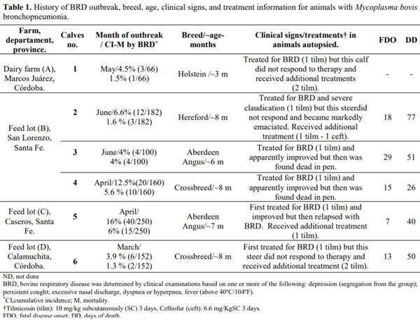
The first outbreak of BRD associated with M.bovis in Argentina occurred in dairy calves at a farm with220 milking cows in May 2012 (Farm A). In 2013, five other outbreaks took place in three different industrial feedlots (Farms B, C, and D) where animals had arrived for fattening from the north and central Provinces of Argentina. Other relevant information collected from the farms and veterinarians included fatal disease onset (FDO), defined as the period between feedlot arrival and first treatment for pneumonia; the type and frequency of antibiotic treatment; and the time interval between feedlot arrival and the day of death (DD).
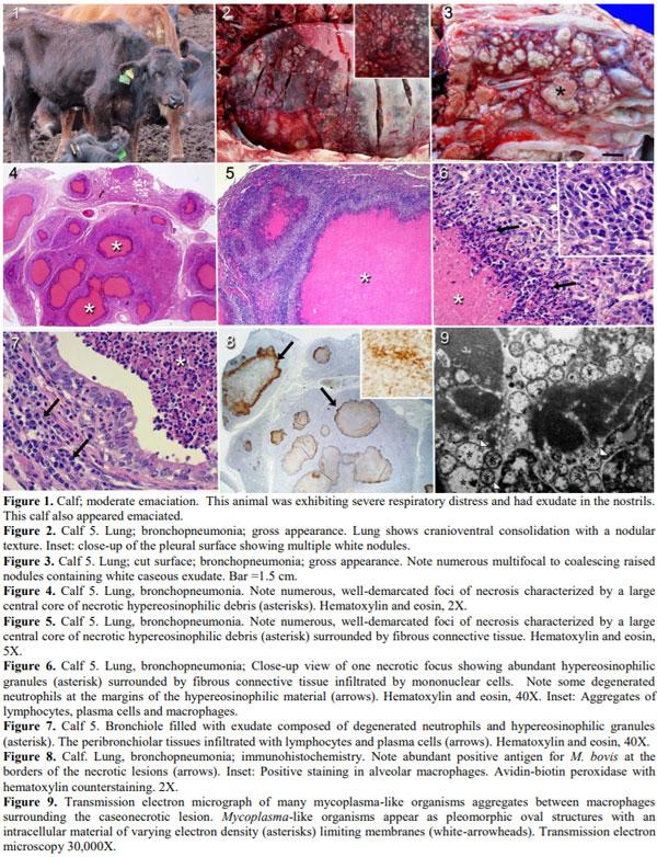
Three calves that were unresponsive to antimicrobial treatment for pneumonia (calves 1, 4, and 5) were euthanized (Fig. 1), while three others (calves 2, 3, and 6) were found dead. All animals were necropsied, and lung samples were collected for bacteriological culture, virus isolation, histopathology and immunohistochemistry (IHC), and electron microscopy.
The bacteriological analysis was done in duplicates, inoculating lung tissue on blood agar and MacConkey agar plates. One set of the culture plates was incubated aerobically, and the duplicate under microaerophilic conditions (5% CO2). All plates were incubated at 37ºC for 48 h. The isolates were subcultured for purity and identified using colony morphology, detection of hemolysis, Gram stain, and conventional biochemical tests (7).
Lung samples for virus isolation were stored in cryovials at -70ºC and submitted to Institute of Virology, National Institute of Agriculture Technology, Morón, Buenos Aires, Argentina. Tissue samples were homogenized in Eagle´s minimal essential medium (D-MEM) supplemented with 10% fetal bovine serum, and inoculated on MDBK (Madin-Darby Bovine Kidney) cells. Cell cultures were incubated at 37ºC and 5% CO2 for 5 days, and examined daily. After nine consecutive passages were carried out, cultures were tested for BRSV and BVDV by indirect fluorescent antibody (IFA) and PCR.
For histology and IHC, six lung samples obtained from the anterior, posterior cranial and caudal lobes of both left and right lungs were fixed in 10% buffered formalin, embedded in paraffin, sectioned at 3 µm, and stained with both hematoxylin and eosin, and Gram stains. Selected paraffin-embedded tissues were submitted for immunohistochemical detection of M. bovis (Prairie
Diagnostic Services Inc. Saskatoon Saskatchewan, Canada). Immunohistochemistry was conducted using a commercial staining platform (Ventana Medical System, Tucson AZ). Epitope retrieval was done by applying protease solution, and a mouse anti-M. bovis monoclonal antibody (mAb-5A10.2) was applied for 32 min at a dilution of 1: 800 (16).
Duplicate sections of each paraffin block were tested for M. bovis using a monoclonal antibody at 1:400 and 1:800 dilutions using positive and negative controls. No other Mycoplasma spp. were tested by IHC in this study.
Lung tissue of calves 1, 4 and, 5 were fixed in glutaraldehyde and post-fixed in osmium tetroxide for transmission electron microscopy (TEM). After fixation, tissues were dehydrated and embedded in epoxy resin. Semi-thin sections cut at 1-2 µm were mounted on slides and stained with toluidine blue. Slides were examined by light microscopy to select the areas of necrosis which were subsequently cut at 60 nm and contrasted with uranyl acetate and lead citrate. Thin sections were evaluated ultrastructurally with a JEM 1200 EX II (JEOL) transmission electron microscope.
Results
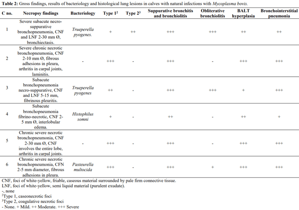
One of the affected calves in this study showed a clinical course that reached up to 59 days of illness. Table 1 summarizes farm locations, rates of infection, breed, age, and clinical signs of affected animals. There was no cytopathic effect observed in cell cultures, and all samples of lung tissue tested negative for BVDV and BRSV by IFA and PCR. Trueperella pyogenes was isolated from the lungs of two calves, H. somni from one calf, and P.multocida from another calf (Table 2).
On postmortem examination, the lungs showed cranioventral consolidation involving 40-70% of pulmonary parenchyma and the affected lung had a nodular texture and numerous focal to confluent white foci (Fig. 2). On the cut surface, the consolidated lung exhibited focal to confluent areas of necrosis composed largely of white, thick caseonecrotic exudate (Fig. 3).
Microscopically, the consolidated lung showed well-demarcated areas of necrosis surrounded by a thick layer of fibroblasts infiltrated with macrophages, lymphocytes, and few plasma cells (Figs. 4 and 5). These necrotic foci contained in the center abundant hypereosinophilic and granular material (Fig. 6), while the periphery comprised bronchial walls some of which were dilated or effaced by the inflammatory reaction (Fig. 7). There was also thickening of the alveolar septa and suppurative bronchitis and bronchiolitis (Fig. 7), as well as hyperplasia of the bronchus-associated lymphoid tissue (BALT). The bronchiolar lumens of three calves were obliterated by aggregates of fibroblast, collagen and newly formed blood capillaries (bronchiolitis obliterans) (Table 2). The lungs of calves 2, 3, 5, and 6 exhibited moderate pleural fibrosis. Calf 1 also had irregular and distinctive areas of coagulative necrosis surrounded by a rim of degenerated leukocytes with nuclear streaming resembling "oat-shaped cells" (Table 2). Viral inclusions were not observed in any of these lungs.
Mycoplasma bovis pneumonia in feedlot cattle and dairy calves in Argentina - Image 1
Immunohistochemistry showed abundant M. bovis antigen in the lungs of all six calves. The positive staining was observed mainly at the margin of the necrotic lesions, and to a lesser extent in the center of the necrotic foci (Fig. 8). M. bovis antigen was also detected in alveolar macrophages (Fig. 8).
Observation of lungs sections by electron microscopy revealed round to oval to pleomorphic structures with an approximated diameter ranging from 0.40 to 0.90 µm covered by a trilaminar membrane and varying electron density in the cytoplasm (Fig. 9). These structures were arranged in small aggregates or larger clusters throughout the caseonecrotic foci or admixed with the exudate present in the bronchial lumens (Fig. 9). These structures were also observed primarily in the interstitial space between inflammatory cells such as lymphocytes, macrophages and neutrophils surrounding the necrotic areas.
Discussion
Pathological, ultrastructural, and immunostaining findings confirmed that the etiological agent incriminated in these six cases of chronic BRD in Argentina was M.bovis. To the best of our knowledge, this is the first reportthat unequivocally identifies M bovis as the etiology of BRD in this country. Some researchers in Argentina had previously incriminated this organism as the putative agent in mastitis, but not related to pneumonia or respiratory disease (11).
The gross, microscopic and immunohistochemical findings in the lungs were identical to those previously reported in naturally occurring M. bovis pneumonia in other countries (5, 15, 20, 25, 26, 32). The same lesions have also been reproduced experimentally in calves (1, 2, 27).
Mycoplasma bovis pneumonia in feedlot cattle and dairy calves in Argentina - Image 2
 
Mycoplasma bovis pneumonia in feedlot cattle and dairy calves in Argentina - Image 3
Feedlot systems appeared in Argentina around the 1990s and pneumonia has been the most common disease, as reported in the feedlot industry of North America (8, 22). The protracted clinical signs for all six cases in these BRD outbreaks in Argentina mimicked those for M. bovis pneumonia reported by others (26). M. bovis should always be suspected when a calf or steer has a history of an unresponsive BRD or when post mortem examination reveals bronchopneumonia with caseonecrotic nodules in the cranioventral lung lobes (9).
Although the caseonecrotic nodules resemble abscesses, these pulmonary structures are not pulmonary abscesses (sensu stricto), but rather segmentally distended bronchi filled with inspissated exudate (bronchiectasis) (8, 22). The pathogenesis of M. bovis-associated bronchiectasis is incompletely understood but presupposes long-term pulmonary inflammation that leads to the destruction of the bronchial walls (18, 22). This bronchial and bronchiolar changes are irreversible, and thus, explains why animals rarely respond to treatment and die, or are euthanized in extremis (5, 17, 26). Through experience and close observation, veterinarians and pathologists should be able to distinguish pulmonary abscesses frequently seen in cattle as an embolic sequel to liver abscesses, from caseonecrotic nodules and bronchiectasis, the long-term sequela of M. bovis bronchopneumonia. Nonetheless, it should also be noted that in some animals caseonecrotic nodules can progress into abscesses if there is a concurrent infection with T. pyogenes (10).
Bronchiolitis obliterans was another microscopic lesion seen in the M. bovis positive lungs of the calves in Argentina. This form of chronic obliterative bronchiolitis is a non-specific lesion resulting from severe injury and necrosis to the bronchiolar epithelium, where organized fibrovascular tissue progressively replaces the fibrin and neutrophils attached to the walls of the bronchioles (10, 22). The pathogenesis involved in bronchiolar necrosis and fibrosis in the bronchiolar lumen is still to be determined (18). Bronchiolitis obliterans is yet another contributing factor of respiratory distress in calves infected with M.bovis which is unlikely to resolve with antimicrobialtreatment (17).
Calves 1 and 3 had Gram-positive coccobacilli within the M. bovis-IHC-positive caseonecrotic foci. This microscopic finding was not unexpected since co-infections with T. pyogenes, P. multocida and other mycoplasmas are common in natural and experimental M.bovis pneumonia (4, 6, 14, 15, 18).
The caseonecrotic nodules were also microscopically similar to those seen in M. bovis pneumonia elsewhere (10). Interestingly, the lungs of one calf (C1) showed not only hypereosinophilic granular lesions but also irregular areas of coagulative necrosis, a change most typically found with M. haemolytica infection (8, 22). There are two possible explanations for these co-existing microscopic findings: 1. M. bovis infection could have been a sequel or comorbidity to mannheimiosis as reported by others (22). 2. M. bovis could have caused in the same lung the two patterns of pulmonary necrosis as previously proposed by others (20).
All calves of the present study were unresponsive to antimicrobial treatment or suffered relapses of BRD which likely resulted from the combination of M. bovis capacity of evading the pulmonary defense mechanisms and immune responses (14, 15, 18) and that some strains of M. bovis are resistant to the antimicrobials used in feedlotsto treat pneumonia, particularly tetracyclines and tilmicosin (3, 13, 28, 31). The same studies also suggested that tilmicosin was an ineffective therapeutic option for this agent, but extrapolating in vitro findings to the live animal conditions may not always be applicable.
Mycoplasma bovis is frequently found in thenasopharyngeal flora (10) and viral infections such as BoHV-1, BRSV, and PI-3may predispose cattle to M.haemolytica infection, but the role of these viruses in thepathogenesis of M. bovis still needs elucidation. Since clinical signs of pneumonia appear at the late stages of M.bovis infection, it is difficult to know if there was aproceeding viral infection in the calves of our study. This time difference between viral and M. bovis infections could also be the reason why the lungs of all calves in Argentina tested negative for respiratory viruses.
TEM and immunogold electron microscopy conducted in the lungs of experimentally infected cattle showed that M. bovis resides at the periphery of the necrotic foci, but it can also be found in the exudate within the bronchial lumen (21). The lungs from the affected animals in our study revealed comparable ultrastructural findings.
Polyarthritis is another condition frequently associated with M. bovis infection in cattle, and not surprisingly Calves 2 and 5 had in addition to pneumonia severe joint inflammation (1, 15). This joint infection is presumably a sequel to pneumonia where M. bovis disseminates hematogenously from the lung to the synovial membrane (23).
This first report should alert producers, veterinarians, and diagnosticians of the possible economic importance of M. bovis pneumonia in Argentina. Early detection and appropriate treatment are critical since this disease both clinically or silently progresses to irreversible lung damage and treating animals at the late stages of the diseases becomes expensive and futile. Further studies should determine the prevalence, morbidity, mortality and economic relevance of M. bovis pneumonia in the BRD complex in Argentina.
Acknowledgements
The authors would like to thank Gabriela Toledo, staff at the animals help group of the Agricultural Experiment Station Marcos Juarez, and Roxana Peralta of Central Electron Microscopy, School of Veterinary Sciences Service of National University of La Plata. The work of Ms. Kathy Jones, Atlantic Veterinary College, is also appreciated.
This work was funded by project INTA PNSA-1115054, INTA AUDEAS CONADEV-940148 and ArgenINTA Foundation.
This article was originally published in Brazilian Journal of Veterinary Pathology 2017, 10(2), 79 – 86. DOI: 10.24070/bjvp.1983-0246.v10i2p79-86
  1. Adegboye DS, Halbur PG, Nutsch RG, Kadlec RC, Rosembusch RF. Mycoplasma bovis-associated pneumonia and arthritis complicated with pyogranulomatous tenosynovitis in calves. J Am Vet Med Assoc. 1996;209:647-9.
  2. Arcangioli MA, Duet A, Meyer G, Dernburg A, Be´zille P, Poumarat F, LE GRAND D. The role of Mycoplasma bovis in bovine respiratory disease outbreaks in veal calf feedlots. Vet J. 2008;177:89-93.
  3. Ayling RD, Baker, SE, Peek ML, Simon AJ., Nicholas RAJ. Comparison of in vitro activity of danofloxacin, florfenicol, oxytetracycline, spectinomycin and tilmicosin against recent field isolates of Mycoplasmabovis. Vet Rec. 2000;146:745-7.
  4. Bell C.J, Blackburn P, Elliot M, Patterson TIAP, Ellison S, Lahuerta-marin A, Ball HJ. Investigation of polymerase chain reaction assays to improve detection of bacterial involvement in bovine respiratory disease. J Vet Diag Invest. 2014;26:631-4.
  5. Booker CW, Abutarbush SM, Morley PS, Jim G.K, Pittman TJ, Schunicht OC, Perrett T, Wildman BK, Fenton RK, Guichon PT, Janzen ED. Microbiological and histopathological findings in cases of fatal bovine respiratory disease of feedlot cattle in Western Canada. Can Vet J. 2008;49:473-81.
  6. Bürki S, Frey J, Pilo P. Virulence, persistence and dissemination of Mycoplasma bovis. Vet Microbiol. 2015;179:15-22.
  7. Carter GR. Diagnostic procedures in veterinary bacteriology and mycology. 4th ed. Springfield: C.C. Thomas; 1984. 515 p.
  8. Caswell JL, Williams KJ. Respiratory system. In: Maxie MG, editor. Jubb, Kennedy and Palmer’s Pathology of Domestic Animals. 6th ed., vol. 2. St. Louis: Elsevier; 2016. p. 465-591.
  9. Caswell JL, Archambault M. Mycoplasma bovis pneumonia in cattle. Anim Hlth Res Rev. 2008;8:161-86.
  10. Caswell JL, Bateman KG, Hugh YC, Castillo-Alcala F. Mycoplasma bovis in respiratory disease of feedlot cattle. Vet Clin North Am Food Animal Pract. 2012;26:365-79.
  11. Cerdá R, Xavier J, Sansalone P, De La Sota R, Rosembusch R. Aislamiento de Mycoplasma bovis a partir de un brote de mastitis bovina en una vaquería de la provincia de Buenos Aires. Primera comunicación en la República Argentina. Rev La Am Microbiol. 2001;42:7-11.
  12. Ellis JA. The immunology of the bovine respiratory disease complex. Vet Clin North America Food Anim Pract. 2001;17:535-50.
  13. Francoz D, Fortin M, Fecteau G, Messier SE. Determination of Mycoplasma bovis susceptibilities against six antimicrobial agents using the E test method. Vet Microbiol. 2005;105:57-64.
  14. Fulton, RW, Blood SK, Panciera RJ, Payton ME, Ridpath JF, Confer AW, Saliki JT, Burge TL, Welsh RD, Johnson BJ, Reck A. Lung pathology and infectious agents in fatal feedlot pneumonias and relationship with mortality, disease onset, and treatments. J Vet Diagn Invest. 2009;21:464-77.
  15. Gagea MI, Bateman KG, Van Dreumel T, Macewen BJ, Carman S, Archambault M, Shanahan RA, Caswell JL. Diseases and pathogens associated with mortality in Ontario beef feedlots. J Vet Diagn Invest. 2006;18:18-28.
  16. Haines DM, Chelack BJ. Technical considerations for developing enzyme immunohistochemical staining procedures on formalin-fixed, paraffin-embedded tissues for diagnostic pathology. J Vet Diagn Invest. 1991;3:101-12.
  17. Haines DM, Martin KM, Clark EG, Jim J, Janzen ED. The immunohistochemical detection of Mycoplasmabovis and bovine viral diarrhea virus in tissues of feedlot cattle with chronic, unresponsive respiratory disease and/or arthritis. Can Vet J. 2001;42:857-60.
  18. Hermeyer K, Buchenau I, Thomasmeyer A, Baum B, Spergser J, Rosengarten R, Hewicker-Trautwein M. Chronic pneumonia in calves after experimental infection with Mycoplasma bovis strain 1067: characterization of lung pathology, persistence of variable surface protein antigens and local immune response. Acta Vet Scand. 2012;54:1-11.
  19. Horwood PF, Schibrowski ML, Fowler EV, Gibson JS, Barnes TS, Mahony TJ. Is Mycoplasma bovis a missing component of the bovine respiratory disease complex in Australia? Aust Vet J. 2014;92:185-91.
  20. Khodakaram-Tafti A, López A. Immunohistopathological findings in the lungs of calves naturally infected with Mycoplasma bovis. J Vet Med A Physiol Pathol Clin Med. 2004;51:10-4.
  21. Kleinschmidt S, Spergser J, Rosengarten R, Hewicker-Trautwein M. Long-term survival of Mycoplasmabovis in necrotic lesions and in phagocytic cells as demonstrated by transmission and immunogold electron microscopy in lung tissue from experimentally infected calves. Vet Microbiol. 2013;162:949-53.
  22. López A, Martinson SA. Respiratory System, Mediastinum, and Pleurae. In: Zachary JF, editor. Pathologic Basis of Veterinary Diseases. 6th ed. St. Louis: Elsevier; 2017. p. 471-560.
  23. Nicholas RAJ, Ayling RD. Mycoplasma bovis: disease, diagnosis, and control. Res Vet Sci. 2003;74:105-12.
  24. Panciera R, Confer AW. Pathogenesis and pathology of bovine pneumonia. Vet Clin North America Food Anim Pract. 2010;26:191-214.
  25. Radaelli E, Luini M, Loria GR, Nicholas RAJ, Scanziani E. Bacteriological, serological, pathological and immunohistochemical studies of Mycoplasma bovis respiratory infection in veal calves and adult cattle at slaughter. Res Vet Sci. 2008;85:282-90.
  26. Ramírez-Romero R, Chavarría-Martínez B, Nevárez-Garza, AM, Rodriguez-Tovar LE, Dávila-Martinez C, Hernandez-Vidal G, Hernandez-Escañero JJ, Lopéz-Mayagoitia A. Demostración histoquímica de Mycoplasma bovis en lesiones neumónicas crónicas en ganado de corral de engorda.Vet Mex. 2010;41:289-96.
  27. Rodriguez F, Bryson DG, Ball HJ, Forster F. Pathological and immunohistochemical studies of natural and experimental Mycoplasma bovis pneumonia in calves. J Comp Pathol. 1996;115:151-62.
  28. Rodriguez F, Kennedy S, Bryson TD, Fernandez A, Rodirguez JL, Ball HJ. Immunohistochemical method of detecting Mycoplasma species antigen by use of monoclonal antibodies on paraffin sections of bovine and caprine lungs. Zentralbl Veterinarmed B. 1996;43:429-38.
  29. Rosenbusch, RF, Kinyon JM, Apley M, Funk ND, Smith S, Hoffman JL. In vitro antimicrobial inhibition profiles of Mycoplasma bovis isolates recovered from various regions of the United States from 2002 to 2003. J Vet Diagn Invest. 2005;17:436-41.
  30. Terlaak EA, Wentink GH, Zimmer GM. Increased prevalence of Mycoplasma bovis in the Netherlands. Vet Quart. 1992;14:100-4.
  31. Thomas A, Nicolas C, Dizier I, Mainil J, Linden, A. Antibiotic susceptibilities of recent isolates of Mycoplasma bovis in Belgium. Vet Rec. 2003;153:428-31.
  32. Yilmaz R, Cangul IT, Onat K, Akkoc A, Ozyigit MO, Akdesir E. Histopathological, immunohistochemical and bacteriological characterization of Mycoplasma bovis pneumonia in cattle. Pakistan Vet J. 2016;36:316-21.
 
Related topics:
Authors:
Dr. Alfonso López Mayagoitia
University of Prince Edward Island
Recommend
Comment
Share
Profile picture
Would you like to discuss another topic? Create a new post to engage with experts in the community.
Join Engormix and be part of the largest agribusiness social network in the world.