Introduction
Bovine trichomoniasis is a venereal disease caused by Tritrichomonas foetus, a flagellated protozoan that lodges in the cavities of the gastrointestinal and reproductive tracts of infected animals [1]. In bulls, T. foetus is found on the epithelial surface of the penis and preputial crypts, and although it can produce nodules in the area, it typically does not show clinical signs, causing males to act as asymptomatic carriers [2,3]. Conversely, infected females display clinical signs including vaginitis, endometritis, birth of weak calves, fetal maceration, and abortions, thereby affecting calf production and causing significant economic losses for producers [4].
In Mexico, the first report of bovine trichomoniasis was made more than 60 years ago [5]. In recent years, the importation of bulls into Mexico from other countries where T. foetus is endemic, along with the increase in cases showing clinical signs suggestive of the parasite, has generated concern among animal health professionals and producers, giving rise to the need for diagnostic tools that allow the control of this disease. A study conducted in Chihuahua, a northern Mexican state, using a commercial culture system followed by PCR as a diagnostic method, reported that 21.8% of the sampled bulls were positive for T. foetus, while 60 to 65% of the evaluated herds had at least one positive animal [6]. In turn, [7] employed classic microscopy and reported that 36% of the cows and 14.3% of the bulls sampled in Veracruz (eastern Mexico) were positive for this protozoan.
The standard diagnostic method for trichomoniasis is the microscopic identification of the protozoan from diverse biological samples such as preputial washings, uterine fluid, and cervicovaginal secretions. It is important to highlight that samples for this method need to be cultured in a medium suitable for the replication and growth of T. foetus [8]. Limitations of this technique include variations due to sampling conditions, sensitivity, specificity, and overall accuracy. Moreover, the human factor must also be considered, since trained personnel are required to perform an accurate diagnosis. To overcome such challenges, molecular methods have been developed for the specific detection of T. foetus in cattle. In recent years, the polymerase chain reaction (PCR) has become one of the most popular methods, given that it can exclude false-positive cultures [9]; therefore, it remains a reliable test that can be used as a reference for the development of other molecular assays.
Controlling trichomoniasis in Mexico is challenging due to the scarcity of data related to its prevalence and impact. Obtaining such information relies on efficient diagnostic tests capable of accurately identifying the etiological agent. Thus, the effort toward the development and standardization of alternative diagnostic tests for T. foetus has recently begun in the country. Importantly, for any test standardization, it is required to ensure that the genetic material analyzed corresponds specifically to the pathogen of interest. In this regard, sequencing and phylogenetic analysis can contribute to confirming the identity and phylogeny of the genetic material identified as positive for T. foetus.
Therefore, the aim of the present study was to confirm the presence of T. foetus by sequencing and phylogenetic analyses of molecular isolates obtained from positive animals raised in northwestern Mexico.
Materials and methods
Ethical considerations
The study protocol for this project was approved by the Animal Welfare and Research Ethics Committee of the Instituto Tecnológico de Sonora (report No. 2024-04).
Animals and study location
The study was conducted in northwestern Mexico. A total of forty animals from eight herds were included, specifically 30 bulls of different breeds with a history of at least two breeding seasons, as well as 10 adult cows with previous records of abortions.
Sample collection
Smegma samples were collected from the preputial cavities [8]. First, each bull was immobilized in a cattle chute. Then, excess dirt was removed from the prepuce with disposable paper. Subsequently, the zone was disinfected with 2% iodine and the hairs of the distal end of the prepuce were trimmed, removing excess moisture with paper. After cleaning and disinfection, an external massage was applied for two to three minutes to relax the preputial mucosa. A plastic infusion pipette with a blunt tip was introduced into the prepuce up to the sigmoid flexure, where 20 mL of phosphatebuffered saline (PBS; Sigma-Aldrich, St. Louis, MO) solution was deposited while stirring by hand. Suction was then applied with the syringe while the pipette was gently moved up and down along the preputial mucosa, recovering the largest amount of PBS with preputial cellular tissue. Finally, the sample was placed in a sterile disposable 20-mL test tube and labeled with the animal's information [10].
In cases of abortion (no more than 14 days after the abortion event), purulent vaginal secretion samples were obtained from the uterine content of cows with clinical metritis. Highviscosity samples were diluted with 10 mL of PBS and centrifuged at 2200 × g for 15 min. The supernatant was removed, and the pellet was resuspended again in a volume of 1 mL. Finally, 200 µL were used for DNA extraction, and the rest was frozen at –60 °C until further use.
Automated nucleic acid extraction
Samples of diluted smegma and purulent vaginal secretion were first homogenized by vortexing and inversion. DNA extractions were then performed using the taco™ Nucleic Acid Automatic Extraction System (GeneReach USA) with the taco™ DNA/RNA Extraction Kit, following the manufacturer's protocol. The extracted nucleic acid was quantified with UV spectrophotometry (BioSpec-Nano, Shimadzu®). The integrity of the extracted DNA was verified by electrophoresis in a 1.5% agarose gel stained with ethidium bromide. All extractions were stored in sterile 1.5-mL vials at –20 °C to be subsequently processed for the PCR technique.
PCR detection of T. foetus in biological samples
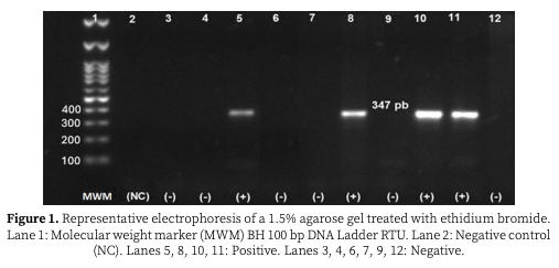
DNA samples were analyzed by PCR using the primer set TFR3: 5′-CGGGTCTTCCTATATGAGA CAGAACC-3′ and TFR4: 5′-CGGGTCTTCCTATAT GAGACAGAACCGGAGCTGAATG-3′, amplifying a 347 bp region of the gene encoding the 5.8S rRNA, as well as the ITS1 and ITS2 regions of T. foetus [11]. For the reactions, the pre-loaded GoTaq® Flexi DNA Polymerase PCR kit (Promega®) was used, containing Green GoTaq®, which serves as a reaction buffer and gel loading solution, allowing reactions to be loaded directly for rapid and efficient analysis. Reactions were performed in a final volume of 25 µL, with a concentration of 1× Green GoTaq Buffer 5×, 1.5 mM MgCl₂, 0.2 mM for each dNTP, 0.4 µM of each primer, 1.25 µL of GoTaq DNA Polymerase, 5 µL of DNA, and nuclease-free H₂O to 25 µL. Analytical conditions for PCR were one cycle of 95 °C for 3 min, 32 cycles of 95 °C for 15 s, 62 °C for 30 s, and 72 °C for 30 s, and a final extension step of 72 °C for 5 min. The product was identified on a 1.5% agarose gel with ethidium bromide, considering positive bands with the size of the agent amplicon.
Phylogenetic analysis
Five positive PCR products were selected and sequenced using the Sanger dideoxy sequencing method at the Institute of Biotechnology of the Universidad Nacional Autónoma de México (UNAM). The sequences obtained were analyzed using the MEGA 11 software [12], employing the Clustal W and UPGMA alignment methods based on multiple sequence alignment analysis. A phylogenetic analysis was performed to determine the identity between Mexican local T. foetus isolates (MX-TFI) and T. foetus isolates registered within NCBI (National Center for Biotechnology Information) through BLAST. Finally, all local isolates were registered in GenBank-NCBI.
Results
With an average quantification of 58.32 ng/µl, according to spectrophotometry, and a purity of 1.85 in the 260/280 OD ratio, the electrophoresis results showed good integrity of the extracted DNA and confirmed the presence of adequate amounts of high-quality DNA free of contaminants such as guanidine, phenols, chaotropic salts, or carbohydrates. Regarding PCR detection, nine out of the 30 bulls (30% of the males) and two out of the 10 cows (20% of the females) were positive for T. foetus. In the positive samples, the specific 347 bp band corresponding to the ribosomal DNA region of T. foetus was observed (Figure 1).
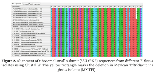
Data generated from sequencing of purified PCR amplicons were analyzed and named for publication within the NCBI database as new Mexican isolates of T. foetus: CEAL-1 (PQ474277), CEAL-2 (PQ474278), CEAL-3 (PQ474279), CEAL-4 (PQ474280), and CEAL-5 (PQ474281). According to NCBI-BLASTn, the alignments of the nucleotide sequences of the MX-TFI and the isolates from GenBank revealed a similarity of 99.65% to isolates previously reported in China, Spain, and the United States. This variation in the percentage of similarity resulted from a deletion of a thymine in the Mexican sequences, unlike all the isolates reported in NCBI-BLASTn and aligned in the MEGA 11 software (Figure 2).
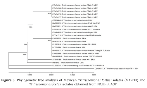
The phylogenetic tree compares T. foetus sequences isolated from different geographic regions and hosts, showing two main groups with a high level of bootstrap support (99%), indicating confidence in the grouping of these clades (Figure 3). The first five isolates at the top (CEAL-1 to CEAL-5) correspond to Mexican T. foetus isolates (MX-TFI), and the 99% support demonstrates the close relationship among them. These isolates are more closely related to other T. foetus isolates from countries such as China (OP856640.1), Spain (MK770850.1), and the United States (MK250821.1), forming a subgroup within the same clade. Another strong cluster with 99% bootstrap support includes isolates from Brazil (KX267765.1, HM400076.1, KU680816.1) and Turkey (HM490937.1). These are more distant from the MX-TFI and form a cluster of their own, which could suggest geographic or evolutionary divergence. Isolates from different regions such as Namibia (KX425878.1), Australia (MW322817.1), Japan (LC054294.1), and the United States (AY349189.1) formed independent branches in the tree, but all within the same major clade. Finally, at the bottom of the tree, important reference isolates were included, such as T. foetus USA (M81842.1) and the T. foetus isolate from Iran (OL455037.1), showing that these isolates are more distant from the others, which could reflect more marked genetic differences among them.
Discussion
In molecular studies, the quality and quantity of extracted DNA are crucial factors for PCR amplification tests and sequencing. The quantification obtained by spectrophotometry was 58.32 ng/µl, suitable for most molecular applications since a minimum concentration of 50 ng/µl is required for PCR [13]. The 260/280 OD ratio was 1.85, confirming good purity with a low presence of contaminants, according to the Microbiological Biosafety Laboratory Manual [14]. These results are similar to those reported by [15], who obtained 45.7 ng/μl and a purity of 1.73 in a study using purulent vaginal secretion from cows with abortions. The slight difference in DNA concentration may be due to variations in the extraction protocol, sample type, or specific study conditions. However, both studies highlighted that adequate DNA quality was achieved, ensuring the success of subsequent molecular applications.
The comparison of the incidence and detection results of T. foetus and the scarcity of data available in different states of Mexico over time shows a worrying trend in the lack of attention to establishing the incidence or prevalence of this disease in national livestock. In our study, 30% of the bulls and 20% of the cows were detected as PCR positive, indicating a considerable presence of the disease in the region. This reflects consistency with the findings of [5], who reported the disease in dairy cattle from the Valley of Mexico more than 60 years ago, suggesting that T. foetus has been a silent and persistent problem in the industry. More recently, [6] reported a prevalence of 21.8% in sampled bulls, and that 60–65% of the farms evaluated in Chihuahua had at least one positive animal. This figure, although lower than the prevalence observed in our study, indicates that trichomoniasis continues to be a significant challenge in northwestern Mexico. Nonetheless, evidence that this could represent a nationwide problem exists, since in Veracruz (eastern Mexico), a prevalence of 36% in cows and 14.3% in bulls was reported [7]. These findings underline the importance of implementing effective diagnostic and control strategies. As bulls move from different regions and countries, the risk of disease spread increases, making early and accurate diagnosis essential. The use of molecular techniques such as PCR has improved detection capacity, allowing infections to be identified at earlier stages and contributing to more effective management of reproductive health in livestock.
Furthermore, phylogenetic studies of T. foetus can provide relevant information about the genetic variability and potential relationships between different strains isolated from different hosts and geographical regions. In our phylogenetic analysis, some strains were grouped into two large clades, indicating a close relationship between the Mexican isolates and strains from countries such as China, Spain, and the United States. This strong grouping suggests that the isolates from Mexico share a recent common ancestor with strains from those countries and could imply transmission routes or genetic exchange between populations.
On the other hand, [16-18] indicated that, despite the moderate genetic distinction between feline, bovine, and porcine genotypes of T. foetus, these might represent a single species. Similarly, [15] reported that T. foetus genotypes in cattle and cats show a degree of homogeneity, supporting the idea of a close identity between isolates from different host species. This study also suggests that host exchange might be a real phenomenon, although cases of interspecies transmission in natural settings have not yet been documented. Conversely, a multilocus analysis by [19] identified more marked genetic differences between T. foetus from cats and cattle, suggesting that, although there are similarities, there may also be genetic differences that reflect adaptations to their respective hosts.
In our phylogenetic tree, independent branches of isolates from Namibia, Australia, Japan, and the United States were highlighted, which could suggest a scenario in which different populations of T. foetus have evolved in isolation. This contrasts with the close relationship observed between Mexican isolates and other strains from the American and European continents, which could indicate greater connectivity or genetic exchange in those regions. Finally, reference isolates of T. foetus such as the one from the United States and the one from Iran appear more distant from the rest, suggesting that there are significant genetic differences that may be relevant for the development of diagnostics and treatments in different cattle populations. This pattern of genetic variability in T. foetus underscores the need for continued surveillance and phylogenetic studies to better understand the epidemiology of the disease and its impact on animal health. The similarities and differences observed between our results and those presented by [1] and [20], who presented a draft assembly of the complete genome of a bovine strain, highlight the importance of continuing to investigate the genetic variability and epidemiology of this protozoan in a global context.
Although available in Mexico, some reliable molecular techniques such as PCR can be expensive to employ on a large scale because their use depends on the importation of reagents that cannot in some cases be replaced due to prior standardization of the test. In addition, the cost of the test is also influenced by the need for sophisticated equipment and infrastructure. In this sense, some producers have even opted to send samples to other countries (e.g., USA) for analysis; however, not all cattle owners can afford such an expense. The present study will allow the development of an alternative test to PCR. That is, the sequences of Mexican isolates will provide the genetic basis for the design of a simpler, less expensive, and even portable molecular test. This is essential for the development of effective control strategies, the prevention of outbreaks, and the improvement of reproductive health in livestock, thus contributing to the sustainability and profitability of the national livestock industry.
Conclusion
We confirmed the presence of T. foetus in northwestern Mexico and characterized the genetic structure of the pathogen. This is the first sequencing and phylogenetic analysis of T. foetus in the country, providing a solid basis for the accurate identification of the disease and characterization of the genetic variability of local strains compared to international isolates. The Mexican strains identified in this study represent a crucial step toward understanding the epidemiology and new diagnostic alternatives for bovine trichomoniasis in Mexico.
This article was originally published in Revista Colombiana de Ciencias Pecuarias; 39(Jan-Dec):e358794. https://doi.org/10.17533/udea.rccp.e358794. This is an Open Access article distributed under the terms and conditions of the Creative Commons Attribution (CC BY) license.