Explore
Communities in English
Advertise on Engormix

Haemorrhagic septicaemia outbreaks in cattle with high mortality following wrong vaccinations in Adamawa and Taraba States, Nigeria

Published: March 20, 2019
By: Garba Ahmed 1, Mohammed A. Qadeer 2, Moses O. Odugbo 3, Christianah I. Odita 1, Olabisi A. Makanju 1, Abdulazeez O. Najeem 4, Lami Galadima 5, Philips A. Okewole 1 and Mohammed S. Ahmed 1.
Summary

Author details

1 Diagnostics and Extension Department, National Veterinary Research Institute, Vom, Plateau State, Nigeria; 2 Department of Zoology, School of Pure and Applied Sciences, Modibbo Adama University of Technology, Yola, Adamawa State, Nigeria; 3 Bacterial Research Department, National Veterinary Research Institute, Vom, Plateau State, Nigeria; 4 Zonal Veterinary Clinic Funtua, Ministry of Agriculture and Natural Resources, Katsina State – Nigeria; 5 Solomon Memorial Veterinary Services, Jalingo, Taraba State, Nigeria.


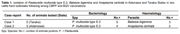
Two outbreaks of haemorrhagic septicaemia (HS) following wrong administration of contagious bovine pleuropneumonia (CBPP) vaccine (CBPPV) combined with black quarter vaccine (BQV) and administration of CBPPV alone, respectively were investigated in cattle herds in Adamawa and Taraba States of Nigeria. Two of the nomadic Fulani cattlemen independently purchased the CBPP and BQ vaccines in October, 2006 at the Zonal Investigation Laboratory, National Veterinary Research Institute (NVRI), Yola, Adamawa State. One of the farmers employed the services of a livestock attendant who diluted each lyophilized CBPP vaccine in a 100 ml of BQV and inoculated 181 cattle each with 1 ml of the formulation in the Mayo lope village of Taraba State. Thereafter, the animals began to show signs of respiratory complications and swelling at the site of inoculation. A total of 125 cattle died 1 to 5 days post inoculation. The second farmer kept a vial of the CBPPV at room temperature for 24 h, then diluted and administered twice the normal dose each to 50 cattle in Yadim area of Adamawa State. A day later, 17 cattle died and in 3 days a total of 35 have died or salvaged. Clinical and laboratory investigations confirmed the presence of Pasteurella multocida Type E2 in both cases. Administration of a broad spectrum antibiotic ‘PENSTREP’ (a combination of Procaine Penicillin and Streptomycin HCL) for three days in both outbreaks brought the death toll to halt. Sub-clinical or underlying HS disease exacerbated by stress due to multiple and improperly inoculated vaccines has been identified as the most probable cause of death in these animals. The use of PENSTREP has proved efficacious in the treatment of field HS outbreaks. Adherence of cattle owners to their local veterinarians for assessment and guidance before vaccination or under his supervision is highly recommended.

Key words: Haemorrhagic septicaemia outbreak, cattle, vaccination, Nigeria.

INTRODUCTION
Haemorrhagic septicaemia (HS) is a disease of cattle and buffalos (Abdulkadir, 1989) caused by 1 of 2 serotypes of Pasteurella multocida, designated B:2 and E:2 (FAO, 2015; IIL, 2015). The disease in cattle is prevalent in the tropics, particularly in the equatorial region of South East Asia and Africa, where outbreaks mostly occur during the rainy season (Dhanda, 1966; Anosa and Esoun, 1975). In Nigeria, HS in cattle was first reported in Sokoto, Bauchi, Plateau, and Adamawa provinces in 1925 (Anon, 1925- 1951). Subsequently, the disease was recognized in the eastern and western provinces in 1948 and 1951, respectively (Anon, 1925-1951).
Haemorrhagic septicaemia outbreaks in cattle with high mortality following wrong vaccinations in Adamawa and Taraba States, Nigeria - Image 1
Since then various reports of HS in African buffalo (Kasali, 1972), in cattle (Anosa and Esoun, 1975; Akpavie et al., 1991), in zoo animals (Okoh, 1980; Okoh and Ocholi, 1986), small ruminants (Ikede, 1977; Odugbo et al., 2006; Ugochukwu, 2008), as well as experimental infections in calves (Odugbo et al., 2005) have been documented in Nigeria. Some epidemiological and environmental factors like climatic stress due to heavy rainfall, a marked drop in environmental temperatures, marked an increase in relative humidity and combination of chronic, debilitating infections could exacerbate the occurrence of HS (Anosa and Esoun, 1975; Abdulkadir, 1989). Stress due to vaccination may be added to this list, especially when vaccination is done wrong. This paper investigated the occurrence of HS outbreaks following vaccination of some cattle with contagious bovine pleuropneumonia (CBPP) combined with black quarter vaccines (BQV) and CBPP alone from Northeastern Nigeria.
METHODOLOGY
Cases history
Case 1
On the 13-10-2006, a report on cattle deaths was received at the National Veterinary Research Institute (NVRI), Yola, Adamawa State, Nigeria. A total of 87 cattle of different ages and sexes had died in two days and over 50 were sick and recumbent on the third day (the day of the report) in the Mayo lope area of Lau local government in Taraba State. The herd of 181 cattle had been vaccinated against CBPP and BQ three days earlier with vaccines that had been purchased from NVRI Zonal Laboratory, Yola four days earlier. A vial of the lyophilised CBPP vaccine was dissolved in 100 ml of the BQ vaccine by an attendant and 1 ml of the formulation administered to each animal intramuscularly in the neck region. Within 12 to 15 h of vaccination, the cattle became ill and in 18 to 24 h high mortality was recorded in the herd. A team of veterinarians was dispatched and on arrival at a village called Mayo lope about 100 km away from Yola, additional 38 cattle had been dead or culled making a total loss of 125 cattle in the herd.
Clinical and laboratory findings: Clinical signs shown by the affected cattle include respiratory distress, drooling of saliva, weakness, recumbency, swelling in the neck region (site of vaccine inoculation), and high temperature of between 39 to 39.5°C. Necropsy findings showed generalized septicaemia evident by congestion and haemorrhages of the internal organs, particularly the liver, spleen, myocardium, lungs, trachea, and that of the musculature. Although poisoning was one of the differentials, it was later ruled out. Due to absence of diarrhea and absence of similar sings in the neighbouring herds that grazed in the same vicinity with the herd in question. HS was the tentative diagnosis, then and a broad spectrum antibiotic PENSTREP (a combination of Procaine Penicillin 200,000 iu/ml and Dihydrostreptomycin 250 mg/ml) at a dose rate of 2.5 ml/50 kg was administered intramuscularly for three days. Tissues from five representative animals were immediately taken for bacteriology culture and histopathology as described (Odugbo et al., 2005), while serum and whole blood were also collected for serology and haematology/serum biochemistry, respectively. All these were sent to the Central Diagnostic Laboratory, National Veterinary Research Institute, Vom, Plateau State, Nigeria, while treatment with PENSTREP was continued. Laboratory analyses (bacteriology and haematology) consequently confirmed P. multocida serotype E:2 in the tissues and Babesia bigemina in the whole blood (Table 1).
Case 2
On the 15-10-2006, another nomadic Fulani herdsman from Yadim village of Fufore local government in Adamawa State, Nigeria reported the death of 17 out of 50 animals in the herd. Investigation revealed that the herdsman purchased 2 vials of the CBPP vaccine from the NVRI, Yola, Adamawa State two days prior to the incident. The herdsman dissolved a vial of the lyophilized CBPP vaccine within 100 ml of sachet water and administered 2 ml each of the diluted vaccine to each cow at the neck region as against the recommended 0.5 to 1 ml/cow. There was no adverse effect observed by the herdsman in this herd. The following day, he reconstituted the second vial of the CBPP kept at room temperature, as in the first vial and inoculated the second herd totaling 50 cattle at the same dosage of 2 ml/cow. Twenty-four hours later the animals in the second herd of 50 cattle became ill out of which 17 died. At the time of the visit by the team of veterinarians from NVRI, a total of 35 animals were either dead or salvaged.
Clinical and laboratory findings: The clinical findings were similar to what was seen in case 1 reported earlier. HS was the tentative diagnosis and a broad spectrum antibiotic PENSTREP (a combination of Procaine Penicillin 100,000 IU/ml and Streptomycin HCl 250 mg/ml) at a dose rate of 2.5 ml/50 kg was also given for three days. No further mortality was seen following the antibiotic treatment. Laboratory analyses (bacteriology and haematology) of the tissues and serum/whole blood samples collected from five representative animals; sent to NVRI, Vom confirmed the presence of P. multocida serotype E: 2 in the tissues and Anaplasma centrale in the whole blood (Table 1).
Follow up information
In October 2013, the herd from the first case in Taraba State was located and the animals were apparently healthy. Further questioning revealed that no such incidence occurred again and that the number of animals has risen from the 56 cattle left in 2006 to 117 cattle in 2013 (7 year interval). The second case from Adamawa State was however not located as of the year 2013, probably due to the nomadic lifestyle of the owner. Hence, due to their nomadic nature of grazing, no current health and production status of that herd was received.
RESULTS AND DISCUSSION
Too often, vaccines are used without sufficient thought and technical expertise. However, with proper attention and supervision of the type of vaccine, strategic use, in terms of proper handling, correct route of administration/dosage, management, and consideration of influencing factors, successful vaccination can be achieved. The HS outbreaks being reported in this study were arguably exacerbated due to the stress caused by improper vaccines reconstitution and administration. From our knowledge of vaccines and vaccination, the two vaccines (CBPPV and BQV) are live attenuated vaccines produced by the NVRI, Vom, Nigeria. As live vaccines can cause immunosuppression, they have the potential to be an additional stress factor in already immunosuppressed animals (Abdulkadir, 1989). This is particularly of more concern where animals are immunized with multiple vaccines, which is a common practice and is the case in these outbreaks (Abdulkadir, 1989).
Ideally, the lyophilized CBPP vaccine was supposed to be reconstituted with 100 ml (or 50 ml) of phosphate buffered saline (PBS) or distilled water and inoculate subcutaneously 1 ml (or 0.5 ml) per animal. BQV was to be reconstituted with PBS or distilled water in 1 to 9 part ratio and be administered at the rate of 2 ml/animal intramuscularly. In the case report 1, the BQV (which is a 100 ml product) was used as the diluent for CBPPV and were both administered intramuscularly instead of subcutaneous route for CBPP. This may constitute stress to the animal and stress has been reported to aggravate subclinical HS (Anosa and Esoun, 1975) which is the situation in case 1 reported (FAO, 2015). The consequence of this might be a poor immune response to simultaneously administer vaccines or an increase in susceptibility to concurrent infection. The presence of B. bigemina in these animals (in case 1) may constitute yet another stress factor that could trigger or exacerbate the underlying infection/HS disease as manifested.
Although in case 2 reported, CBPP was reconstituted with the correct volume of the diluents, the administration of 2 ml/cow (twice the dosage) and intramuscularly (not subcutaneously) may cause some stress to the animals. However, it was noted that the first batch of the 50 cattle vaccinated on the day of the purchase of the vaccines (when it was fresh and under the proper cold chain) did not show any adverse effect. This may suggest that the overdose was not a major stress factor to the cattle or probably this group were not harbouring the causative agent of the subclinical HS disease. But the second group vaccinated with the second vial of the CBPPV was kept overnight (at room temperature) before inoculation was the group in which illnesses and death occurred. This has redirected our thought to include that the keeping of the CBPPV at room temperature despite inoculated in the same dosage and route as in the first 50, might have been a source of additional stress to the animals in the second group. This second group also harboured an existing subclinical HS disease, hence the outbreak as observed. The ambient temperature of Adamawa State is between 35 and 40°C and the vaccine was supposed to be kept at 4°C. The presence of Anaplasma centrale in this group of animals might not have caused some stress, because the infection by the organism is mild and non-pathogenic to cattle (Soulsby, 1982).
From the foregoing discussion and the clinical, postmortem, and laboratory findings, it is obvious that the disease entity HS is capable of producing such fatal outcome alone (Anosa and Esoun, 1975; Odugbo et al., 2006). The multiple and wrong vaccination might have aided its effect due to immunosuppression. The presence of HS in the area has previously been reported in the old Gongola State where Adamawa and Taraba states were created (Anon, 1925 - 1951). Furthermore, since no agent of CBPP and BQ were isolated in either of the tissues investigated, it is obvious that P. multocida type E:2 might be the principal culprit in this outbreak. Anaphylactic shock associated with some vaccines could be another possible source of death.
However, it was observed that in both cases, broad spectrum antibiotic PENSTREP was administered and was able to stop the ailment and death. This might suggest that Penicillin/Streptomycin combined can be another drug of choice in field outbreaks of HS. In an antibiogram study, it was observed that individually, penicillin has 87.5% efficacy in inhibiting P. multocida, while Streptocymin has up to 85.7% on P. multocida (Okewole and Olubunmi, 2008). These drugs have a strong pharmacological effect individually on P. multocida, it is our belief that the combination of these drugs has a synergic effect as seen in its usage in these outbreaks. From the follow-up information, it can be deduced that cattle under such attack can have a prolonged immunity and can reproduce effectively after treatment. The inability to make contact with the herd under the second case for follow up may not be unconnected to the nomadic nature of rearing cattle in Nigeria.
Conclusion
From our knowledge of vaccines and vaccination, the clinical examination of the affected herds, the postmortem lesions as well as haematological/tissue culture and identification, it can be concluded that the animals were not vaccinated by a veterinarian or under his supervision and the vital parameters of the animals were not monitored prior to vaccination. Furthermore, there were errors in the dilution and administration of the multiple vaccines (CBPPV and BQV) and that there was inadequate handling of vaccines, especially in case report 2 where the vaccine was not used immediately, but kept at room temperature until the next day. Additionally, the nomadic nature of grazing constitutes a major stress factor and that in both cases, P. multocida serotype E:2 was isolated in the specimens collected. Most importantly, the animals were already incubating a disease (HS) which independently has the capacity to produce such severe outcomes and therefore were just exacerbated by lapses as enumerated above. Since these animals have a subclinical infection, they were already immunocompromised and hence the vaccination served to trigger off the sub-clinical infection. And therefore the vaccines could not have been responsible for these unfortunate losses.
RECOMMENDATIONS
  1. Herdsmen should ensure that vaccines handling and administration are done only by a veterinarian or under his supervision.
  2. Animals should be screened by veterinarians before vaccination.
  3. Herdsmen should contact the nearest veterinarian to design the vaccination schedules for their animals.
  4. Herdsmen should report any ill health of their animals to their local veterinarians
  5. Herdsmen should seek advice of their local veterinarian concerning record keeping of all veterinary health care provided to their animals.
  6. Once vaccination schedules are designed they should strictly be adhered to.
  7. Ensure routine examination of their animals by veterinarians to detect any subclinical and/or clinical conditions. Conflict of interests The authors have not declared any conflict of interest.
ACKNOWLEDGEMENT
The authors sincerely appreciate the management of the National Veterinary Research Institute, Vom, Nigeria, particularly the following scientists of the institute Dr. P. R. Kumbish, Dr. S. N. Damina, Dr. A. H. Bakari, A. I. Jamo as well as Dr. AS Edgar from Ministry of Animal Health and Nomadic Settlement, Adamawa State, for their follow up and technical support.
 
This article was originally published in International Journal of Livestock Production, Vol. 7(1), pp. 1-4, January 2016 DOI: 10.5897/IJLP2015.0272. This is an Open Access article under the terms of the Creative Commons Attribution License 4.0 International License.

Abdulkadir IA (1989). Infectious Diseases of Livestock in Nigeria. Ahmadu Bello University Press Limited. pp. 129-132.

Akpavie SO, Okoro HO, Salaam NA, Ikheloa JO (1991). The pathology of adult bovine lung in Nigeria. Trop. Vet. 9:151-161.

Anon (1925-1951). Reports of the Veterinary Department of Nigeria 1925 to 1951.

Anosa VO, Esoun TT (1975). An outbreak of haemorrhagic septicaemia in Holstein cattle in Nigeria: Possible role of associated factors. Bull. Anim. Health Prod. Afr. 23:337-340.

Dhanda WR (1966). In: International Encyclopedia of Veterinary Medicine. Sir Thomas D. (Ed). 1st Edition. The Eastern Press Ltd. pp. 1221-1227.

Food and Agricultural Organization - FAO (2015). Haemorrhagic Septicaemia. Emergency Prevention System. Available at: http://www.fao.org/ag/againfo/programmes/en/empres/disease_haem o.asp.

Ikede BO (1977). The pattern of respiratory diseases in goats and sheep in Nigeria. Bull. Anim. Health Prod. Afr. 25:49-59.

Indian Immunologicals Ltd, IIL (2015). Haemorrhagic Septicaemia. Animal Health, Veterinarian Resources. Available at: https://www.indimmune.com/ahhaemo.html.

Kasali OB (1972). A case of haemorrhagic septicaemia in an African Buffalo (Syncerus nanus) in Nigeria. Bull. Epizoot. Dis. Afr. 20:203- 204.

Odugbo MO, Odama LE, Umoh JU, Lamorde AG (2006). Pasteurella multocida pneumonic infection in sheep: Prevalence, clinical and pathological studies. Small Rumin. Res. 66:273-277.

Odugbo MO, Turaki UA, Itodo AE, Okwori AEJ, Yakubu RA (2005). Experimental Hemorrhagic septicemia of calves with Pasteurella multocida Serotype E:2: Clinical, Pathologic and Microbiologic Studies. Rev. Elev. Med. Vet. Pays Trop. 58(3):133-137.

Okewole EA, Olubunmi PA (2008). Antibiograms of pathogenic bacteria isolated from laboratory rabbits in Ibadan, Nigeria. Lab. Anim. 42:511-514.

Okoh AEJ (1980). An outbreak of Pasteurellosis in Kano Zoo. J. Wildl. Dis. 16(1):3-5.

Okoh AEJ, Ocholi RA (1986). A fatal case of pasteurellosis in a paus monkey in Jos Zoo, Nigeria. J. Zool. Wildl. Med. 17: 55-56.

Soulsby EJL (1982). Helminths, Arthropods and Protozoa of Domesticated Animals. Bailli?re Tindall, 1st Anne’s Road, Eastbourne, East Sussex BN21 3UN. P 754

Ugochukwu EI (2008). Isolation and characterization of Pasteurella multocida from caprine pneumonic lungs. Anim. Res. Int. 5(2):880- 882.

Related topics
Authors:
Dr. Garba Ahmed
Recommend
Comment
Share
Vinodsingh Jadon
27 de mayo de 2019

What treatment should be done in typical H.S.?

Recommend
Reply
Faruk Aral
27 de mayo de 2019

Store vaccines at ideal temperature: 5°C refrigerated vaccines. Different single-components of combination vaccines should never be mixed in the same syringe by an end-user unless specifically licensed for such use. Single-dose vials and manufacturer-filled syringes are designed for single-dose administration and should be discarded if vaccine has been withdrawn or reconstituted and subsequently not used within the time frame specified by the manufacturer. For live vaccines that require reconstitution, manufacturers typically recommend the vaccine be used as soon as possible after reconstitution and be discarded if not used within 30 minutes after reconstitution. In certain circumstances in which a single vaccine type is being used, filling a small number (10 or fewer) of syringes may be considered.
The doses should be administered as soon as possible after filling, by the same person who filled the syringes. Unused syringes that are prefilled by the manufacturer and activated (i.e., syringe cap removed or needle attached) should be discarded at the end of the clinic day.
The method of administration of injectable vaccines is determined, in part, by the inclusion of adjuvants in some vaccines. An adjuvant is a vaccine component distinct from the antigen that enhances the immune response to the antigen, but might also increase risk of adverse reactions. If multiple vaccines are administered at a single visit, administer each preparation at a different anatomic site. The location of all injection sites with the corresponding vaccine injected should be documented in each animal record. all veterinary health care should consider using a vaccination site map so that all veterinarian and veterinarian tech. administering vaccines routinely use a particular anatomic site for each particular vaccine. General Best Practice Guidelines for Immunization from CDC "Vaccine Administration"

Recommend
Reply
Profile picture
Would you like to discuss another topic? Create a new post to engage with experts in the community.
Join Engormix and be part of the largest agribusiness social network in the world.