Autogenous Escherichia coli Vaccine Application as an Innovative Antimicrobial Therapy in Poultry Farming — A Case Report
Escherichia coli (E. coli) is one of the most common bacterial causes of infection in poultry farming. Whether the infection is localized or systemic, a primary or secondary disease, it is most frequently treated through the application of wide-spectrum antimicrobials. Excessive use of antimicrobials in agriculture is significantly contributing to the worldwide rise of antimicrobial resistance, but is also very expensive and often ineffective in the long term. Here, we present a case where a colibacillosis outbreak on a family farm of laying hens was treated using an autogenous vaccine. The birds had septicemia, cellulitis, and severe skin wounds. They were not vaccinated against E. coli, and did not receive any antimicrobials previously. E. coli strains were isolated from the daily mortalities on the farm and used for preparation of the vaccine. Each bird was given an intramuscular injection of the autogenous vaccine. The immunogenicity of the vaccine was tested by the determination of specific antibody levels in the sera of the birds using the in-house ELISA. Shortly after vaccination, the morbidity and mortality rates significantly decreased, and egg production was improved. The application of the autogenous vaccine served as a curative and preventive measure, and has proven to be a very efficient method of antimicrobial therapy.
Keywords: Escherichia coli; colibacillosis; poultry; vaccination; autogenous vaccine; innovative antimicrobial therapy
1. Introduction
2. Material and Methods
2.1. Case History
2.2. Sampling and Laboratory Testing
2.3. Antimicrobial Susceptibility Testing
2.4. DNA Extraction and Phylogenetic Group Analysis
2.5. Vaccine Preparation and Vaccination
2.6. Enzyme-Linked Immunosorbent Assay
2.7. Statistical Analysis
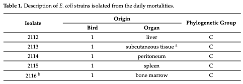
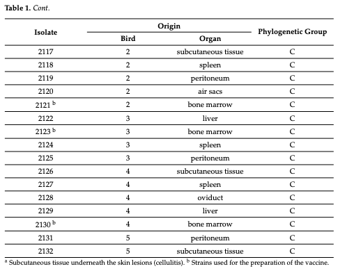
3. Results and Discussion
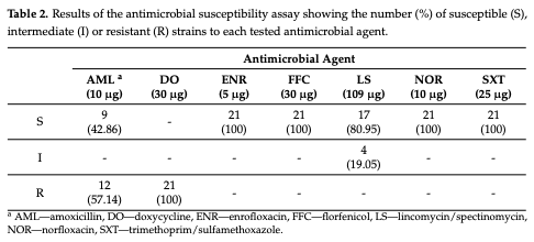
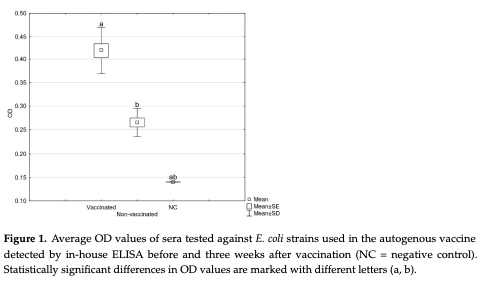
4. Conclusions
- Nolan, L. K., Vaillancourt, J.-P., Barbieri, N. L., & Logue, C. M. (2020). Colibacillosis. In D. E. Swayne (Ed.), Diseases of Poultry (14th ed., pp. 770–860). Wiley-Blackwell.
- Mageiros, L., Méric, G., Bayliss, S. C., Pensar, J., Pascoe, B., Mourkas, E., Calland, J. K., Yahara, K., Murray, S., Wilkinson, T. S., et al. (2021). Genome evaluation and the emergence of pathogenicity in avian Escherichia coli. Nature Communications, 12, 765–767.
- Pires dos Santos, T., Bisgaard, M., & Christensen, H. (2013). Genetic diversity and virulence profiles of Escherichia coli causing salpingitis and peritonitis in broiler breeders. Veterinary Microbiology, 162, 876–880.
- Li, L., Thøfner, I., Christensen, J. P., Ronco, T., Pedersen, K., & Olsen, R. H. (2017). Evaluation of the efficacy of an autogenous Escherichia coli vaccine in broiler breeders. Avian Pathology, 46, 300–308.
- Koutsianos, D., Gantelet, H., Franzo, G., Lecoupeur, M., Thibault, E., Cecchinato, M., & Koutoulis, K. C. (2020). An assessment of the level of protection against colibacillosis conferred by several autogenous and/or commercial vaccination programs in conventional pullets upon experimental challenge. Veterinary Sciences, 7, 80.
- Ievy, S., Islam, M. S., Sobur, M. A., Talukder, M., Rahman, M. B., Khan, M. F. R., & Rahman, M. T. (2020). Molecular detection of avian pathogenic Escherichia coli (APEC) for the first time in layer farms in Bangladesh and their antibiotic resistance patterns. Microorganisms, 8, 1021.
- Christensen, H., Bachmeier, J., & Bisgaard, M. (2021). New strategies to prevent and control avian pathogenic Escherichia coli (APEC). Avian Pathology, 50, 370–381.
- Kairmi, S. H., Taha-Abdelaziz, K., Yitbarek, A., Sargolzaei, M., Spahany, H., Astill, J., Shojadoost, B., Alizadeh, M., Kulkarni, R. R., Parkinson, J., et al. (2022). Effects of therapeutic levels of dietary antibiotics on the cecal microbiome composition of broiler chickens. Poultry Science, 101, 101864.
- Oliveira, A., Sereno, R., & Azaredo, J. (2010). In vivo efficiency evaluation of a phage cocktail in controlling severe colibacillosis in confined conditions and experimental poultry houses. Veterinary Microbiology, 146, 303–308.
- Abd-El-Ghany, W. A., & Ismail, M. (2014). Tackling experimental colisepticaemia in broiler chickens using phytobiotic essential oils and antibiotic alone or in combination. Iranian Journal of Veterinary Research, 15, 110–115.
- Ceccarelli, D., Van-Essen-Zandbergen, A., Smid, B., Veldman, K. T., Boender, G. J., Fischer, E. A. J., Mevius, D. J., & Van der Goot, J. A. (2017). Competitive exclusion reduces transmission and excretion of extended-spectrum-β-lactamase-producing Escherichia coli in broilers. Applied and Environmental Microbiology, 83, e03439-16.
- Wernicki, A., Nowaczek, A., & Urban-Chmiel, R. (2017). Bacteriophage therapy to combat bacterial infections in poultry. Virology Journal, 14, 1–13.
- Chantziaras, I., Smet, A., Filippitzi, M. E., Damiaans, B., Haesebrouck, F., Boyen, F., & Dewulf, J. (2018). The effect of a commercial competitive exclusion product on the selection of enrofloxacin resistance in commensal E. coli in broilers. Avian Pathology, 47, 443–454.
- Methner, U., & Rösler, U. (2020). Efficacy of a competitive exclusion culture against extended-spectrum β-lactamase-producing Escherichia coli strains in broilers using a seeder bird model. BMC Veterinary Research, 16, 1–7.
- Guabiraba, R., & Schouler, C. (2015). Avian colibacillosis: Still many black holes. FEMS Microbiology Letters, 362, fnv118.
- Landman, W. J. M., & Van Eck, J. H. H. (2017). The efficacy of inactivated Escherichia coli autogenous vaccines against the E. coli peritonitis syndrome in layers. Avian Pathology, 46, 658–665.
- Kromann, S., Olsen, R. H., Bojesen, A. M., Jensen, H. E., & Thøfner, I. (2021). Protective potential of an autogenous vaccine in an aerogenous model of Escherichia coli infection in broiler breeders. Vaccines, 9, 1233.
- Gottstein, Ž., Lozica, L., Lukač, M., & Horvatek Tomić, D. (2022). Production parameters after ad hoc application of autogenous vaccine during production following clinical colibacillosis in layer flocks. In Proceedings of the XIV Symposium Poultry Days 2022, Poreč, Croatia, 11–14 May 2022 (pp. 45–50).
- Clermont, O., Christenson, J. K., Denamur, E., & Gordon, D. M. (2013). The Clermont Escherichia coli phylo-typing method revisited: Improvement of specificity and detection of new phylo-groups. Environmental Microbiology Reports, 5, 58–65.
- Lozica, L., Ekert Kabalin, A., Dolenčić, N., Vlahek, M., & Gottstein, Ž. (2021). Phylogenetic characterization of avian pathogenic Escherichia coli strains longitudinally isolated from broiler breeder flocks vaccinated with autogenous vaccine. Poultry Science, 100, 101079.
- Leitner, G., Melamed, D., Drabkin, N., & Heller, E. D. (1990). An enzyme-linked immunosorbent assay for detection of antibodies against Escherichia coli: Association between indirect hemagglutination test and survival. Avian Diseases, 34, 58–62.
- Zanella, A., Alborali, G. L., Bardotti, M., Candotti, P., Guadagnini, P. F., Martino, P. A., & Stonfer, M. (2000). Severe Escherichia coli O111 septicaemia and polyserositis in hens at the start of lay. Avian Pathology, 29, 311–317.
- Clermont, O., Dixit, O. V. A., Vangchhia, B., Condamine, B., Dion, S., Bridier-Nahmias, A., Denamur, E., & Gordon, D. (2019). Characterization and rapid identification of phylogroup G in Escherichia coli, a lineage with high virulence and antibiotic resistance potential. Environmental Microbiology, 21, 3107–3117.
- Logue, C. M., Wannemuehler, Y., Nicholson, B. A., Doetkott, C., Barbieri, N. L., & Nolan, L. K. (2017). Comparative analysis of phylogenetic assignment of human and avian ExPEC and fecal commensal Escherichia coli using the previous and revised Clermont phylogenetic typing methods and its impact on avian pathogenic Escherichia coli (APEC) classification. Frontiers in Microbiology, 8, 283.
- Starčič Erjavec, M., Predojević, L., & Žgur-Bertok, D. (2017). Commentary: Comparative analysis of phylogenetic assignment of human and avian ExPEC and fecal commensal Escherichia coli using the previous and revised Clermont phylogenetic typing methods and its impact on avian pathogenic Escherichia coli (APEC) classification. Frontiers in Microbiology, 8, 1904.
- Lozica, L., Repar, J., & Gottstein, Ž. (2021). Longitudinal study on the effect of autogenous vaccine application on the sequence type and virulence profiles of Escherichia coli in broiler breeder flocks. Veterinary Microbiology, 259, 109159.
- Hess, C., Troxler, S., Jandreski-Cvetkovic, D., Zloch, A., & Hess, M. (2022). Escherichia coli isolated from organic laying hens reveal a high level of antimicrobial resistance despite no antimicrobial treatments. Antibiotics, 11, 467.
- Turner, S. P. (2011). Breeding against harmful social behaviours in pigs and chickens: State of the art and the way forward. Applied Animal Behaviour Science, 134, 1–9.
- Crespo, R. (2020). Developmental, metabolic, and other noninfectious disorders. In D. E. Swayne (Ed.), Diseases of Poultry (14th ed., pp. 1307–1329). Wiley-Blackwell.
- El-Lethey, H., Aerni, V., Jungi, T. W., & Wechsler, B. (2000). Stress and feather pecking in laying hens in relation to housing conditions. British Poultry Science, 41, 22–28.
- Gray, P., Jenner, R., Norris, J., Page, S., Browning, G., Australian Veterinary Association Ltd., & Animal Medicines Australia. (2021). Antimicrobial prescribing guidelines for poultry. Australian Veterinary Journal, 99, 181–235.
- Giersberg, M. F., Spindler, B., Rodenburg, B., & Kemper, N. (2020). The dual-purpose hen as a chance: Avoiding injurious pecking in modern laying hen husbandry. Animals, 10, 16.
- Lozica, L., Villumsen, K. R., Li, G., Hu, X., Maurić Maljković, M., & Gottstein, Ž. (2022). Genomic analysis of Escherichia coli longitudinally isolated from broiler breeder flocks after application of an autogenous vaccine. Microorganisms, 10, 377.









Candidate Genes Associated with Survival Following Highly Pathogenic Avian Influenza Infection in Chickens

Adisseo – a Bluestar Company





Norel Animal Nutrition

Evonik Animal Nutrition


United States