Introduction
The poultry industry plays a vital role in global food security, providing an affordable and efficient source of animal protein (Castro et al., 2023). However, maintaining flock health and productivity remains a major challenge, with intestinal health playing a central role in poultry performance (Korver, 2023). With the rise of antibiotic resistance across the globe, the potential restriction of in-feed antibiotic growth promoters (AGPs) in animal food production could have a significant impact on the growth, health, and welfare of poultry (McEwen et al., 2017). The removal of AGPs can lead to an increase in enteric infections, such as necrotic enteritis (NE), within flocks, affecting the sustainability of the poultry industry (Casewell et al., 2003; M’Sadeq et al., 2015). Necrotic enteritis (NE) is a serious intestinal disease in broiler chickens, primarily caused by C. perfringens (mainly type G) (Keyburn et al., 2006). These bacteria produce tissue-degrading and pore-forming toxins, including α-toxin, NetB, and TpeL, which damage the intestinal barrier and contribute to disease progression (Ou et al., 2024). The clinical form of NE can result in high mortality rates (up to 50%) and substantial economic losses. In contrast, the subclinical form causes mild intestinal damage, impairing nutrient absorption and leading to a significant decline in growth and overall performance (Gholamiandehkordi et al., 2007).
Over the past years, various alternative strategies have been explored to control NE in chickens, with varying degrees of success (Dahiya et al., 2006; Abreu et al., 2023). Therefore, the identification and evaluation of novel antimicrobial strategies for controlling necrotic enteritis in the post-antibiotic era remain a critical priority. Manipulation of gut microbiota or establishment of a healthy gut microbiota with probiotics could serve as a strategy to maintain intestinal health and support the development and maturation of a gut-associated immune system in chickens (Yadav and Jha, 2019). Probiotics are considered one of the potential alternatives due to their immunomodulatory and intestinal health benefits (El Jeni, 2021). We have previously demonstrated that oral administration of certain Lactobacillus strains modulates intestinal immune responses and microbiota in chickens, alleviating the severity of necrotic enteritis (NE) (Alizadeh et al., 2023, 2024, 2025). Beyond traditional lactic acid bacteria, next-generation probiotics have gained increasing attention for their potential to modulate gut function and host immunity. In this context, B. thetaiotaomicron is emerging as a promising next-generation probiotic due to its intestinal health-promoting properties (Porter et al., 2018). This anaerobic bacterium, naturally present in the chicken gut, breaks down complex plant polysaccharides into simpler compounds and produces short-chain fatty acids (SCFAs) that support host health (Porter et al., 2018). Studies in humans indicate that B. thetaiotaomicron exhibits remarkable flexibility by producing numerous carbohydrate-degrading enzymes that can break down diverse types of dietary fibers (Lap´ebie et al., 2019). SCFAs serve as an energy source for intestinal epithelial cells and contribute to gut barrier integrity by enhancing tight junction expression and increasing mucus production (Liu et al., 2021; Ali et al., 2022). Additionally, a previous study in mice highlighted that SCFAs promote the production of interleukin-22 (IL-22) by innate lymphoid cells, which contributes to maintaining intestinal homeostasis and protecting against mucosal inflammation (Yang et al., 2020). Experimental evidence also suggests that Bacteroides thetaiotaomicron can stimulate Paneth cell–derived antimicrobial peptides, such as angiogenin-4 (Ang4), enhancing host defenses against enteric pathogens (Xu et al., 2003). Despite growing evidence supporting the beneficial role of B. thetaiotaomicron in mammalian gut health, its effects in poultry remain poorly understood. Therefore, the present study was designed to evaluate the impact of B. thetaiotaomicron supplementation on gut health, innate immune responses, gut microbiota composition, and resistance to NE in chickens. We hypothesized that oral administration of B. thetaiotaomicron could enhance intestinal barrier integrity, favorably modulate gut microbiota and innate immune responses, and thereby reduce the severity of NE in broiler chickens.
Materials and methods
Birds housing
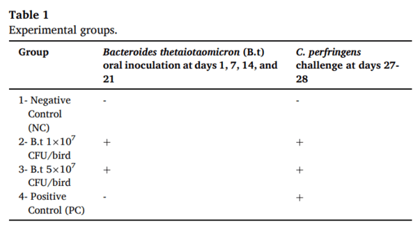
One-day-old male broiler chickens (Ross 708, n = 120) were obtained from a commercial hatchery (Guelph, Canada) and randomly assigned to four treatment groups (Table 1). Chickens were vaccinated at the hatchery according to the standard vaccination schedule for broiler chickens. Birds were group-housed in separate floor pens (≈12 birds/m²) with wood shavings and ad libitum access to feed and water in the isolation facility of the Ontario Veterinary College, University of Guelph. The temperature was maintained at 32 ◦C for the first week, then reduced by 2 ◦C per week until reaching 26 ◦C at week 4. Humidity was kept at 40% throughout the trial. All birds’ experiments were approved by the Animal Care Committee of the University of Guelph and adhered to the guidelines for the use of animals (Animal Utilization Protocol # 4815).
Bacteroides thetaiotaomicron preparation
Brain Heart Infusion (BHI3) broth (BHI supplemented with hemin, vitamin K, and L-cysteine) was prepared according to Dr. Michale Surette’s lab protocol at McMaster University. Using sterile loops, the frozen stock was aseptically transferred from the cryovial and gently streaked onto the surface of a preconditioned BHI3 agar plate. The inoculated plate was then placed inside the anaerobic chamber and incubated at 37 ◦C for 24–48 hours. Cells were collected under anaerobic conditions from the agar plate using a sterile loop, suspended in 5 mL of BHI3 broth, and incubated at 37 ◦C for an additional 24–48 hours. The resulting culture was then passaged into 40 mL of fresh BHI3 broth at a 1:100 dilution and incubated at 37 ◦C for 24–48 hours. The final inoculum was subsequently used to inoculate the birds. The optical density at 600 nm (OD₆₀₀) was measured using a spectrophotometer (Thermo Fisher Scientific, Mississauga, ON, Canada) to achieve the desired bacterial cell densities of 1 × 10⁷ and 5 × 10⁷ colony-forming units (CFU)/mL.
NE challenge model
NE was experimentally induced in chickens as previously described (Shojadoost et al., 2022).The highly virulent Clostridium perfringens strain CP4 used in this experiment was kindly supplied by Dr. John Prescott (University of Guelph, Canada). The strain was cultured anaerobically at 37 ◦C overnight in Cooked Meat Medium (Thermo Fisher Scientific, Mississauga, ON, Canada). Subsequently, 3% (v/v) of the overnight culture was inoculated into Fluid Thioglycollate Medium (FTG; Sigma-Aldrich, Oakville, ON, Canada) and incubated anaerobically for 15 h at 37 ◦C. The optical density (OD) of the culture was measured at 600 nm using a spectrophotometer (Fisher Scientific, Canada) to ensure that each chicken received at least 3 × 10⁸ CFU of bacteria. The prepared inoculum was subsequently used to challenge the birds.
Experimental design and sampling
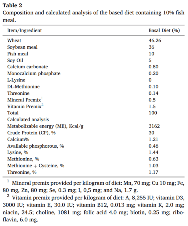
One-day-old male broiler chickens (n = 120) were randomly assigned to four treatment groups, with 30 birds per group. On days 1, 7, 14, and 21 post-hatch, birds in groups 2 and 3 were orally inoculated with 1 × 10⁷ and 5 × 10⁷ CFU/chicken of B. thetaiotaomicron, respectively. Groups 1 and 4 did not receive probiotic treatment and served as the negative and positive control groups, respectively (Table 1). All birds were initially fed a commercial starter diet; however, on day 14 of age, they were switched to a wheat-based diet rich in non-starch polysaccharides, high in protein (30%), and containing 5% fish meal was used as a predisposing factor for the development of clinical NE (Table 2). On days 22 and 27 post-hatch (prior to the C. perfringens challenge), seven birds per group were euthanized, and 3-cm segments of the ileum and cecal tonsils were collected for gene expression analysis (stored in RNAlater at − 80 ◦C). On the same days, cecal contents were also collected for microbiome analysis. On day 27 of age, all birds in groups 2, 3, and 4 were orally challenged with 3 × 10⁸ CFU/mL of C. perfringens twice daily for two consecutive days. Group 1 remained unchallenged. On day 29, all birds were euthanized, and intestinal lesions were scored.
RNA extraction and reverse transcription
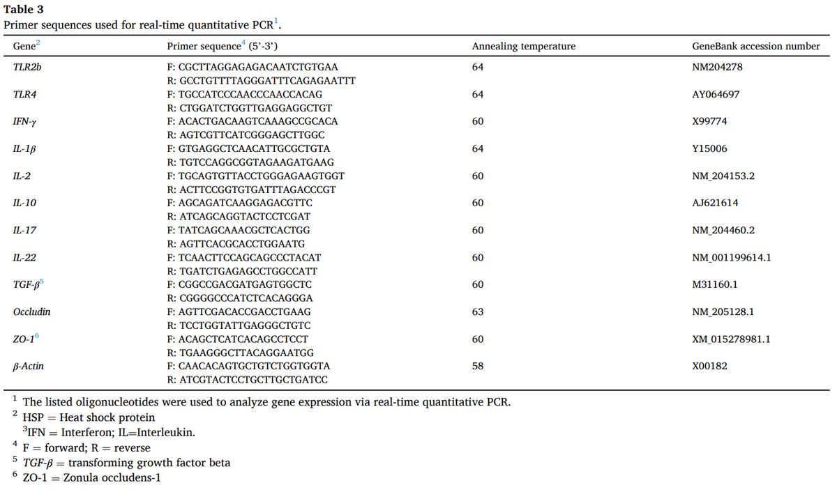
RNA was isolated from the ileum and cecal tonsils using TRIzol reagent (Thermo Fisher Scientific, Mississauga, ON, Canada), following the protocol described by (Alizadeh et al., 2022). To eliminate genomic DNA contamination, the samples were treated with DNase. RNA concentration and purity were determined using a NanoDrop spectrophotometer (Thermo Fisher Scientific, Mississauga, ON, Canada). Complementary DNA (cDNA) was synthesized from RNA using the Superscript® II First Strand Synthesis Kit (Invitrogen, Burlington, ON, Canada), following the manufacturer’s instructions. The primer sequences used for the PCR reactions, along with their respective annealing temperatures, are listed in Table 3.
Reverse transcriptase polymerase chain reaction (RT-PCR)
Quantitative real-time PCR was performed using the LightCycler 480 II system (Roche Diagnostics GmbH, Mannheim, Germany), following the protocol previously described (Alizadeh et al., 2020). Quantitative real-time PCR (qRT-PCR) was carried out using the LightCycler® 480 II system (Roche Diagnostics GmbH, Mannheim, Germany). Each reaction (20 μl total) contained 10 μl of 2X SYBR Green Master Mix (Roche Diagnostics), 1 μl of forward primer (5 μM), 1 μl of reverse primer (5 μM), 3 μl of PCR-grade water, and 5 μl of target cDNA diluted 1:10 in nuclease-free water. The cycling program consisted of an initial denaturation at 95◦C, followed by 40–50 amplification cycles of 95◦C for 10 s, annealing at primer-specific temperatures (as listed in Table 1), and extension at 72◦C for 10 s. The relative expression levels of Toll-like receptors (TLR)2 and TLR4, cytokines including interferon-gamma (IFN-γ), interleukins IL-1β, IL-2, IL-10, IL-17, and IL-22; transforming growth factor-beta (TGF-β); and tight junction proteins zonula occludens-1 (ZO-1) and occludin were quantified. All gene expression levels were normalized to the housekeeping gene β-actin.
Lesion scoring
On day 29 of age, all remaining birds were euthanized, and lesion scoring was conducted following the method previously described (Shojadoost et al., 2012).
DNA extraction and 16S rRNA gene sequencing
Microbial genomic DNA was extracted from the cecal contents of six birds per treatment using the QIAamp Fast DNA Stool Mini Kit (Qiagen, Canada), following the manufacturer’s instructions. The concentration and purity of the extracted DNA were assessed using a NanoDrop 2000 spectrophotometer (Thermo Fisher Scientific, Mississauga, ON, Canada). Sequencing and library preparation were performed at the Integrated Microbiome Resource (IMR) (http://imr.bio) following established internal protocols. Target amplification focused on the bacterial 16S rRNA gene, specifically the V3/V4 variable region, using dual-barcoded primers: 341F (5′-CCTACGGGNGGCWGCAG-3′) and 805R (3′-GACTACHVGGGTATCTAATCC-5′).
Statistical and bioinformatics analysis
Power analyses (with a target power of 0.85) were conducted to determine the appropriate sample size needed to detect statistically significant effects. The data for lesion scoring and relative gene expression were assessed for normality using the Shapiro-Wilk test. Statistical significance among groups was analyzed using a one-way ANOVA followed by Tukey's test, with a significance level of α = 0.05. Statistical analyses were performed using GraphPad Prism (version 9).
Bioinformatics analysis was performed as described previously (Oladokun et al., 2025) using QIIME 2 software (version 2023.7). Rare amplicon sequence variants (ASVs) were excluded if present in less than one sample or if frequency was less than 0.1% of the mean sample depth. Visualization was performed using the MicrobiomeAnalyst 2.0 web-based platform (Lu et al. 2023). Individual alpha diversity was evaluated through rarefaction curves based on the observed operational taxonomic unit (OTU) metric. Differences in alpha diversity were statistically assessed using the Shannon index and the Kruskal–Wallis test. Beta diversity analysis was conducted through principal coordinates analysis (PCoA) plots, utilizing the weighted UniFrac distance metric, with statistical significance assessed via Permutational Multivariate Analysis of Variance (PERMANOVA). Relative abundances at different taxonomic levels were visualized using stacked bar charts, and significant microbiota abundance was determined by Linear Discriminant Analysis Effect Size (LEfSe) and visualized using box plots in the MicrobiomeAnalyst 2.0 web-based platform.
Results
Gross lesion in the intestine
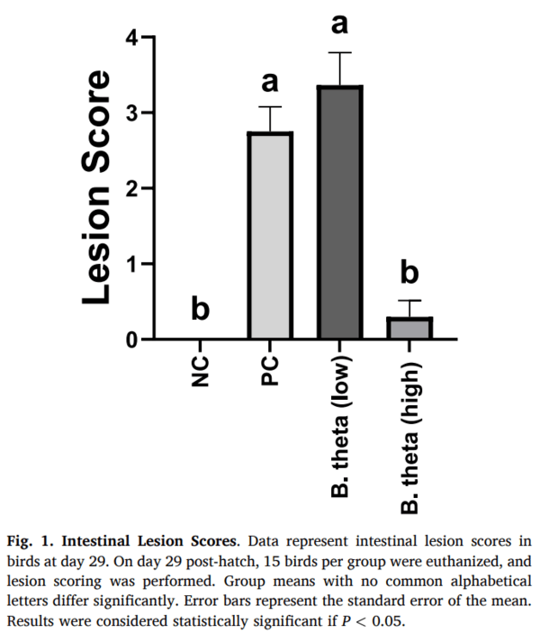
The results of gross pathology (Fig. 1) demonstrated that, although no significant difference was observed for the low dose of B. thetaiotaomicron (1 × 10⁷ CFU/mL) compared to the positive control group (untreated and C. perfringens-challenged), oral administration of birds with a high dose of B. thetaiotaomicron (5 × 10⁷ CFU/mL) reduced (P = 0.0001) the mean lesion score compared to the positive control group (2.75 vs 0.3).
Toll-like receptors (TLR) expression in the intestine
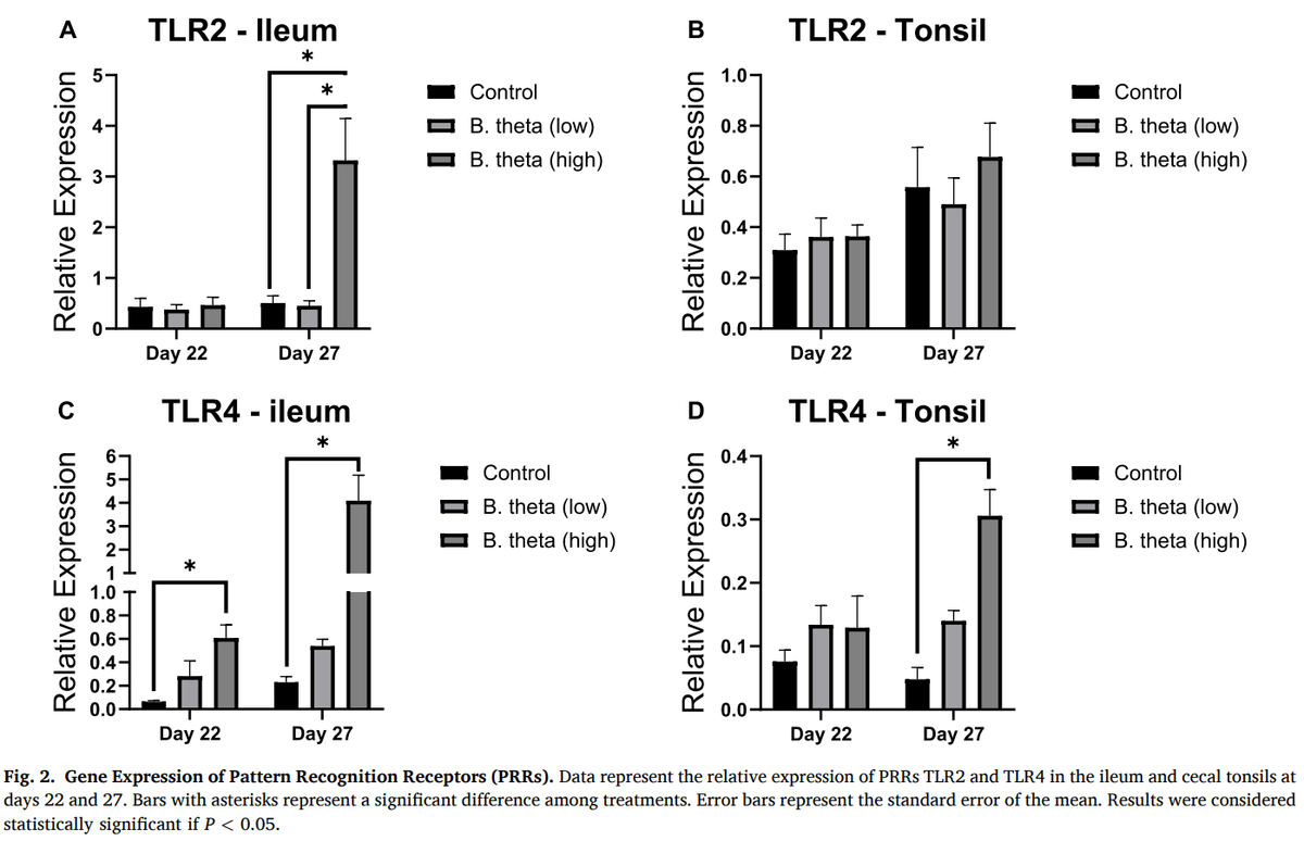
TLR2 expression (Fig. 2A, B) in the ileum was not affected by treatment group on day 22 of age. TLR2 expression in the cecal tonsil also remained unchanged following probiotic treatment on both day 22 and day 27. However, on day 27, high concentrations of B. thetaiotaomicron upregulated (P = 0.028) TLR2 expression in the ileum (~6.5-fold). TLR4 expression in the cecal tonsil (Fig. 2C, D) was unaffected by treatment group on day 22 of age. High concentrations of B. thetaiotaomicron enhanced TLR4 expression in the cecal tonsil (~6.3- fold; P = 0.001) on day 27, and in the ileum on day 22 (~9.5-fold; P = 0.035) and day 27 (~18-fold; P = 0.026).
Cytokine expression in the intestine
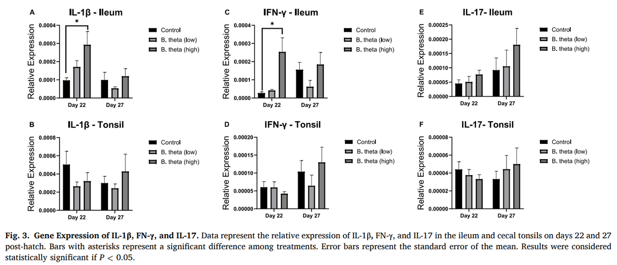
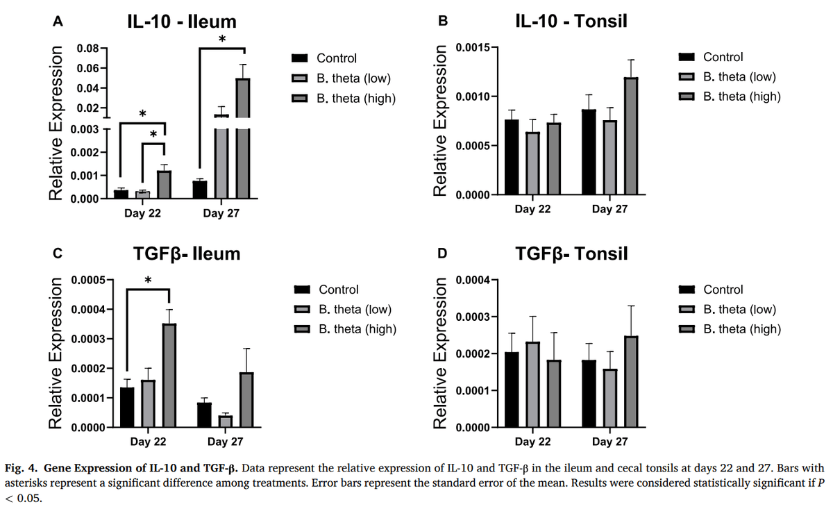
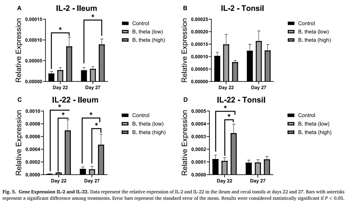
IL-1β expression (Fig. 3A, B) in the ileum was upregulated (P = 0.021) by high concentrations of B. thetaiotaomicron on day 22 of age (~3.3-fold). Nevertheless, no significant difference was observed in the ileum on day 27 or in the cecal tonsil at either time point. Expression of IFN-γ (Fig. 3C, D) in the ileum was upregulated (P = 0.002) by high concentrations of B. thetaiotaomicron on day 22 of age (~9.3-fold). Nevertheless, no significant difference was observed on day 27. Furthermore, in the cecal tonsil, IFN-γ expression remained unchanged following probiotic treatment on both days 22 and 27. IL-17 expression in both the ileum and cecal tonsils was not affected by treatment at either time point (Fig. 3E, F). High-dose administration of B. thetaiotaomicron upregulated IL-10 expression in the ileum at day 22 (≈3.3-fold; P = 0.001) and day 27 (≈65-fold; P = 0.042; Fig. 4A, B). In contrast, IL-10 expression in the cecal tonsil remained unchanged at both time points. Expression of TGF-β (Fig. 4C, D) in the ileum was upregulated (P = 0.002) by high concentrations of B. thetaiotaomicron on day 22 of age (~2.6-fold). However, no significant difference was observed in the cecal tonsil at either time point. Expression of IL-2 (Fig. 5A, B) in the ileum was induced by oral administration of high concentrations of B. thetaiotaomicron at both day 22 (~4.4-fold; P = 0.005) and day 27 (~3.3-fold; P = 0.003), whereas no significant differences were observed in the cecal tonsil at either time point. Expression of IL-22 (Fig. 5C, D) was upregulated in the ileum in the group that received high concentrations of B. thetaiotaomicron at both day 22 (≈60- fold, P = 0.0005) and day 27 (≈5-fold, P = 0.032). Similarly, in the cecal tonsil, IL-22 expression was upregulated (P = 0.004) at day 22 (~2.7- fold), but not at day 27.
Tight junction proteins (TJPs) expression in the intestine
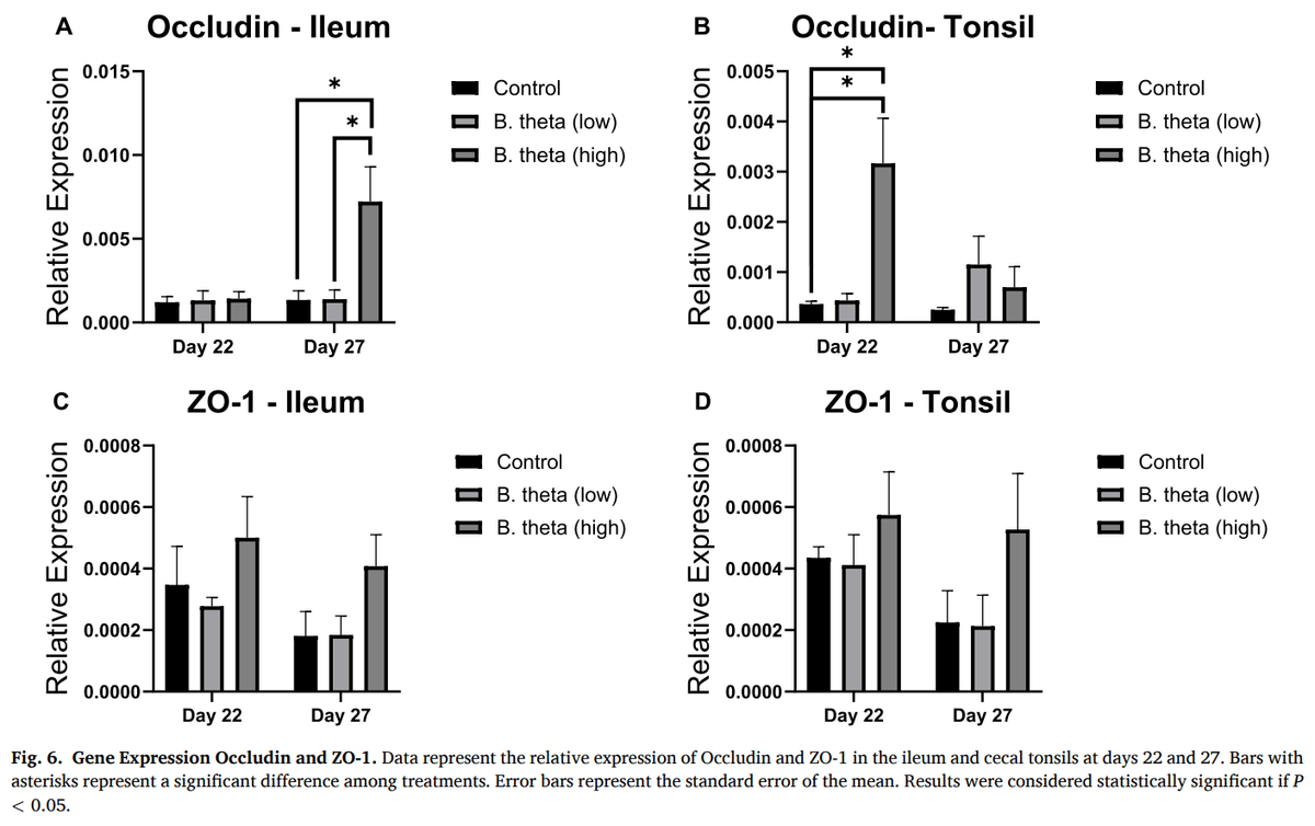
Expression of occludin (Fig. 6A, B) in the ileum was not affected by the treatment groups on day 22 of age. However, on day 27, oral administration of a high concentration of B. thetaiotaomicron significantly upregulated occludin expression in the ileum (~5.3-fold; P = 0.022). High-dose B. thetaiotaomicron treatment increased (P = 0.001) occludin expression in the caecal tonsil on day 22 (~8.7-fold), but not on day 27. Expression of zonula occludens protein 1 (ZO-1) (Fig. 6C, D) was not affected by treatment groups at either time point in either tissue.
Cecal microbiome
Sequencing yielded a total of 2,652,752 high-quality reads, averaging 74,477 reads per sample after quality filtering and demultiplexing. Across all samples, 491 operational taxonomic units (OTUs) were identified at a 97% sequence similarity threshold. Sequencing depth was observed to sufficiently capture the microbial diversity in the analyzed samples, as indicated by the rarefaction curves showing individual alpha diversity at all sampling times (Supplementary Figure 1).
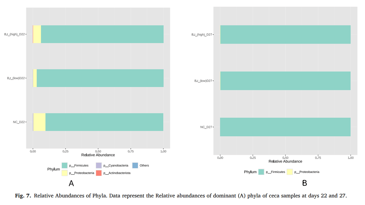
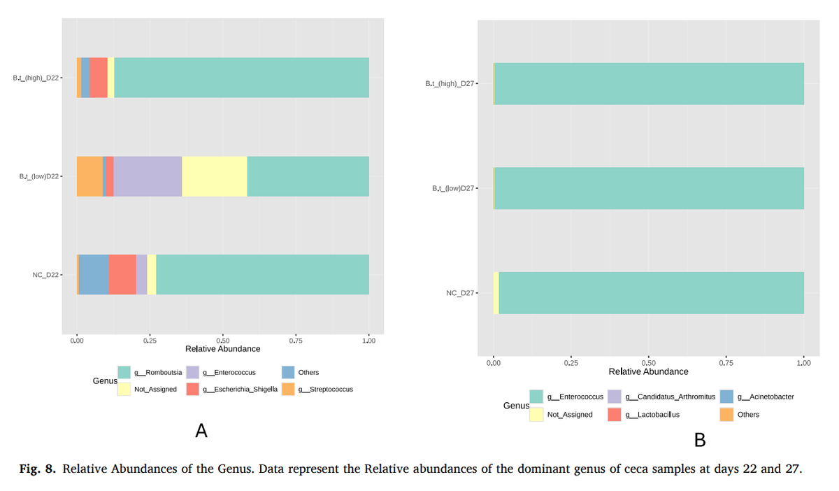
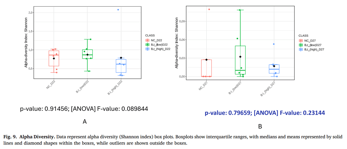
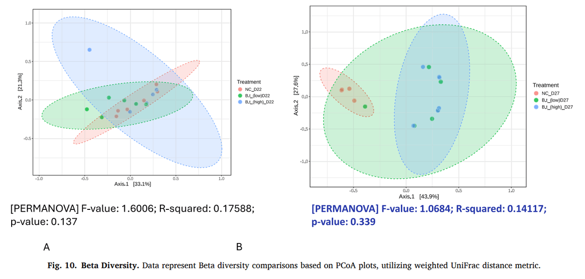
An overview of microbiota relative (percentage) abundances at the phylum level showed that Firmicutes (> 80%) was the most dominant phylum, followed by Proteobacteria (< 12%) across both timepoints (Fig. 7). At the genus taxa, while microbes from the genus Romboutsia accounted for at least 75% of the community, the genus Enterococcus accounted for at least 80% of the community at day 27 (Fig. 8). Additional results on the relative abundances of other dominant taxa are presented in Supplementary Figures 2A–H. Additionally, alpha diversity analysis, measured by Shannon Index (which considers both species richness and evenness), showed no significant effects of treatment groups on microbial diversity, both at days 22 (P = 0.92) and 27 (P = 0.80) (Fig. 9). With respect to beta diversity, treatment groups did not have a statistically significant effect on the microbial community similarities or differences across samples at either time point (d22- P = 0.14; d27- P = 0.34) (Fig. 10A, B). However, on day 27, the negative control samples clustered almost perfectly, in contrast to the other treatment groups (Fig. 10B).
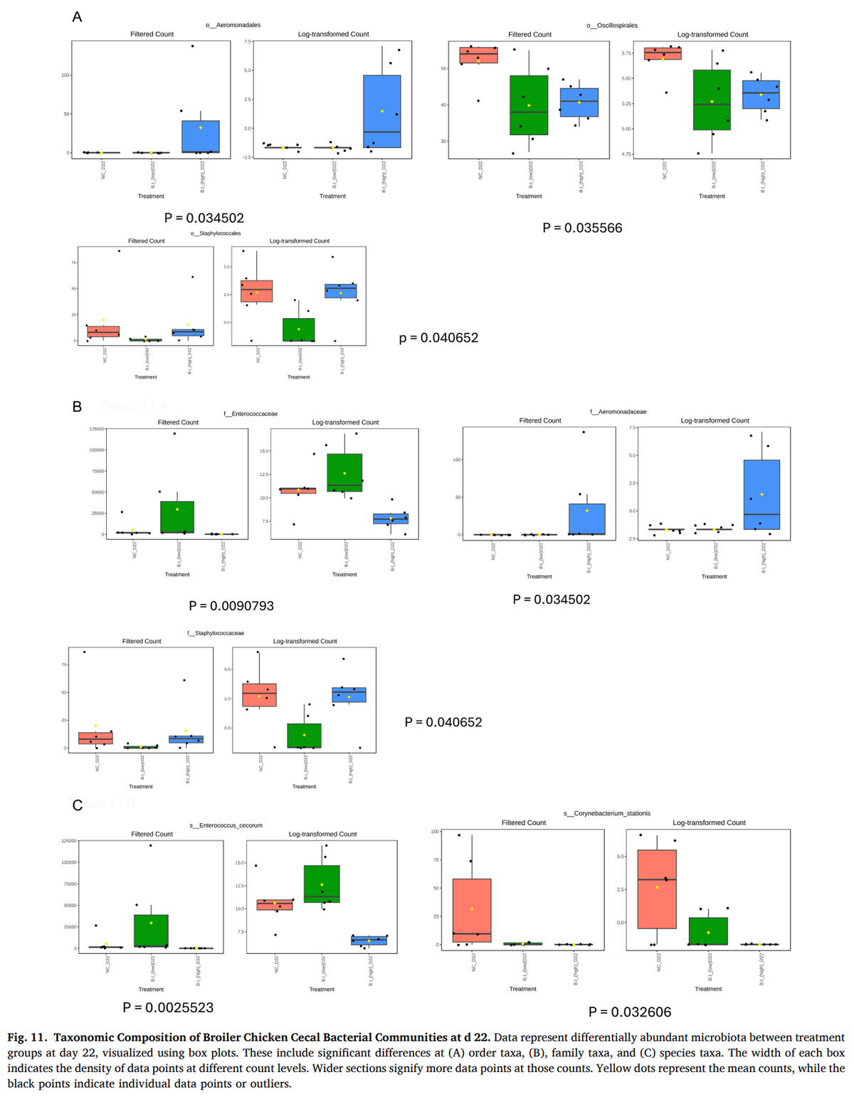
In terms of significant microbiota abundance, visualizations are provided for both filtered counts and log-transformed counts to stabilize variance and achieve a more normal distribution of the data. For samples collected at day 22, birds receiving the high dose of B. thetaiotaomicron showed a significantly (P = 0.03) higher abundance of the order Aeromonadales compared to other treatment groups. In contrast, the low-dose B. thetaiotaomicron group had a significantly (P =0.04) lower mean abundance of the order Staphylococcales than the other groups. Both high- and low-dose B. thetaiotaomicron treatments similarly reduced (P = 0.04) the mean abundance of the order Oscillospirales compared to the negative control group (Fig. 11A). This trend of significant increases and decreases was consistent for the high dose with respect to the family Aeromonadaceae, and for the low dose with respect to the family Staphylococcaceae. In addition, the high-dose treatment also reduced the mean abundance of the family Enterococcaceae at day 22 (Fig. 11B). At the species level, high-dose B. thetaiotaomicron treatment significantly (P < 0.05) reduced the mean abundance of Enterococcus cecorum (P = 0.003) and Corynebacterium stationis (P = 0.03), relative to other treatment groups at day 22 (Fig. 11C).
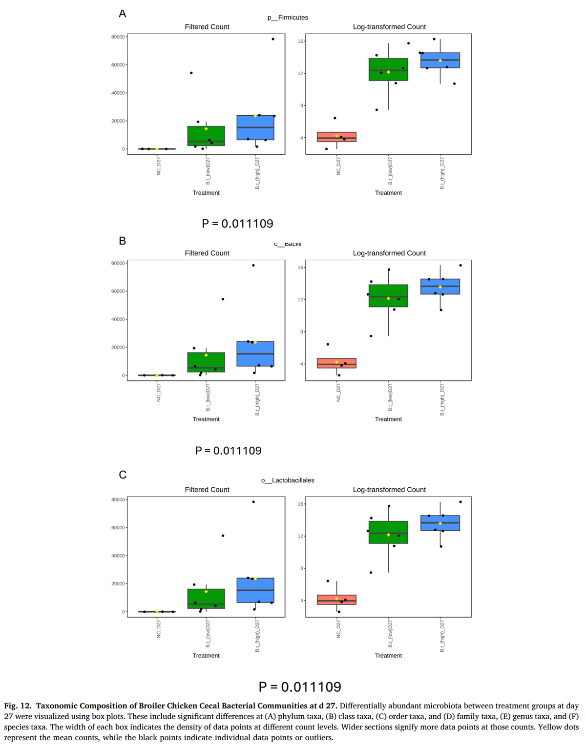
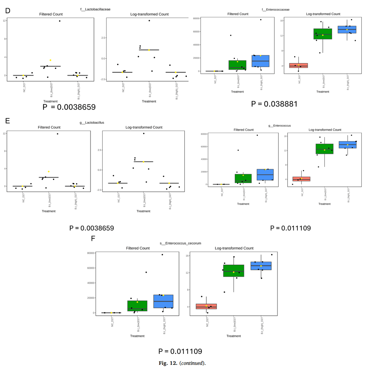
At day 27, both high- and low-dose B. thetaiotaomicron treatments consistently increased the mean abundance of the phylum Firmicutes (P = 0.01), class Bacilli (P = 0.01), order Lactobacillales (P = 0.01), family Enterococcaceae (P = 0.04), genus Enterococcus (P = 0.01), and species Enterococcus cecorum (P = 0.01), compared to the negative control group (Fig. 12A, B, C, D, E, F). In addition, the low-dose treatment significantly increased (P = 0.004), the mean abundance of the family Lactobacillaceae and genus Lactobacillus relative to the other groups (Fig. 12D, E).
Discussion
Necrotic enteritis (NE), caused by C. perfringens, is a major poultry disease that leads to substantial economic losses due to impaired growth and increased mortality (Skinner et al., 2010; Emami and Dalloul, 2021). With growing restrictions on antibiotic use, interest has shifted toward alternative strategies, such as next-generation probiotics (NGPs), for the prevention and control of NE. Among these, B. thetaiotaomicron, a prominent gut commensal, has emerged as a promising candidate due to its immunomodulatory properties and ability to enhance intestinal barrier function (Elahi et al., 2021; Li et al., 2021).
In the present study, oral administration of a high concentration of B. thetaiotaomicron (5 × 10⁷ CFU/mL) significantly alleviated intestinal lesions induced by C. perfringens, suggesting a protective effect against necrotic enteritis. The absence of protection at the lower dose (1 × 10⁷ CFU) indicates that a threshold level of supplementation may be required to elicit sufficient biological activity, supporting a dosedependent mechanism of action. This protective effect was associated with modulation of innate immune responses, particularly increased TLR4 expression in the ileum and cecal tonsils. High-dose B. thetaiotaomicron also enhanced the expression of key proinflammatory (IFN-γ, IL-1β), regulatory (IL-10, TGF-β), and barrierassociated cytokines (IL-22) in a tissue- and time-dependent manner, with the ileum showing greater responsiveness than the cecal tonsils. Furthermore, increased expression of the tight junction protein occludin suggested an improvement in intestinal barrier integrity. In addition to immunomodulatory effects, high-dose B. thetaiotaomicron supplementation reshaped the gut microbiota by enriching beneficial taxa (e.g., Enterococcus, Lactobacillus) while reducing potential pathogens (e.g., Enterococcus cecorum, Corynebacterium stationis). Collectively, these findings suggest that high doses of B. thetaiotaomicron may protect birds against enteric pathogens by modulating the host immune response, enhancing epithelial barrier function, and reshaping the gut microbiome.
The increased expression of TLR2 and TLR4 observed in this study suggests enhanced innate immune sensing in response to B. thetaiotaomicron, potentially promoting early immune activation while maintaining immune balance. Such activation is consistent with a controlled inflammatory response that supports epithelial defense and tissue repair rather than excessive inflammation. Toll-like receptors (TLRs), particularly TLR2 and TLR4, are essential components of the innate immune system, recognizing microbial signals and initiating immune responses that balance inflammation and tissue repair (Akira and Takeda, 2004). B. thetaiotaomicron, a prominent gut commensal, is a Gram-negative anaerobic bacterium characterized by the presence of lipopolysaccharides (LPS) in its outer membrane, which are classic ligands for TLR4. Engagement of TLR4 by B. thetaiotaomicron LPS triggers downstream signaling pathways leading to activation of innate immune responses and production of pro-inflammatory cytokines (Lu et al., 2008). However, unlike many pathogenic Gram-negative bacteria, B. thetaiotaomicron possesses a structurally distinct and less immunostimulatory LPS, which can induce a more balanced and regulated TLR4-mediated response that supports gut homeostasis rather than excessive inflammation (Abreu, 2010). Consistent with this, B. thetaiotaomicron has been shown to interact with TLR4 to modulate host immune responses and enhance intestinal barrier function (Pither et al., 2022).
In addition to TLR4, the observed modulation of TLR2 expression suggests that B. thetaiotaomicron engages multiple innate immune pathways. Although TLR2 is traditionally associated with recognizing lipoproteins, peptidoglycan, and lipoteichoic acid primarily from Grampositive bacteria, it can also recognize atypical or modified microbial components from Gram-negative bacteria. B. thetaiotaomicron produces a variety of surface molecules, including lipoproteins, outer membrane proteins, and polysaccharides, that serve as ligands for TLR2 (Engelhart et al., 2023). Evidence indicates that B. thetaiotaomicron induces IL-10 production in a predominantly TLR2–MyD88-dependent manner (Engelhart et al., 2023). IL-10 is a key regulatory cytokine that helps modulate inflammation and promote tissue repair. Together, these findings underscore the dose-dependent interaction of B. thetaiotaomicron with TLR2 and TLR4, contributing to a balanced inflammatory response and strengthened epithelial barrier, thereby supporting its potential as a probiotic strategy for controlling necrotic enteritis in poultry.
The upregulation of IFN-γ and IL-1β observed in this study indicates an enhanced early pro-inflammatory response in the ileum following high-dose B. thetaiotaomicron administration. This response was evident at day 22 of age, suggesting timely immune activation that may contribute to the host’s ability to limit C. perfringens infection during necrotic enteritis. However, this effect was not sustained at later time points (day 27), and no significant changes were observed in the cecal tonsil at any time. Interferon-gamma (IFN-γ) and interleukin-1 beta (IL1β) are key pro-inflammatory cytokines involved in early host defense mechanisms against enteric pathogens such as C. perfringens (Dinarello, 2000). IFN-γ plays a critical role in activating macrophages and enhancing antigen presentation, while IL-1β contributes to the recruitment of immune cells and amplification of the inflammatory response (Eriguchi et al., 2018; Kaminsky et al., 2021). Modulation of these cytokines by probiotic interventions could therefore significantly influence disease outcomes. Overall, the findings of the present study suggest that B. thetaiotaomicron induces a transient, tissue-specific pro-inflammatory response, primarily in the ileum, which may provide effective immune activation without promoting chronic inflammation, supporting its potential as a targeted immunomodulatory probiotic in early-stage disease intervention.
n the present study, high-dose treatment with B. thetaiotaomicron led to a significant upregulation of TGF-β expression in the ileum on day 22, suggesting early induction of regulatory immune mechanisms prior to C. perfringens challenge. However, this effect was not sustained, as no significant differences were observed in TGF-β expression on day 27 or in the cecal tonsil at either time point. Similarly, IL-10 expression in the ileum was significantly upregulated at both day 22 and day 27, indicating a sustained regulatory immune environment in the gut ahead of infection, whereas IL-10 expression in the cecal tonsil remained unchanged. Regulatory cytokines such as IL-10 and transforming growth factor-beta (TGF-β) play critical roles in maintaining intestinal immune homeostasis by limiting excessive inflammation and promoting mucosal tolerance (Levings et al., 2002; Konkel and Chen, 2011; Wei et al., 2020). Their induction prior to infection can help prepare the host to mount a controlled immune response, minimizing tissue damage upon exposure to enteric pathogens. Together, these findings suggest that B. thetaiotaomicron selectively enhances local regulatory immune responses, particularly in the ileum, through early and sustained expression of IL-10 and transient induction of TGF-β, which may contribute to immune tolerance and reduced inflammatory damage in the event of a pathogenic challenge.
High-dose treatment with B. thetaiotaomicron significantly upregulated IL-2 expression in the ileum at both day 22 and day 27, indicating sustained activation of T cell–mediated immune responses in the gut prior to C. perfringens challenge. IL-22 expression was also significantly increased on day 22 in both the ileum and cecal tonsils, indicating an enhancement of epithelial barrier defenses before infection. However, IL-22 levels returned to baseline by day 27, suggesting that its expression may be tightly regulated and time-dependent. Interleukin-2 (IL-2) and interleukin-22 (IL-22) are cytokines that play essential roles in intestinal immune regulation and epithelial defense. IL-2 is a key regulator of T cell proliferation and activation, contributing to immune surveillance and pathogen clearance (Boyman and Sprent, 2012). IL-22, produced primarily by innate lymphoid cells, Th17, and Th22 cells, supports epithelial repair and enhances mucosal barrier function by inducing antimicrobial peptides and promoting epithelial cell survival (Wei et al., 2020). Overall, these findings suggest that B. thetaiotaomicron prepares the gut’s immune system by maintaining IL-2–driven immune alertness and IL-22–driven support for the gut lining, potentially enhancing the host’s resistance to necrotic enteritis.
In the present study, treatment with B. thetaiotaomicron did not alter zonula occludens-1 (ZO-1) expression in either the ileum or cecal tonsil at any time point, suggesting that this component of the tight junction complex was unaffected by the probiotic. In contrast, occludin expression showed tissue- and time-specific modulation. In the ileum, occludin expression was significantly upregulated on day 27, while in the cecal tonsil, expression was elevated at both day 22 and day 27. Enhancement of TJP function by beneficial microbes has been shown to reinforce mucosal integrity and may help prevent enteric disorders such as necrotic enteritis (Emami et al., 2019). Tight junction proteins (TJPs) such as ZO-1 and occludin are critical components of the intestinal epithelial barrier, preventing pathogen translocation into subepithelial tissues and maintaining gut integrity (Vermette et al., 2018). These protein complexes seal the gaps between adjacent epithelial cells and regulate epithelial permeability, thereby controlling the movement of substances between the intestinal lumen and the bloodstream (Horowitz et al., 2023). These results suggest that B. thetaiotaomicron may contribute to strengthening specific components of the intestinal barrier in a region- and time-dependent manner; however, further studies are needed to confirm its effects on overall mucosal integrity.
High-throughput 16S rRNA sequencing revealed that Firmicutes remained the dominant phylum across all experimental groups, with only subtle shifts in community composition associated with B. thetaiotaomicron treatment. At the genus level, high-dose supplementation led to a notable enrichment of beneficial taxa such as Enterococcus and Lactobacillus, particularly evident at day 27. Conversely, the relative abundance of potentially pathogenic species such as Enterococcus cecorum was significantly reduced following highdose B. thetaiotaomicron administration on day 22, suggesting targeted microbial remodeling ahead of pathogen challenge. Notably, both alpha and beta diversity metrics revealed no statistically significant differences across treatment groups at either day 22 or 27, indicating that overall microbial richness, evenness, and community structure remained largely unchanged.
These findings suggest that B. thetaiotaomicron supplementation modulated the gut microbiota in a dose- and time-dependent manner without disrupting global microbial homeostasis. Rather than inducing broad community restructuring, B. thetaiotaomicron promoted selective shifts in specific bacterial taxa, which may be sufficient to confer functional benefits while maintaining microbiota stability. Such targeted modulation may represent a desirable feature of next-generation probiotics, particularly in the context of disease prevention.
At the functional level, enrichment of genera such as Enterococcus and Lactobacillus is consistent with their well-documented probiotic activities, including competitive exclusion of pathogens, promotion of mucosal health, and production of short-chain fatty acids (SCFAs) that support gut integrity and host metabolism (Rastogi and Singh, 2022; Im et al., 2023). Likewise, the reduction of Enterococcus cecorum, a species associated with enteric and systemic infections in poultry, supports the notion of a protective microbial shift induced by B. thetaiotaomicron prior to C. perfringens challenge (Jung et al., 2018).
Conclusions
In this study, high-dose Bacteroides thetaiotaomicron supplementation in broiler chickens significantly reduced intestinal lesions caused by Clostridium perfringens and modulated local immune responses, including both pro-inflammatory (IFN-γ, IL-1β) and regulatory cytokines (IL-10, TGF-β, IL-22). High-dose supplementation also promoted beneficial shifts in gut microbiota composition, increasing genera such as Lactobacillus and Enterococcus while reducing the abundance of potential pathogens such as Enterococcus cecorum. Together, these findings indicate that B. thetaiotaomicron confers protection against necrotic enteritis through combined immunomodulatory and microbiota-mediated mechanisms, supporting its potential as a next-generation probiotic for improving gut health and disease resilience in poultry. Despite these promising results, this study is limited by the lack of direct measurements of intestinal short-chain fatty acid production and functional barrier integrity. Additionally, the efficacy of B. thetaiotaomicron should be further validated under commercial production conditions and across different challenge models to confirm its broader applicability.