Explore
Communities in English
Advertise on Engormix

Temporal Dynamics of the Gut Bacteriome and Mycobiome in the Weanling Pig

Published: June 7, 2021
By: Ann M. Arfken, Juli Foster Frey and Katie Lynn Summers / Animal Biosciences and Biotechnology Laboratory, U.S. Department of Agriculture, Agricultural Research Service, Beltsville, MD 20705, USA.
Summary

Abstract: Weaning is a period of environmental changes and stress that results in significant alterations to the piglet gut microbiome and is associated with a predisposition to disease, making potential interventions of interest to the swine industry. In other animals, interactions between the bacteriome and mycobiome can result in altered nutrient absorption and susceptibility to disease, but these interactions remain poorly understood in pigs. Recently, we assessed the colonization dynamics of fungi and bacteria in the gastrointestinal tract of piglets at a single time point post-weaning (day 35) and inferred interactions were found between fungal and bacterial members of the porcine gut ecosystem. In this study, we performed a longitudinal assessment of the fecal bacteriome and mycobiome of piglets from birth through the weaning transition. Piglet feces in this study showed a dramatic shift over time in the bacterial and fungal communities, as well as an increase in network connectivity between the two kingdoms. The piglet fecal bacteriome showed a relatively stable and predictable pattern of development from Bacteroidaceae to Prevotellaceae, as seen in other studies, while the mycobiome demonstrated a loss in diversity over time with a post-weaning population dominated by Saccharomycetaceae. The mycobiome demonstrated a more transient community that is likely driven by factors such as diet or environmental exposure rather than an organized pattern of colonization and succession evidenced by fecal sample taxonomic clustering with nursey feed samples post-weaning. Due to the potential tractability of the community, the mycobiome may be a viable candidate for potential microbial interventions that will alter piglet health and growth during the weaning transition.

Keywords: piglet; weaning; microbiome; mycobiome; bacteriome; porcine.
1. Introduction
The gastrointestinal (GI) tract is home to the gut microbiome, trillions of colonizing and transient microbes. These microbes support critical health functions including digestion, immune development, metabolism, and resistance to pathogens. While many studies have focused on the bacterial component of the microbiome, the bacteriome, recent studies have demonstrated the ability of fungal microbiome members, the mycobiome, to alter gut microbial community structure and cause disease [1–5]. Commensal fungi can alter the severity of disease as well as modify host immunological responses during normal health [6–9]. In pigs, the diversity trends of the mycobiome and the complex interactions between the indigenous bacteria and fungi in the gut remain largely unknown, as most studies have investigated pathogenic fungi or mycotoxins, fungal secondary metabolites.
The weaning transition involves drastic changes in diet and environment, resulting in stress and broad alterations in the microbiome. These changes can result in poor growth performance and a predisposition to infections, such as post-weaning diarrhea, making potential health interventions of interest to industry and farmers [10–12]. Previous work from our laboratory investigated the bacterial and fungal members of the piglet mucosal-associated gastrointestinal tract microbiome two weeks post-weaning (day 35) to characterize the healthy microbiome. Bacterial and fungal populations were distinct between GI organs (stomach, duodenum, jejunum, ileum, cecum, and colon) and our data suggested that some fungal species had positive and negative correlations with certain bacterial groups [13]. Kazachstania slooffiae is a commonly found fungus in pigs and is proposed to be a commensal [13–16] while other fungi, such as Aspergillus, have negative inferred interactions with beneficial bacteria like Prevotella [13]. However, this study only provided information on the mycobiome at a single time point (day 35) and the development of these important microbial populations remains poorly understood. The aim of this study was to analyze the temporal development of both the fecal microbiome and mycobiome from birth through two weeks post-weaning in healthy piglets to help gain insight into potential dietary interventions during this critical period.
2. Materials and Methods
2.1. Animals
In total, 23 Large White x Landrace piglets from 3 litters (L.1–3) were assessed from birth through day 35 of age and were weaned at day 21. Piglets and lactating sows were housed in a single farrowing barn located at the USDA-ARS facility in Beltsville, MD. Adult pigs are housed in outdoor housing prior to farrow, when gilts and sows are brought into farrowing facilities. Piglets were not provided milk replacer/supplement or creep feed at any point throughout the experiment. The diet was formulated to meet the National Research Council estimate of nutrient requirements (Table S1). Following weaning at day 21, piglets were moved to pens located in a single nursery room connected to the farrowing barn and separated by litter. From days 21 to 28, piglets received Nursery Diet 1 followed by Nursery Diet 2 from days 29 to 35. Piglets were given free access to feed and water, evaluated daily for health and weights were measured for each sample time point (Figure S1); all piglets used in this study were observed to be healthy. No antibiotics, antifungals, or supplementary additives were administered to the piglets at any time during the experiment. Care and treatment of all pigs were approved by the USDA-ARS Institutional Animal Care and Use Committee of the Beltsville Agricultural Research Center (AUP#18-022).
2.2. Sample Collection
Fresh fecal samples were collected into sterile cryovial tubes from the rectum of individual piglets (n = 23) for the following 8 time intervals: days 1 (post-natal), 3, 7, 14, 21, 24, 28 and 35. Additional fresh fecal samples were collected from indoor lactating sows (n = 2), adult female pigs (n = 5 pigs/pen) housed in outdoor pens at the Beltsville facility (labeled A–F), and nursery feed samples (n = 3). Samples were initially flash frozen in liquid nitrogen and stored at −80 ◦C until further processing. A total of 234 samples were collected.
2.3. DNA Extraction and Sequencing
DNA was isolated from 0.25 g feces using the MagAttract Power Microbiome Kit (Qiagen, Hilden, Germany) by the Microbial Systems Molecular Biology Laboratory at the University of Michigan. Cells were lysed to isolate DNA using mechanical bead beating for 20 total minutes with 20 frequency/second and extracted using magnetic bead technology according to the Qiagen protocol. The V4 region of the 16S rRNA-encoding gene was amplified from extracted DNA using the barcoded dual-index primers developed previously [17]. The ITS region was sequenced utilizing primers ITS3 (5′ GCATCGATGAAGAACGCAGC 3′) and ITS4 (5′ TCCTCCGCTTATTGATATGC 3′) with the Illumina adaptor sequence added to the 5′ end. Both the 16S and ITS regions were sequenced with the Illumina MiSeq Sequencing platform, generating 250 and 300 bp paired-end reads, respectively. All sequences are publicly available in the NCBI’s Sequence Read Archive (SRA) under accession IDs PRJNA613280 and PRJNA610764.
2.4. Bacteriome and Mycobiome Sequence Processing
2.4.1. Bacteria (16S)
Quality filtering, pairing, denoising, amplicon sequence variant (ASV) determination, and chimera removal were conducted with the DADA2 plugin [18] in QIIME2 v. 2019.7 [19]. For quality trimming, paired-end sequences were truncated to 240 and 160 bp for forward and reverse reads, respectively. Taxonomic classification of the ASVs was performed using the pretrained 16S 515F/806R from the Silva 132 database [20]. ASVs identified as Archaea, chloroplast, mitochondria, or unassigned were removed from further analysis.
2.4.2. Fungi (ITS)
Forward and reverse primers were removed from paired-end reads with cutadapt v 1.18 [21]. Due to the biologically relevant length variation found in fungal ITS sequences, Trimmomatic v 0.39 [22] and the sliding window option were used to trim individual sequences where the average quality score was <15 across 4 base pairs. Following primer removal and trimming, QIIME2 plugin DADA2 was used to identify ASVs. Taxonomic classification was trained and conducted on fungal sequences using the UNITE [23] developer’s full-length, dynamic ITS reference sequences in QIIME2. Fungal ASVs without a phylum or higher classification or those identified as unassigned were removed. Additional classification using BLAST (https://blast.ncbi.nlm.nih.gov) (National Center for Biotechnology Information, Bethesda, MD, USA) was performed on unassigned sequences to confirm non-fungal origin.
Separate rarefaction curves for bacterial and fungal samples were produced using the vegan package [24] in R v 3.5.1 (R Core Team, 2018 https://www.R-project.org) and visualized in GraphPad Prism v 7 (La Jolla, CA, USA) to determine minimum sequencing depth (Figure S2A,B). A cutoff of 5000 sequences was determined for bacterial and fungal samples. Samples <5000 sequences were removed (bacteria, n = 23; fungus, n = 8). A total of 5,369,707 sequences from 211 bacterial and 17,339,872 sequences from 226 fungal samples were selected for downstream analysis (Table S2).
2.4.3. Characterization of the Bacteriome and Mycobiome
Calculations of alpha diversity including Shannon diversity, evenness and observed ASVs among piglet fecal samples were performed on rarefied bacterial (n = 5433) and fungal (n = 5144) samples using the phyloseq package [25]. Satisfaction of normality was tested using the Shapiro–Wilk test, and data was transformed using box cox root transformations when necessary. A mixed-effects linear model with piglet as the random variable was used to determine significant trends in diversity over time. Non-metric multidimensional scaling (NMDS) of piglet, adult, and feed communities were conducted using the vegan package on log-transformed bacterial and fungal sequence counts using Bray–Curtis dissimilarity distances. To reduce potential ASV artifacts, ASVs present in <1.0% of samples were removed prior to analysis. NMDS of pre- and post-wean piglet fecal communities were calculated as described above. The envfit function in the R-package vegan was used to fit taxonomic vectors to the pre- and post-wean ordination plots [24]. NMDS plots were visualized using the ggplot2 package [26]. Permutational analysis of variance (PERMANOVA) was used to determine the main effect of growth time points on the piglet fecal microbial communities using the adonis function in vegan. The strata option (strata = piglet) was used to account for repeated sampling of individual piglets. Pairwise comparisons of mean Bray–Curtis distances to group centroids among piglet fecal samples were calculated using the permutational analysis of multivariate dispersion (PERMDISP) function in vegan and plotted in R. Relative abundances of taxa are presented as the mean % value by litter for each fecal sampling time point. Differentially abundant genera between pre- and post-wean piglet fecal communities were determined using the linear regression log (llm2) method implemented in the R-package DAtest [27], with piglet as the paired variable and litter as a covariate. Figures for taxonomy bar plots and differential abundance plots were created with GraphPad Prism 8. Co-occurrence network analysis to test for associations between bacterial and fungal genera were conducted using the Meinhausen–Buhlmann neighborhood selection method in the Sparse and Compositionally Robust Inference of Microbial Ecological Networks (SPIEC-EASI) [28] R-package. Genera present in <20% of the samples for each time point of growth were removed prior to analysis to reduce noise and complexity. Networks were plotted using the R-package igraph [29]. Unless otherwise indicated, errors are given as ± SE and p-values were corrected for multiple testing using FDR, and significance was determined as p < 0.05.
3. Results
3.1. Temporal Diversity Trends in the Bacteriome and Mycobiome
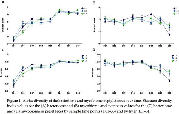
The piglet fecal bacteriome and mycobiome showed different temporal trends in alpha diversity over the eight time points from day 1 through day 35 post-natal (Figure 1 and Figure S3A,B). In the bacteriome, microbial diversity (Shannon index; β1 = 0.11, SE = 0.18, p < 0.001), richness (observed ASVs; β1 = 6.44, SE = 0.30, p < 0.001) and evenness (β1 = 0.002, SE = 0.0001, p < 0.001) all demonstrated overall significant positive increases from day 1 to day 35, with the greatest increases between day 1 to day 7 and day 21 to day 24 (Figure 1A,C and Figure S3A). Alpha diversity plateaued or showed a slight decrease following day 24. In contrast, the mycobiome demonstrated a decrease in diversity (Shannon index; β1 = −0.01, SE = 0.003, p < 0.001) and evenness (β1 = −0.004, SE = 0.0006, p < 0.001) from day 1 to day 35, with most of the decrease occurring post-wean (Figure 1B,D). Richness (observed ASVs; β1 = 0.01, SE = 0.002, p < 0.001) in the mycobiome showed a slight increase from day 1 to day 35, with the greatest increase occurring post-wean from day 21 to day 28 (Figure S3B).
Temporal Dynamics of the Gut Bacteriome and Mycobiome in the Weanling Pig - Image 1
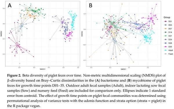
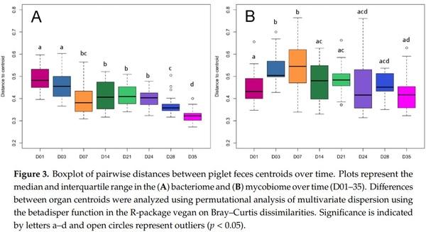
Non-metric multidimensional scaling (NMDS) plots based on Bray–Curtis distance of the piglet fecal microbiomes revealed significant temporal shifts from day 1 to day 35 in both the bacteriome (PERMANOVA; F = 22.25, R2 = 0.49, p < 0.001) and mycobiome (PERMANOVA; F = 8.17, R2 = 0.25, p < 0.001) (Figure 2, Table S3). In the bacteriome, each of the eight time points from day 1 to 35 showed distinct clusters and centroids, with the largest separation between clusters occurring between preand post-wean samples (Figure 2A). The outdoor adult and lactating sow fecal bacteriome was closely clustered with the post-wean samples, but remained distinct from the piglet fecal bacteriome, while the feed sample did not cluster closely with any of the time points. Dispersion of samples showed a decreasing trend from day 1 to day 35, with day 35 having significantly less dispersion (PERMDISP; p < 0.001) than the rest of the time points (Figure 3A, Table S4A). A similar, but slightly less-distinct, temporal clustering pattern was displayed in the piglet fecal mycobiome (Figure 2B). The greatest separation of clusters occurred between pre- and post-wean samples; however, there was more overlap of samples among time points, particularly among pre-wean piglet samples. Unlike the bacteriome, the feed samples clustered near outdoor adult and piglet day 35 fungal samples. Dispersion among the fecal mycobiome samples significantly increased from day 1 to day 7 (PERMDISP; p < 0.001), then decreased to similar dispersion levels found at day 1 (Figure 3B, Table S4B).
Temporal Dynamics of the Gut Bacteriome and Mycobiome in the Weanling Pig - Image 2
Temporal Dynamics of the Gut Bacteriome and Mycobiome in the Weanling Pig - Image 3
3.2. Evaluation of the Bacteriome and Mycobiome Composition
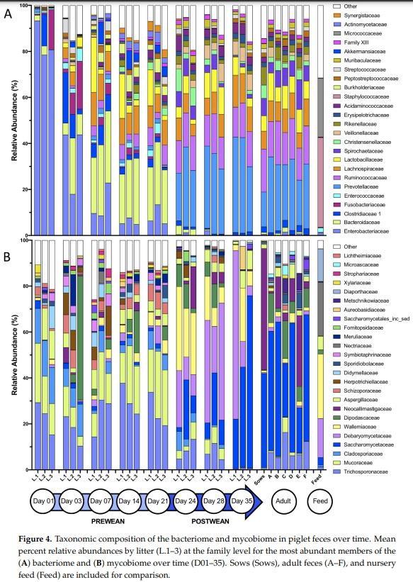
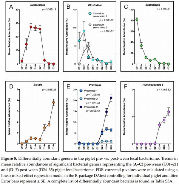
The dynamic shifts in bacteriome and mycobiome taxonomic composition over the eight time points (day 1 to day 35) were examined at the phylum, family and genus classification level. At the phylum level, the bacteriome transitioned from a Proteobacteria- to a Firmicutes- and Bacteroidetes-driven community (Figure S4A). At the family level, a dramatic shift in taxa was demonstrated from the pre-wean (day 1–21) to post-wean (day 24–35) time period, with post-wean family taxa similar to those found in the sows and adult pigs (Figure 4A). The dominant (>60% of sequences) families, Staphylococcaceae and Micrococcaceae, found in nursery feed were found in <0.10% of piglet fecal sequences. Family taxon vectors fitted to an NMDS ordination plot displaying pre- and post-wean bacteriomes revealed that dominant (>50% of sequences) families Enterobacteriaceae, Enterococcaceae, Fusobacteriaceae, and Bacteroidaceae were significantly correlated with pre-wean piglet feces, while Veillonellaceae, Prevotellaceae, Lactobacillaceae, Ruminococcaceae and Lachnospiraceae were significantly correlated with post-wean piglet feces (p < 0.05, Figure S5A). Differentially abundant genera between pre- and post-wean piglet fecal bacteria included highly abundant Bacteroides (p < 0.001), Clostridium (sensu stricto 1 and 2, p < 0.001), and Escherichia (p < 0.001) in pre-wean piglet feces and Blautia (p < 0.001), Prevotella (1, 2 and 9, p < 0.001) and Ruminococcus 1 (p < 0.001) in post-wean piglet feces (Figure 5A–F, Table S5A).
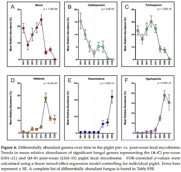
In the mycobiome, there was an overall shift from Mucoromycota and Basidiomycota pre-wean (day 1 to day 21) to Ascomycota post-wean (day 24–day 35) in piglet feces (Figure S4B). At the family level, dominant (>50% of sequences) families Trichosporonaceae, Symbiotraphinaceae, Mucoraceae, and Cladosporaceae significantly correlated (p < 0.05) with pre-wean piglet feces (Figure S5B) and Saccharomycetaceae, Debaryomycetaceae and Wallemiaceae significantly correlated (p < 0.05) with post-wean piglet feces (Figure S5B). Families Saccharomycetaceae, Dipodascaceae, Aspergillaceae, Debaryomycetaceae, and Wallemiaceae, making up >43% of the sequences found in nursery feed samples, were also present in >79% of sequences in post-wean piglet feces and <18% of pre-wean piglet feces (Figure 4B). Sows and adult pigs showed slightly different taxonomic profiles than post-wean piglets, with an overall greater mean relative abundance of Dipodascaceae(11.7 ± 3.5% vs. 4.6 ± 0.9%) and Neocallimastigaceae(10.3 ± 1.4% vs. 3.1 ± 1.4%) and a lower mean relative abundance of Wallemia (2.6 ± 0.4% vs. 17.0 ± 2.0%). Genera Mucor (p < 0.001), Cladosporium (p < 0.05), and Trichosporon (p < 0.001) were significantly more abundant in pre-wean piglet feces, while Wallemia (p < 0.001), Kazachstania (p < 0.001), and Hyphopichia (p < 0.001) were significantly more abundant in post-wean piglet feces (Figure 6A–F, Table S5B).
Temporal Dynamics of the Gut Bacteriome and Mycobiome in the Weanling Pig - Image 4
Temporal Dynamics of the Gut Bacteriome and Mycobiome in the Weanling Pig - Image 5
Temporal Dynamics of the Gut Bacteriome and Mycobiome in the Weanling Pig - Image 6
3.3. Interactions between the Bacteriome and Mycobiome
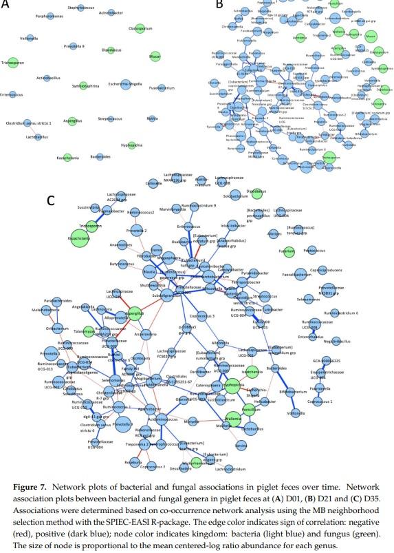
A co-occurrence network was constructed to determine the interactions between bacterial and fungal genera in piglet fecal microbiomes at days 1, 21, and 35 (Figure 7A–C). At day 1, only negative association was found in piglet feces between bacteria Clostridium sensu stricto 1 and Lactobacillus (Figure 7A). No fungal-bacterial interactions were identified. At day 21, 93 different interactions were determined between the microbiota (Figure 7B). Of the 11 identified fungi present day 21 feces, four fungal genera showed interactions with bacteria, including a negative association between Aspergillus and Ruminococcaceae UCG-004 and between Cladosporium and Alistipes. At day 35, 142 interactions were found in the pig fecal microbiome (Figure 7C). Of the fungal genera, Aspergillus had the highest number of interactions with bacteria (n = 4 each), Hyphopichia showed positive associations with Eubacterium rumaniantium group, Ruminococcaceae UCG-003, and Succiniclasticum and a negative association with Escherichia-Shigella. Aspergillus showed a positive association with Anaerovibrio and negative associations with Lachnospiraceae UCG-010, Prevotellaceae UCG-003 and Subdoligranulum. Positive fungi-bacteria associations Penicillium-Lactobacillus, Talaromyces-Prevotellaceae UCG-003, Trichosporon-Pygmaiobacter, and negative fungi-bacteria association Issatchenkia-Lachnospiraceae UCG-001 were also found.
Temporal Dynamics of the Gut Bacteriome and Mycobiome in the Weanling Pig - Image 7
4. Discussion
The colonization, development, and interactions of the piglet gut microbiota from birth through weaning are critical to piglet heath and growth performance. In both bacterial and fungal populations, there is a dynamic shift in community structure and composition during this time, with adult-like microbiomes developing by day 35. Our longitudinal examination of the piglet fecal bacteriome and mycobiome from day 1 (post-natal) through day 35 revealed different patterns in temporal diversity among bacteria and fungus as well as potential interactions in the fecal community.
The temporal alpha diversity patterns found in the piglet fecal bacteriome (Figure 1A) were similar to those found in previous fecal studies [30–33], with increasing Shannon diversity from birth through the weaning. This same pattern was also evident in evenness and richness over the same time period (Figure 1C and Figure S3A). After day 24, diversity in the bacteriome plateaued, suggesting that the progressive early colonization of bacteria coupled with a sudden transition from sow milk to nursery feed resulted in the concurrent, rapid expansion of different bacterial populations, able to exploit novel niches within the piglet GI tract. However, by day 35, these populations appear to have reached stability or equilibrium in the piglet.
In contrast, the fecal mycobiota showed a reduction in Shannon diversity and evenness from birth through weaning (Figure 2B,D). Interestingly, richness remained relatively steady with a slight increase post-weaning (Figure S3B). This indicates that Shannon diversity decreased, as a result of reduced evenness, within the mycobiome. Data from our study showed dramatic increases in the presence of two yeast genera, Kazachstania and Hyphopichia, post-wean (Figure 6), which is likely driving down community evenness by outcompeting other fungal species. In general, high microbial diversity is deemed beneficial to a community or host by providing functional redundancy and resiliency [34,35]. However, in the piglet gut environment, the establishment of dominant yeast species and reduction in community evenness and diversity in healthy, post-weaned piglets may be an exception. In humans, low fungal diversity has been consistently identified in healthy adult feces [36,37], while increased fungal diversity has been linked to gastrointestinal disease [3,38]. We hypothesize that the same may be true in piglets, where the presence of low fungal diversity may indicate health and high fungal diversity may suggest the presence of disease or dysbiosis.
The effect of time on the developing microbiota was clearly evident in both bacterial and fungal populations in the piglet feces, with time being a more important factor in the developing bacteriome than in the mycobiome (Table S3). In the bacteriome, the effect of time was especially evident in the transition from day 21 to day 24, when piglets were removed from the sow and introduced to nursery feed; this transition was primarily driven by compositional shifts in the bacterial community from milk oligosaccharide-utilizing Bacteroidaceae to plant polysaccharide-utilizing Prevotellaceae, which has been well documented in other piglet weaning studies [10,31,39,40]. Another important transition in the bacteriome was the immediate reduction in Enterobacteriaceae (genus Escherichia/Shigella) following birth that occurred between day 1 and day 3 (Figure 5). This trend has also been consistently detected in other piglet studies [32,39,41], and is likely part of the similar dynamic transition from aerobes/facultative anaerobes to obligate anaerobes in developing human infant guts [42].
In addition to trends in composition, the bacteriome demonstrated reduced dispersion among communities over time, which was not found in the mycobiome (Figure 3). This indicates that the piglet fecal bacteriome likely follows a defined pattern of colonization and succession in healthy developing piglets, whereas in the mycobiome a large portion of the community may be transient and driven by other factors, such as environmental exposure, diet, or host immunity, which may vary by piglet. Other studies have purported similar hypotheses regarding the transient nature of the mycobiome based on compositional variability among sample subjects and association of fungal genera with food or environmental sources [36,43,44]. In the pre-wean piglet mycobiome, environmental influence is suggested by the dominance of naturally ubiquitous fungal families Trichosporonaceae, Mucoraceae, and Cladosporiacea, all of which are found in a variety of environments including organic debris, soils, and indoor/outdoor air [45–47]. Furthermore, post-wean piglet feces mycobiomes clustered near nursery feed samples (Figure 2) and showed a sharp increase in abundance of fungal taxa Wallemiaceae and Debaryomycetaceae, highly abundant in nursery feed samples, directly following weaning. The role of these transient species on host development and resident bacteria remain to be elucidated. The piglet fecal bacteriomes, in contrast, did not cluster near feed samples nor resemble any of the abundant bacterial taxa found in the nursery feed.
Despite receiving the same feed and housing, there were marked compositional differences between litter mycobiomes by day 35, indicating potential involvement of host genetic or immunity factors in shaping the fungal community [48,49]. One of the most striking differences included a high level of variation in the relative abundances of fungal families Saccharomycetaceae (K. slooffiae) and Debaryomcetaceae (H. burtonii). K. slooffiae, a persistent fungus in post-weaned piglets [30,44], is hypothesized to be a potential protein source to healthy pigs [14] and may behave similarly to commensal Candida species in humans [13]. In a study on human diets, Candida was positively correlated with high carbohydrate consumption [50] and has been shown to degrade starches [51]. In piglets, the transition from a milk diet to high carbohydrate nursery feed at weaning may explain the increase in K. slooffiae in post-weaned piglets. In comparison, H. burtonii is a common fungal contaminant of corn [52] and has not been found to be abundant in other piglet fecal studies regardless of piglet age [30,44]. However, H. burtonii, like Candida, is also able to degrade starch [53] and may compete with K. slooffiae in the gut environment. No differences in piglet growth rate or in bacterial composition were seen among the litters with high abundances of H. burtonii, indicating that high abundances of H. burtonii did not adversely affect piglet health. Further compositional, transient differences were seen between litters at early time points. One example is the temporary increase in Dipodascaceae in L.3 at day 3 (Figure 4B), which was present in high abundances in all pigs from that litter (data not shown). We hypothesize, that this temporary spike in Dipodascaceae unique to one litter reflects the highly transient nature of the early mycobiome as microbes compete for space and nutrients in the gastrointestinal tract as well as a potentially acute environmental exposure event by L.3 to Dipodascaceae, even though all litters were housed in the same environment.
Development had a strong effect on the associations among bacterial and fungal genera. Day 1 piglet fecal communities showed no interactions between fungus and bacterial genera, but as the fecal communities developed, there was a progressive increase in number and complexity of interactions between bacteria and fungus (Figure 7). This increase in interactions may be partially driven by a greater bacterial production of short-chain fatty acids following weaning. Firmicutes bacteria are known to produce a variety of SCFA via fermentation of complex carbohydrates that can inhibit fungal growth [54,55]. In particular, a negative correlation between SCFAs and potential fungal pathogen Aspergillus has been demonstrated in human diets [50] and several SCFAs have been shown to negatively correlate with relative abundances of Aspergillus in piglet digesta samples [5]. A similar negative association was seen in day 35 piglet feces in this study between Aspergillus and SCFA producers Subdoligranulum, Prevotellaceae UCG-003, and Lachnospiraceae UCG-010. Furthermore, previously published data on the mucosal microbiota of these piglets’ lower GI organs (colon and cecum) also showed significant negative correlations between Aspergillus and several SCFA producers, suggesting that SCFAs are likely inhibiting Aspergillus growth in the piglet gut [13]. While inferred interactions are seen, a lack of supporting literature on these fungal species makes further interpretation speculative and more studies are needed, but the effect of SCFA producers in the gut on the health of the piglet cannot be overlooked.
From birth through weaning, healthy piglet feces in this study showed a dramatic shift in the bacterial and fungal communities, as well as an increase in network connectivity between the two kingdoms. While the piglet fecal bacteriome followed a relatively stable and predictable pattern of development, the development of the mycobiome was more variable and likely more transient and environmentally driven, particularly prior to day 35 piglets. Due to the flexibility of mycobiome in early piglets and evidence from this study on the potential influence of fungal taxa found in feed, successful manipulation of the mycobiome with fungal probiotics may be beneficial during the weaning transition. Furthermore, potential fungal pathogens like Aspergillus may be suppressed in piglets by enhanced SCFAs. These SCFAs could be introduced through supplementation of SCFAs in the piglet diet or through the augmentation of bacterial populations that enhance SCFA production.
Our findings provide a significant contribution to the understanding of the developing fecal mycobiome and potential bacterial-fungal interactions in healthy piglets. The ability of microbes to alter the environment and nutrient availability of other microbes is accepted, but these environmental changes can also alter the host health and growth. While studies in piglets are lacking, the direct role of microbes and their interactions and their ability to enhance piglet growth need to be assessed. Incorporating the mycobiome in future piglet microbiome studies will further elucidate the importance and role of fungi in porcine health.
This article was originally published in Microorganisms 2020, 8, 868; doi:10.3390/microorganisms8060868. This is an Open Access article distributed under the terms and conditions of the Creative Commons Attribution (CC BY) License (http://creativecommons.org/licenses/by/4.0/).

1. Erb Downward, J.R.; Falkowski, N.R.; Mason, K.L.; Muraglia, R.; Huffnagle, G.B. Modulation of post-antibiotic bacterial community reassembly and host response by Candida albicans. Sci. Rep. 2013, 3, 2191. [CrossRef]
[PubMed]
2. Mason, K.L.; Erb Downward, J.R.; Falkowski, N.R.; Young, V.B.; Kao, J.Y.; Huffnagle, G.B. Interplay between the gastric bacterial microbiota and Candida albicans during postantibiotic recolonization and gastritis.
Infect. Immun. 2012, 80, 150–158. [CrossRef] [PubMed]
3. Ott, S.J.; Kuhbacher, T.; Musfeldt, M.; Rosenstiel, P.; Hellmig, S.; Rehman, A.; Drews, O.; Weichert, W.;
Timmis, K.N.; Schreiber, S. Fungi and inflammatory bowel diseases: Alterations of composition and diversity.
Scand. J. Gastroenterol. 2008, 43, 831–841. [CrossRef] [PubMed]
4. Iliev, I.D.; Leonardi, I. Fungal dysbiosis: Immunity and interactions at mucosal barriers. Nat. Rev. Immunol.
2017, 17, 635–646. [CrossRef] [PubMed]
5. Li, X.V.; Leonardi, I.; Iliev, I.D. Gut mycobiota in immunity and inflammatory disease. Immunity 2019, 50,
1365–1379. [CrossRef] [PubMed]
6. Mukherjee, P.K.; Sendid, B.; Hoarau, G.; Colombel, J.F.; Poulain, D.; Ghannoum, M.A. Mycobiota in gastrointestinal diseases. Nat. Rev. Gastroenterol. Hepatol. 2015, 12, 77–87. [CrossRef]
7. Kureljusic, B.; Weissenbacher-Lang, C.; Nedorost, N.; Stixenberger, D.; Weissenbock, H. Association between
Pneumocystis spp. and co-infections with Bordetella bronchiseptica, Mycoplasma hyopneumoniae and
Pasteurella multocida in Austrian pigs with pneumonia. Vet. J. 2016, 207, 177–179. [CrossRef]
8. Weissenbacher-Lang, C.; Kureljusic, B.; Nedorost, N.; Matula, B.; Schiessl,W.; Stixenberger, D.; Weissenbock, H.
Retrospective analysis of bacterial and viral co-infections in pneumocystis spp. positive lung samples of austrian pigs with pneumonia. PLoS ONE 2016, 11, e0158479. [CrossRef]
9. Niederwerder, M.C. Fecal microbiota transplantation as a tool to treat and reduce susceptibility to disease in animals. Vet. Immunol. Immunopathol. 2018, 206, 65–72. [CrossRef]
10. Guevarra, R.B.; Hong, S.H.; Cho, J.H.; Kim, B.R.; Shin, J.; Lee, J.H.; Kang, B.N.; Kim, Y.H.; Wattanaphansak, S.;
Isaacson, R.E.; et al. The dynamics of the piglet gut microbiome during the weaning transition in association with health and nutrition. J. Anim. Sci. Biotechnol. 2018, 9, 54. [CrossRef]
11. Guevarra, R.B.; Lee, J.H.; Lee, S.H.; Seok, M.J.; Kim, D.W.; Kang, B.N.; Johnson, T.J.; Isaacson, R.E.; Kim, H.B.
Piglet gut microbial shifts early in life: Causes and effects. J. Anim. Sci. Biotechnol. 2019, 10, 1. [CrossRef]
[PubMed]
12. Campbell, J.M.; Crenshaw, J.D.; Polo, J. The biological stress of early weaned piglets. J. Anim. Sci. Biotechnol.
2013, 4, 19. [CrossRef] [PubMed]
13. Arfken, A.M.; Frey, J.F.; Ramsay, T.G.; Summers, K.L. Yeasts of burden: Exploring the mycobiome-bacteriome of the piglet GI tract. Front. Microbiol. 2019, 10, 2286. [CrossRef] [PubMed]
14. Urubschurov, V.; Busing, K.; Freyer, G.; Herlemann, D.P.; Souffrant, W.B.; Zeyner, A. New insights into the role of the porcine intestinal yeast, Kazachstania slooffiae, in intestinal environment of weaned piglets.
FEMS Microbiol. Ecol. 2017, 93. [CrossRef] [PubMed]
15. Urubschurov, V.; Busing, K.; Souffrant, W.B.; Schauer, N.; Zeyner, A. Porcine intestinal yeast species,
Kazachstania slooffiae, a new potential protein source with favourable amino acid composition for animals.
J. Anim. Physiol. Anim. Nutr. (Berl.) 2018, 102, e892–e901. [CrossRef]
16. Urubschurov, V.; Janczyk, P.; Pieper, R.; Souffrant, W.B. Biological diversity of yeasts in the gastrointestinal tract of weaned piglets kept under different farm conditions. FEMS Yeast Res. 2008, 8, 1349–1356. [CrossRef]
17. Kozich, J.J.; Westcott, S.L.; Baxter, N.T.; Highlander, S.K.; Schloss, P.D. Development of a dual-index sequencing strategy and curation pipeline for analyzing amplicon sequence data on the MiSeq Illumina sequencing platform. Appl. Environ. Microbiol. 2013, 79, 5112–5120. [CrossRef]
18. Callahan, B.J.; McMurdie, P.J.; Rosen, M.J.; Han, A.W.; Johnson, A.J.; Holmes, S.P. DADA2: High-resolution sample inference from Illumina amplicon data. Nat. Methods 2016, 13, 581–583. [CrossRef]
19. Caporaso, J.G.; Kuczynski, J.; Stombaugh, J.; Bittinger, K.; Bushman, F.D.; Costello, E.K.; Fierer, N.; Pena, A.G.;
Goodrich, J.K.; Gordon, J.I.; et al. QIIME allows analysis of high-throughput community sequencing data.
Nat. Methods 2010, 7, 335–336. [CrossRef]
20. Yilmaz, P.; Parfrey, L.W.; Yarza, P.; Gerken, J.; Pruesse, E.; Quast, C.; Schweer, T.; Peplies, J.; Ludwig, W.;
Glockner, F.O. The SILVA and “All-species Living Tree Project (LTP)” taxonomic frameworks. Nucleic Acids Res.
2014, 42, D643–D648. [CrossRef]
21. Martin, M. Cutadapt removes adaptor sequences from high-throughput sequencing reads. Emb. J. 2011, 17,
10–12. [CrossRef]
22. Bolger, A.M.; Lohse, M.; Usadel, B. Trimmomatic: A flexible trimmer for Illumina sequence data. Bioinformatics
2014, 30, 2114–2120. [CrossRef] [PubMed]
23. Koljalg, U.; Nilsson, R.H.; Abarenkov, K.; Tedersoo, L.; Taylor, A.F.; Bahram, M.; Bates, S.T.; Bruns, T.D.;
Bengtsson-Palme, J.; Callaghan, T.M.; et al. Towards a unified paradigm for sequence-based identification of fungi. Mol. Ecol. 2013, 22, 5271–5277. [CrossRef]
24. Oksanen, J.; Guillaume, B.F.; Michael, F.; Roeland, K.; Pierre, L.; Dan, M.; Peter, R.; Minchin, R.; O’Hara, B.;
Gavin, L.; et al. Vegan: Community Ecology Package. Available online: https://cran.r-project.org (accessed on 15 April 2020).
25. McMurdie, P.J.; Holmes, S. Phyloseq: An R package for reproducible interactive analysis and graphics of microbiome census data. PLoS ONE 2013, 8, e61217. [CrossRef] [PubMed]
26. Wickham, H. Ggplot2: Elegant Graphics for Data Analysis, 2nd ed.; Springer International Publishing: Cham,
Switzerland, 2016. [CrossRef]
27. Russel, J.; Thorsen, J.; Brejnrod, A.D.; Bisgaard, H.; Sørensen, S.J.; Burmølle, M. DAtest: A framework for choosing differential abundance or expression method. bioRxiv 2018. [CrossRef]
28. Kurtz, Z.D.; Muller, C.L.; Miraldi, E.R.; Littman, D.R.; Blaser, M.J.; Bonneau, R.A. Sparse and compositionally robust inference of microbial ecological networks. PLoS Comput. Biol. 2015, 11, e1004226. [CrossRef]
29. Csardi, G.; Nepusz, T. The igraph software package for complex network research. InterJ. Complex Syst. 2006,
1695, 1–9.
30. Summers, K.L.; Frey, J.F.; Ramsay, T.G.; Arfken, A.M. The piglet mycobiome during the weaning transition:
A pilot study1. J. Anim. Sci. 2019, 97, 2889–2900. [CrossRef]
31. Frese, S.A.; Parker, K.; Calvert, C.C.; Mills, D.A. Diet shapes the gut microbiome of pigs during nursing and weaning. Microbiome 2015, 3, 28. [CrossRef]
32. Chen, L.; Xu, Y.; Chen, X.; Fang, C.; Zhao, L.; Chen, F. The maturing development of gut microbiota in commercial piglets during the weaning transition. Front. Microbiol. 2017, 8, 1688. [CrossRef]
33. Vo, N.; Tsai, T.C.; Maxwell, C.; Carbonero, F. Early exposure to agricultural soil accelerates the maturation of the early-life pig gut microbiota. Anaerobe 2017, 45, 31–39. [CrossRef] [PubMed]
34. Konopka, A. What is microbial community ecology? ISME J. 2009, 3, 1223–1230. [CrossRef] [PubMed]
35. Turnbaugh, P.J.; Hamady, M.; Yatsunenko, T.; Cantarel, B.L.; Duncan, A.; Ley, R.E.; Sogin, M.L.; Jones, W.J.;
Roe, B.A.; Affourtit, J.P.; et al. A core gut microbiome in obese and lean twins. Nature 2009, 457, 480–484.
[CrossRef]
36. Raimondi, S.; Amaretti, A.; Gozzoli, C.; Simone, M.; Righini, L.; Candeliere, F.; Brun, P.; Ardizzoni, A.;
Colombari, B.; Paulone, S.; et al. Longitudinal Survey of Fungi in the Human Gut: ITS Profiling, Phenotyping, and Colonization. Front. Microbiol. 2019, 10, 1575. [CrossRef]
37. Nash, A.K.; Auchtung, T.A.; Wong, M.C.; Smith, D.P.; Gesell, J.R.; Ross, M.C.; Stewart, C.J.; Metcalf, G.A.;
Muzny, D.M.; Gibbs, R.A.; et al. The gut mycobiome of the human microbiome project healthy cohort.
Microbiome 2017, 5, 153. [CrossRef]
38. Kuhbacher, T.; Ott, S.J.; Helwig, U.; Mimura, T.; Rizzello, F.; Kleessen, B.; Gionchetti, P.; Blaut, M.; Campieri, M.;
Folsch, U.R.; et al. Bacterial and fungal microbiota in relation to probiotic therapy (VSL#3) in pouchitis. Gut
2006, 55, 833–841. [CrossRef]
39. Pajarillo, E.A.; Chae, J.P.; Balolong, M.P.; Kim, H.B.; Seo, K.S.; Kang, D.K. Pyrosequencing-based analysis of fecal microbial communities in three purebred pig lines. J. Microbiol. 2014, 52, 646–651. [CrossRef]
40. Alain, B.P.E.; Chae, J.P.; Balolong, M.P.; Bum Kim, H.; Kang, D.K. Assessment of fecal bacterial diversity among healthy piglets during the weaning transition. J. Gen. Appl. Microbiol. 2014, 60, 140–146. [CrossRef]
41. Mach, N.; Berri, M.; Estelle, J.; Levenez, F.; Lemonnier, G.; Denis, C.; Leplat, J.J.; Chevaleyre, C.; Billon, Y.;
Dore, J.; et al. Early-life establishment of the swine gut microbiome and impact on host phenotypes.
Environ. Microbiol. Rep. 2015, 7, 554–569. [CrossRef]
42. Kostic, A.D.; Gevers, D.; Siljander, H.; Vatanen, T.; Hyotylainen, T.; Hamalainen, A.M.; Peet, A.; Tillmann, V.;
Poho, P.; Mattila, I.; et al. The dynamics of the human infant gut microbiome in development and in progression toward type 1 diabetes. Cell Host Microbe 2015, 17, 260–273. [CrossRef]
43. Hallen-Adams, H.E.; Suhr, M.J. Fungi in the healthy human gastrointestinal tract. Virulence 2017, 8, 352–358.
[CrossRef] [PubMed]
44. Ramayo-Caldas, Y.; Prenafeta, F.; Zingaretti, L.M.; Gonzales, O.; Dalmau, A.; Quintanilla, R.; Ballester, M.
Gut eukaryotic communities in pigs: Diversity, composition and host genetics contribution. bioRxiv 2020, 2.
[CrossRef]
45. Colombo, A.L.; Padovan, A.C.; Chaves, G.M. Current knowledge of Trichosporon spp. and Trichosporonosis.
Clin. Microbiol. Rev. 2011, 24, 682–700. [CrossRef] [PubMed]
46. Richardson, M. The ecology of the Zygomycetes and its impact on environmental exposure.
Clin. Microbiol. Infect. 2009, 15 (Suppl. 5), 2–9. [CrossRef]
47. Bensch, K.; Braun, U.; Groenewald, J.Z.; Crous, P.W. The genus cladosporium. Stud. Mycol. 2012, 72, 1–401.
[CrossRef]
48. Forbes, J.D.; Bernstein, C.N.; Tremlett, H.; Van Domselaar, G.; Knox, N.C. A fungal world: Could the gut mycobiome be involved in neurological disease? Front. Microbiol. 2018, 9, 3249. [CrossRef]
49. Cui, L.; Morris, A.; Ghedin, E. The human mycobiome in health and disease. Genome Med. 2013, 5, 63.
[CrossRef]
50. Hoffmann, C.; Dollive, S.; Grunberg, S.; Chen, J.; Li, H.; Wu, G.D.; Lewis, J.D.; Bushman, F.D. Archaea and fungi of the human gut microbiome: Correlations with diet and bacterial residents. PLoS ONE 2013, 8, e66019.
[CrossRef]
51. McCann, A.K.; Barnett, J.A. The utilization of starch by yeasts. Yeast 1986, 2, 109–115. [CrossRef]
52. Kurtzman, C.P. Phylogeny of the ascomycetous yeasts and the renaming of Pichia anomala to
Wickerhamomyces anomalus. Antonie Van Leeuwenhoek 2011, 99, 13–23. [CrossRef]
53. Abe, C.A.; Faria, C.B.; de Castro, F.F.; de Souza, S.R.; dos Santos, F.C.; da Silva, C.N.; Tessmann, D.J.;
Barbosa-Tessmann, I.P. Fungi Isolated from Maize (Zea mays L.) Grains and Production of Associated
Enzyme Activities. Int. J. Mol. Sci. 2015, 16, 15328–15346. [CrossRef] [PubMed]
54. Sam, Q.H.; Chang, M.W.; Chai, L.Y. The fungal mycobiome and its interaction with gut bacteria in the host.
Int. J. Mol. Sci. 2017, 18, 330. [CrossRef] [PubMed]
55. Nogueira, F.; Sharghi, S.; Kuchler, K.; Lion, T. Pathogenetic impact of bacterial-fungal interactions.
Microorganisms 2019, 7, 459. [CrossRef]

Related topics
Authors:
Ann Arfken
USDA - United States Department of Agriculture
Follow
Juli Foster Frey
USDA - United States Department of Agriculture
Follow
Katie Lynn Summers
USDA - United States Department of Agriculture
Follow
Join to be able to comment.
Once you join Engormix, you will be able to participate in all content and forums.
* Required information
Would you like to discuss another topic? Create a new post to engage with experts in the community.
Create a post
Join Engormix and be part of the largest agribusiness social network in the world.
LoginRegister