Explore
Communities in English
Advertise on Engormix

Structure and immune recognition of the porcine epidemic diarrhea virus spike protein

Published: September 22, 2021
By: Robert N. Kirchdoerfer 1,3; Mahesh Bhandari 2; Olnita Martini 1; Leigh M. Sewall 1; Sandhya Bangaru 1; Kyoung-Jin Yoon 2; Andrew B. Ward 1.
Summary

Author details:

1 Department of Integrative Structural and Computational Biology, The Scripps Research Institute, La Jolla, California 92037, United States; 2 Department of Veterinary Diagnostic and Production Animal Medicine, Iowa State University, Ames, Iowa 50011, United States; 3 Current address: Department of Biochemistry, University of Wisconsin-Madison, Madison, Wisconsin 53706, United States.

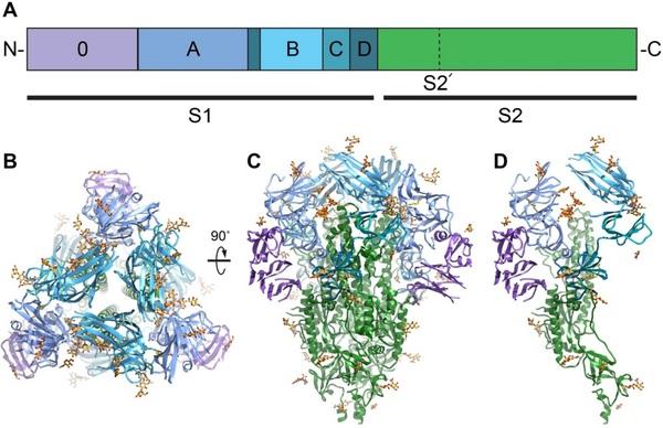
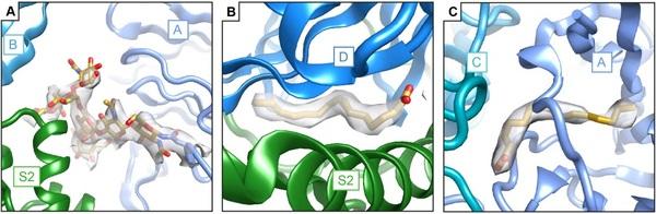
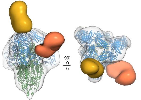
Porcine epidemic diarrhea virus is an alphacoronavirus responsible for significant morbidity and mortality in pigs. A key determinant of viral tropism and entry, the PEDV spike protein is a key target for the host antibody response and a good candidate for a protein-based vaccine immunogen. We used electron microscopy to evaluate the PEDV spike structure, as well as pig polyclonal antibody responses to viral infection. The structure of the PEDV spike reveals a configuration similar to that of HuCoV-NL63. Several PEDV protein-protein interfaces are mediated by non-protein components including a glycan at Asn264 and two bound palmitoleic acid molecules. The polyclonal antibody response to PEDV infection shows a dominance of epitopes in the S1 region. This structural and immune characterization provides new insights into coronavirus spike stability determinants and explores the immune landscape of viral spike proteins.

Introduction
Porcine epidemic diarrhea virus is a coronavirus of the alphacoronavirus genus. Identified as a viral agent distinct from transmissible gastroenteritis virus (Wood, 1977) and as a coronavirus (Pensaert and de Bouck, 1978), this virus is responsible for an enteric infection in pigs. Originally identified in England (Wood, 1977), PEDV is now a global pathogen. PEDV was first identified in the United States in 2013 (Stevenson et al., 2013) where it swept through pig populations causing several million piglet deaths (Sawyer and Sherwell, 2014). Mortality due to viral infection varies with age, with mortalities of 1-3 day old piglets in naïve herds approaching 100% (Lee, 2015). While mortality due to PEDV infection is lower in older pigs, infection can still result in decreased growth performance.
Like all coronaviruses, PEDV possesses a spike glycoprotein (S) responsible for cell attachment and virus-host membrane fusion mediating viral entry into host cells. Coronavirus spikes are class I viral fusion proteins (Bosch et al., 2003; Chambers et al., 1990), possessing structural and functional parallels to influenza hemagglutinin (Wilson et al., 1981) and HIV-1 Env (Julien et al., 2013; Lyumkis et al., 2013). This class of proteins proceeds from a metastable prefusion conformation to a highly stable postfusion conformation (Bullough et al., 1994). For coronavirus spikes, this transition of conformations has been proposed to be triggered by progressive destabilization of the prefusion structure through receptor binding and host proteolytic cleavage (Belouzard et al., 2009; Millet and Whittaker, 2015; Taguchi and Matsuyama, 2002). As the major surface glycoprotein on the enveloped virions, spikes are also the target of neutralizing antibodies (Okda et al., 2017). These antibodies primarily target the PEDV S1 region and some epitopes have been found to be neutralizing (Li et al., 2017a; Okda et al., 2017; Sun et al., 2007). However, a structural mapping of PEDV antibody epitopes targeted during viral infection is lacking.
Coronavirus spikes adopt a shared domain arrangement where the N-terminal S1 regions of the homotrimeric spike surround and cap the C-terminal S2 regions. In betacoronavirus spikes, the S1 and S2 regions are often demarcated by a protease cleavage site (Millet and Whittaker, 2015). However, this site is lacking in many alphacoronavirus spikes including PEDV. Coronavirus S1 regions of the spike contain domains contributing to receptor binding. Receptors include both host glycans and proteins and vary widely between coronaviruses. PEDV has been shown to bind to sialic acid glycans using its S1 domain 0 (Li et al., 2016). By analogy with TGEV, PEDV spikes were also proposed to recognize porcine aminopeptidase N (pAPN) (Li et al., 2007). However, the use of pAPN as a receptor for PEDV has been called into question (Li et al., 2017b).
High-resolution structural studies of coronavirus spike proteins have the potential to shed light on molecular mechanisms of spike prefusion stability and the lessons learned can be utilized in the production of stabilized spike proteins as vaccine immunogens. Here we used cryoelectron microscopy (cryo-EM) to determine the structure of the PEDV spike protein at 3.5 Å resolution. This structure has allowed us to compare our findings with the structure of the human alphacoronavirus NL63 spike as well as another recently published PEDV spike structure (Walls et al., 2016b; Wrapp and McLellan, 2019). We found several protein-protein interfaces mediated by non-protein components suggesting roles for these glycans and ligands in enhanced prefusion spike stability. We also used negative-stain electron microscopy to illuminate the recognition of the PEDV spikes by the host polyclonal antibody response and map dominant antibody epitopes on the spike surface. Together, these data supply insights for the production of prefusion stabilized spikes as vaccine immunogens.
Structure and immune recognition of the porcine epidemic diarrhea virus spike protein - Image 1
Figure 1: Overall structure of the PEDV spike protein. A) The primary sequence of the PEDV spike can be divided into S1 receptor-binding and S2 fusion machinery regions based on homology with betacoronaviruses. The S1 region can be further subdivided into domains 0, A, B, C and D (purple, light blue, cyan, teal and dark teal respectively. The S2 fusion machinery contains the S2¢ cleavage site N-terminal to the fusion peptide. B) Viewing the trimeric PEDV spike protein from the membrane distal apex shows that all three copies of the domain B are in a downwards conformation. C) A 90˚ view of the spike trimer and D) and the corresponding view of the spike monomer demonstrate that PEDV adopts a conformation highly similar to other coronavirus spikes particularly HuCoV-NL63 (Walls et al., 2016b).
Results
Structural description
Coronavirus spikes are homotrimeric glycoproteins which can be separated into an N-terminal S1 region containing the receptor binding regions and a C-terminal S2 region containing the membrane fusion machinery (Fig. 1A). The overall structure of the PEDV spike strongly resembles that of the human coronavirus NL63 spike (Walls et al., 2017) (Fig 1B-D, Supplementary Table 1 and Supplementary Fig. 1). Like the NL63 spike, PEDV spike contains two structurally homologous S1 N-terminal domains, domain 0 and domain A. The arrangement of these S1 N-terminal domains is similar to that of NL63 where domain A occupies a position at the apex of the spike, distal to the viral membrane, while domain 0 is positioned beneath domain A, packing against the side of the spike protein and contacting both S1 domain D (also called sub-domain 2) and S2. The observed PEDV domain arrangement is in contrast to the recently published PEDV spike structure where domain 0 has flipped up and away from the main portion of the spike (Wrapp and McLellan, 2019) (Supplementary Fig. 2). Though sequence differences do exist between this previous PEDV spike structure (strain CV777) and that presented here (strain USA/Colorado/2013), these differences are small compared to the sequence differences with HuCoV-NL63 which is structurally more consistent with the PEDV spike described here. Indeed, even in our attempts at 3D classification of the PEDV spike particles, we do not see evidence for this flipped up conformation of domain 0. To test the previously proposed hypothesis that the difference in domain 0 conformations may be due to differences in expression system, we expressed the PEDV spike in both insect and mammalian cells including expression conditions to produce high-mannose glycosylation patterns similar to the methods used for the previous PEDV spike structure (Wrapp and McLellan, 2019). In all cases, we observe domain 0 in the beneath-domain A conformation (Supplementary Table 2 and Supplementary Fig. 2). The reasons for the altered conformation observed in the previous PEDV spike structure remain to be determined and 
Structure and immune recognition of the porcine epidemic diarrhea virus spike protein - Image 2
Figure 2: Comparison of S2¢ cleavage sites in coronavirus spike structures. S2¢cleavage occurs after a coronavirus conserved arginine residue. This position is preceded by protein loop and an N-linked glycan which appear to occlude this cleavage site in the prefusion conformation. Structures used for comparison include spikes from HuCoV-NL63 (5SZS.pdb (Walls et al., 2016b)), Porcine deltacoronavirus (6BFU.pdb, (Xiong et al., 2018)), MERS-CoV (5W9I.pdb, (Pallesen et al., 2017)), SARS-CoV (6CRV.pdb, (Kirchdoerfer et al., 2018)) and Infectious bronchitis virus (6CV0.pdb, (Shang et al., 2018a)).
will require additional experimental studies of alphacoronavirus spike structure.
Despite superimposing well onto the S2 fusion subunit and containing a similar overall S1 domain arrangement, the PEDV spike differs in several regards to the previously determined NL63 spike structure. Unlike the NL63 spike, the reconstructed density for domain 0 is more poorly defined than other domains indicating a higher degree of motion relative to the rest of the spike. Superimposition of the coordinate models of NL63 spike and the PEDV spike presented here reveals that both domain 0 and domain A have shifted approximately 15 Å towards the S2 region in the PEDV spike while domain B (also referred to as the S1 CTD) has remained in approximately the same position. The three B domains of the trimer are also more tightly packed around the spike three-fold axis with domain B making fewer contacts to domain A than observed in NL63 spike. All three B domains are in the downwards or lying conformation similar to that observed for available alpha-, gamma-, and deltacoronavirus spike structures (Shang et al., 2018a; Shang et al., 2018b; Walls et al., 2016b) as well as several betacoronavirus spike structures (Kirchdoerfer et al., 2016; Tortorici et al., 2019; Walls et al., 2016a).
PEDV spike is known to bind sialic acid and hemagglutinate red blood cells. This activity has been attributed to the S1 domain 0 (Hou et al., 2017; Li et al., 2016). However, comparison of the PEDV S1 domain 0 with the equivalent domain of the recently determined betacoronavirus OC43 spike in complex with sialic acid (Tortorici et al., 2019) suggests that the OC43 spike sialic acid binding site is not shared with PEDV despite structural homology between the two glycan-binding domains indicating an altered mode of glycan recognition in PEDV. In a portion of field isolates and tissue culture adapted viruses, PEDV S1 domain 0 has been deleted, leading to an apparent loss of sialic acid binding activity (Diep et al., 2017; Hou et al., 2017; Masuda et al., 2015). Though originally suggested to result in viruses with reduced mortality in piglets (Hou et al., 2017; Masuda et al., 2015), spike domain 0 deletion viruses have been found to be capable of causing high mortality (Diep et al., 2017). Moreover, it has been suggested that the deletion of domain 0 may contribute to viral persistence and reoccurrence of PEDV on pig farms (Diep et al., 2017). To examine the effect of this domain deletion on the overall structure of the PEDV spike, we used negative-stain EM to determine the structure of the PEDV spike with a 197 amino acid deletion, 
Structure and immune recognition of the porcine epidemic diarrhea virus spike protein - Image 3
Figure 3: Non-protein components mediate protein-protein interactions in the PEDV spike. A) An N-linked glycan at Asn264 is sandwiched between domain A (light blue) and the S2 region of an adjacent protomer (green), capping the S2 central helices. B) Density for palmitoleic acid binding between S1 domain D (blue) and the S2 subunit of an adjacent protomer (green). B) A second site for palmitoleic acid was located in S1 domain A where the fatty acid head group reaches out to interact with S1 domain C of an adjacent protomer (teal).
removing domain 0 (Diep et al., 2017). This low-resolution structure displays a similar structure to the full-length spike indicating that the deletion of this domain does not impart any macroscopic changes in spike protein conformation (Supplement Fig. 1).
The S2¢ protease cleavage site in the S2 region of the spike is conserved across coronavirus spikes (Millet and Whittaker, 2015) and is believed to only be exposed for cleavage by host proteases during viral entry (Belouzard et al., 2009; Park et al., 2016). Examination of the PEDV S2 fusion machinery indicates a high structural homology with other coronavirus spike proteins including the S2¢ cleavage site. This S2¢ cleavage site at Arg894 is presented on the side of the spike following a short loop. The conformation of this preceding loop along with an upstream glycan at Asn873, blocks recognition of the S2¢ cleavage site by host proteases in the prefusion conformation (Fig. 2). A comparison of available coronavirus spike structures (Kirchdoerfer et al., 2018; Pallesen et al., 2017; Shang et al., 2018a; Tortorici et al., 2019; Walls et al., 2016b; Xiong et al., 2018) shows the positioning and conformation of the preceding loop (875-893) to be conserved. In addition, the presence of a glycan 18-22 amino acids N-terminal to the S2¢ cleavage site also appears to be shared across coronavirus genera.
Sugar and fatty acids mediate protein chain interfaces
In modeling coordinates into the reconstructed EM density, we observed three non-protein densities at different domain interfaces. The first of these is a glycan density emanating from Asn264. This glycan is also present in the NL63 spike structure previously determined (Walls et al., 2016b) and occupies a hole observed in coronavirus spikes. However, due to the lower positioning of the S1 domain A relative to S2, this glycan in PEDV spike is sandwiched between the S1 domain A and the S2 region of an adjacent protomer. In coronavirus spikes, the S1 domain B caps the S2 central helices. So far unique to PEDV is the additional coordination of these S2 central helices by the Asn264 glycan (Fig. 3A) suggesting a contributing role for this glycan in stabilizing the prefusion spike conformation. Indeed, the sequon coding for this glycan position is 99.8% conserved across full-length PEDV spike sequences in the NCBI protein database (Pickett et al., 2012). As our PEDV spike protein used for high-resolution cryo-EM was produced in insect cells which possess alternative glycosylation patterns (Thirstrup et al., 1993), we sampled the overall conformations of PEDV spikes possessing a variety of glycosylation patterns by expressing an identical PEDV spike DNA construct using mammalian HEK-293F, with kifunensine, and HEK-293S Gnt-/- expression systems and examining the protein with negative-stain EM (Supplementary Fig. 1). The low-resolution reconstructions reveal identical conformations of the PEDV spike despite the differing glycosylation states, including the lower positioning of the S1 domain A relative to the S2 region. We also removed the sequon for the Asn264 glycan by mutating the Asn to Asp. This mutation strongly reduced PEDV spike protein expression levels in HEK-293F cells by approximately 10-fold, consistent with a stabilizing effect. Nonetheless, this spike protein was still capable of adopting the pre-fusion conformation (Supplementary Fig. 2) indicating that the presence of the Asn264 glycan is not strictly required.
The second non-protein density that we observed at an inter-domain interaction site lies between S1 domain D (also called sub-domain 2) and the S2 region of an adjacent protomer (Fig. 3B). The elongated, unbranched appearance of this density and a close proximity to neighboring Arg842 in S2 led us to model this density as a 16-carbon, unbranched fatty acid. An identical unmodeled density is present in the previously determined PEDV spike structure (Wrapp and McLellan, 2019). We confirmed the presence of this ligand using mass spectrometry, which indicated the presence of a molecule with a molecular weight matching that of palmitoleic acid (Supplementary Fig. 3). This fatty acid occupies a hydrophobic pocket formed between domain D and S2 and has limited surface exposure leading us to hypothesize that this fatty acid was incorporated during the folding of the protein domains. Comparison with published structures of NL63, 229E and Feline infectious peritonitis spikes shows that despite some amino acid sequence differences, the hydrophobic fatty acid pocket is shared between these alphacoronavirus spikes (Li et al., 2019; Walls et al., 2016b; Yuan et al., 2017). Moreover, examination of the NL63 spike reconstructed density (Walls et al., 2016b) reveals unmodeled density of a nearly identical ligand at this position (Supplementary Fig. 3) suggesting that the binding of palmitoleic acid to this site may be shared across alphacoronaviruses. It should be noted that while the reconstructed densities of 229E and feline infectious peritonitis virus spikes do contain some unmodeled density in this region, these smaller densities are less indicative of a stoichiometrically bound ligand. This binding pocket for palmitoleic acid is so far a feature only for available alphacoronavirus spike structures as spike structures from beta-, gamma- and deltacoronavirus spikes possess neither an intact hydrophobic pocket between domain D and S2 nor density for a bound ligand nearby. While the shared palmitoleic acid binding site between NL63 and PEDV spikes suggests an important role for this ligand, the importance of this binding site is enigmatic and remains to be explored experimentally. One possibility may be the burial of additional hydrophobic surfaces to stabilize interactions between S1 and S2.
A third non-protein density was observed in S1 domain A near its contact with the S1 domain C from an adjacent protomer (Fig. 3C). This density was also modeled as palmitoleic acid due to its positioning within a hydrophobic pocket and its unbranched appearance. A similar unmodeled density is present in the previously determined PEDV spike, albeit in altered conformation possibly owing to the different conformation of domains 0 and A in this structure (Wrapp and McLellan, 2019). In contrast to the palmitoleic acid binding pocket described above, this hydrophobic pocket is composed entirely of S1 domain A. However, the palmitoleic acid head group faces the S1 domain C from an adjacent protomer. Neighboring basic residues on the surface of domain C may provide favorable electrostatic interactions for the palmitoleic acid head group. Also, in contrast to the first palmitoleic acid site described above, density for this second palmitoleic acid is not present in the previously determined NL63 spike structure and is so far unique to the PEDV spike.
Reactivity of PEDV spike with polyclonal antibody sera
To structurally characterize the antibody responses to PEDV infection, we examined serum samples from infected pigs for the presence of antibodies targeting PEDV spike proteins. Fab derived from the IgG of pigs experimentally infected with PEDV or a negative control animal was combined with recombinant PEDV spike and single particle negative-stain electron microscopy was performed as recently described (Bianchi et al., 2018). Extensive 3D classification revealed that all three of the sera from experimentally infected pigs had spike-specific antibodies that targeted the same two epitopes in S1 (Fig. 4, Supplementary Table 3 and Supplementary Fig. 4). The first epitope is located at the spike apex primarily contacting the membrane distal loops of S1 domain A as well as possibly contacting select loops of domain B. The second epitope lies on the side of the PEDV spike bridging S1 domains C and D. This side epitope of PEDV spike corresponds to the previously mapped “S1 D” neutralizing epitope, amino acids 636-789 (Okda et al., 2017; Sun et al., 2007). Antibodies recognizing this epitope were shown to contain much of the neutralizing activity of anti-PEDV polyclonal sera. Interestingly, we did not observe Fabs binding to other previously identified epitopes including domain 0 or domain B (Chang et al., 2002; Li et al., 2017a) possibly owing to a lower prevalence of these antibodies or the unique truncated immunogens used to originally elicit antibodies against these epitopes rather than intact trimeric spikes presented during infection.
Discussion
These structures of the PEDV spike visualized by electron microscopy present new opportunities for structural comparisons against the collection of available coronavirus spike structures as well as indicating unique features such as glycan and fatty acid ligands mediating domain interfaces. We also determined the dominant antibody epitopes on the PEDV spike, one of which overlaps with a known neutralizing antibody epitope.
Similar to NL63 spike, the PEDV spike domain B are all in downwards conformations. This is in contrast to the betacoronavirus spike domain B of SARS- and MERSCoV which adopt upwards conformations to recognize host protein receptors (Kirchdoerfer et al., 2018; Song et al., 2018). It has previously been proposed that the PEDV spike may use porcine APN as a host protein receptor for viral entry. However, recent evidence suggests APN is not necessary for PEDV entry into host cells (Li et al., 2017b). The observed poor accessibility of the PEDV spike domain B for receptor recognition supports the notion that this domain may recognize host receptors more poorly than the domain B of spikes which transition to upwards conformations more readily.
Spike conformational changes like those induced by host receptor binding (Walls et al., 2019), initiate the transition of coronavirus spikes from a pre-fusion to a postfusion state during viral entry. Key to these conformational transitions is the stability between the S1 and S2 regions of the viral spike as S1 shedding has been suggested as a 
Structure and immune recognition of the porcine epidemic diarrhea virus spike protein - Image 4
Figure 4: Polyclonal antibody recognition of PEDV spike epitopes. Antibody Fabs from three experimentally PEDV infected pigs recognize two distinct epitopes. The first epitope (yellow) is at the spike apex and is composed primarily of S1 domain A. The second epitope (orange) is on the side of the spike protein and is composed of S1 domains C and D and corresponds to a known neutralizing epitope (Okda et al., 2017; Sun et al., 2007).
prerequisite for this transition (Walls et al., 2017; Yuan et al., 2017). Our observation of glycan and fatty acid ligands in S1-S2 interfaces suggests a role for these ligands in modulating these interfaces and we hypothesize that these glycan and fatty acid interactions serve to stabilize the viral spike. These non-protein components add new considerations to protein engineering efforts to stabilize the prefusion conformations of alphacoronavirus spikes to take into account the effect of these non-protein ligands and their contribution to S1-S2 interactions.
In considering vaccine immunogens, it is also important to consider those epitopes which are dominantly recognized by the host immune response. The analysis of sera from pigs experimentally infected with PEDV revealed the same two epitopes in all three experimentally infected pigs studied, demonstrating a remarkably convergent immune response to infection. The epitope at the junction of domains C and D is a known neutralizing epitope (Okda et al., 2017; Sun et al., 2007). Moreover, previous work on this epitope demonstrated that immunizing with just this region of the PEDV spike was sufficient to produce sera with significant protection against viral infection (Sun et al., 2007). In addition, the paucity of diverse epitopes seen in the PEDV infected pig sera samples suggests significant opportunity for the specific presentation of spike protein regions as vaccine immunogens to explore and exploit novel epitopes not dominant in natural infection particularly against the S2 region of the protein.
These analyses of the PEDV spike reveal the ectodomain protein structure, non-protein ligands contributing to protein stability and dominant spike antibody epitopes arising during PEDV infection. Each of these observations will find future utility in not only understanding PEDV entry and immune responses, but also aid in the design of vaccine immunogens across the alphacoronavirus family.
This article was originally published in bioRxiv preprint on February 19, 2020 (doi: https://doi.org/10.1101/2020.02.18.955195), under a CC-BY-NC-ND 4.0 International license. A later version (April 1, 2021) was published in the journal Structure: https://doi.org/10.1016/j.str.2020.12.003.

Adams, P.D., Afonine, P.V., Bunkoczi, G., Chen, V.B.,
Davis, I.W., Echols, N., Headd, J.J., Hung, L.W., Kapral,
G.J., Grosse-Kunstleve, R.W., et al. (2010). PHENIX: a comprehensive Python-based system for macromolecular structure solution. Acta Crystallogr D Biol Crystallogr 66,
213-221.
Barad, B.A., Echols, N., Wang, R.Y., Cheng, Y., DiMaio,
F., Adams, P.D., and Fraser, J.S. (2015). EMRinger: side chain-directed model and map validation for 3D cryoelectron microscopy. Nat Methods 12, 943-946.
Belouzard, S., Chu, V.C., and Whittaker, G.R. (2009).
Activation of the SARS coronavirus spike protein via sequential proteolytic cleavage at two distinct sites. Proc
Natl Acad Sci U S A 106, 5871-5876.
Berndsen, Z., Bowman, C., Jang, H., and Ward, A.B. (2017). EMHP: an accurate automated hole masking algorithm for single-particle cryo-EM image processing.
Bioinformatics 33, 3824-3826.
Bianchi, M., Turner, H.L., Nogal, B., Cottrell, C.A., Oyen,
D., Pauthner, M., Bastidas, R., Nedellec, R., McCoy,
L.E., Wilson, I.A., et al. (2018). Electron-MicroscopyBased Epitope Mapping Defines Specificities of
Polyclonal Antibodies Elicited during HIV-1 BG505
Envelope Trimer Immunization. Immunity 49, 288-
300.e288.
Bosch, B.J., van der Zee, R., de Haan, C.A., and Rottier, P.J. (2003). The coronavirus spike protein is a class I virus fusion protein: structural and functional characterization of the fusion core complex. J Virol 77, 8801-8811.
Bullough, P.A., Hughson, F.M., Skehel, J.J., and Wiley,
D.C. (1994). Structure of influenza haemagglutinin at the pH of membrane fusion. Nature 371, 37-43.
Chambers, P., Pringle, C.R., and Easton, A.J. (1990).
Heptad repeat sequences are located adjacent to hydrophobic regions in several types of virus fusion glycoproteins. J Gen Virol 71 ( Pt 12), 3075-3080.
Chang, S.H., Bae, J.L., Kang, T.J., Kim, J., Chung, G.H.,
Lim, C.W., Laude, H., Yang, M.S., and Jang, Y.S. (2002).
Identification of the epitope region capable of inducing neutralizing antibodies against the porcine epidemic diarrhea virus. Mol Cells 14, 295-299.
Diep, N.V., Norimine, J., Sueyoshi, M., Lan, N.T., and
Yamaguchi, R. (2017). Novel Porcine Epidemic Diarrhea
Virus (PEDV) Variants with Large Deletions in the Spike (S) Gene Coexist with PEDV Strains Possessing an Intact
S Gene in Domestic Pigs in Japan: A New Disease
Situation. PLoS One 12, e0170126.
Emsley, P., Lohkamp, B., Scott, W.G., and Cowtan, K. (2010). Features and development of Coot. Acta
Crystallogr D Biol Crystallogr 66, 486-501.
Hoang, H., Killian, M.L., Madson, D.M., Arruda, P.H., Sun,
D., Schwartz, K.J., and Yoon, K.J. (2013). Full-Length
Genome Sequence of a Plaque-Cloned Virulent Porcine
Epidemic Diarrhea Virus Isolate (USA/Iowa/18984/2013) from a Midwestern U.S. Swine
Herd. Genome Announc 1.
Hou, Y., Lin, C.M., Yokoyama, M., Yount, B.L., Marthaler,
D., Douglas, A.L., Ghimire, S., Qin, Y., Baric, R.S., Saif,
L.J., et al. (2017). Deletion of a 197-Amino-Acid Region in the N-Terminal Domain of Spike Protein Attenuates
Porcine Epidemic Diarrhea Virus in Piglets. J Virol 91.
Julien, J.P., Cupo, A., Sok, D., Stanfield, R.L., Lyumkis, D.,
Deller, M.C., Klasse, P.J., Burton, D.R., Sanders, R.W.,
Moore, J.P., et al. (2013). Crystal structure of a soluble cleaved HIV-1 envelope trimer. Science 342, 1477-1483.
Kirchdoerfer, R.N., Cottrell, C.A., Wang, N., Pallesen, J.,
Yassine, H.M., Turner, H.L., Corbett, K.S., Graham, B.S.,
McLellan, J.S., and Ward, A.B. (2016). Pre-fusion structure of a human coronavirus spike protein. Nature
531, 118-121.
Kirchdoerfer, R.N., Wang, N., Pallesen, J., Wrapp, D.,
Turner, H.L., Cottrell, C.A., Corbett, K.S., Graham, B.S.,
McLellan, J.S., and Ward, A.B. (2018). Stabilized coronavirus spikes are resistant to conformational changes induced by receptor recognition or proteolysis.
Sci Rep 8, 15701.
Lee, C. (2015). Porcine epidemic diarrhea virus: An emerging and re-emerging epizootic swine virus. Virol J
12, 193.
Li, B.X., Ge, J.W., and Li, Y.J. (2007). Porcine aminopeptidase N is a functional receptor for the PEDV coronavirus. Virology 365, 166-172.
Li, C., Li, W., Lucio de Esesarte, E., Guo, H., van den Elzen,
P., Aarts, E., van den Born, E., Rottier, P.J.M., and Bosch,
B.J. (2017a). Cell Attachment Domains of the Porcine
Epidemic Diarrhea Virus Spike Protein Are Key Targets of Neutralizing Antibodies. J Virol 91.
Li, W., Luo, R., He, Q., van Kuppeveld, F.J.M., Rottier,
P.J.M., and Bosch, B.J. (2017b). Aminopeptidase N is not required for porcine epidemic diarrhea virus cell entry.
Virus Res 235, 6-13.
Li, W., van Kuppeveld, F.J.M., He, Q., Rottier, P.J.M., and
Bosch, B.J. (2016). Cellular entry of the porcine epidemic diarrhea virus. Virus Res 226, 117-127.
Li, Z., Tomlinson, A.C., Wong, A.H., Zhou, D., Desforges,
M., Talbot, P.J., Benlekbir, S., Rubinstein, J.L., and Rini,
J.M. (2019). The human coronavirus HCoV-229E Sprotein structure and receptor binding. Elife 8.
Lyumkis, D., Julien, J.P., de Val, N., Cupo, A., Potter, C.S.,
Klasse, P.J., Burton, D.R., Sanders, R.W., Moore, J.P.,
Carragher, B., et al. (2013). Cryo-EM structure of a fully glycosylated soluble cleaved HIV-1 envelope trimer.
Science 342, 1484-1490.
Madson, D.M., Magstadt, D.R., Arruda, P.H., Hoang, H.,
Sun, D., Bower, L.P., Bhandari, M., Burrough, E.R.,
Gauger, P.C., Pillatzki, A.E., et al. (2014). Pathogenesis of porcine epidemic diarrhea virus isolate (US/Iowa/18984/2013) in 3-week-old weaned pigs. Vet
Microbiol 174, 60-68.
Masuda, T., Murakami, S., Takahashi, O., Miyazaki, A.,
Ohashi, S., Yamasato, H., and Suzuki, T. (2015). New porcine epidemic diarrhoea virus variant with a large deletion in the spike gene identified in domestic pigs.
Arch Virol 160, 2565-2568.
Millet, J.K., and Whittaker, G.R. (2015). Host cell proteases: Critical determinants of coronavirus tropism and pathogenesis. Virus Res 202, 120-134.
Okda, F.A., Lawson, S., Singrey, A., Nelson, J., Hain, K.S.,
Joshi, L.R., Christopher-Hennings, J., Nelson, E.A., and
Diel, D.G. (2017). The S2 glycoprotein subunit of porcine epidemic diarrhea virus contains immunodominant neutralizing epitopes. Virology 509, 185-194.
Pallesen, J., Wang, N., Corbett, K.S., Wrapp, D.,
Kirchdoerfer, R.N., Turner, H.L., Cottrell, C.A., Becker,
M.M., Wang, L., Shi, W., et al. (2017). Immunogenicity and structures of a rationally designed prefusion MERSCoV spike antigen. Proc Natl Acad Sci U S A 114, E7348- e7357.
Park, J.E., Li, K., Barlan, A., Fehr, A.R., Perlman, S.,
McCray, P.B., Jr., and Gallagher, T. (2016). Proteolytic processing of Middle East respiratory syndrome coronavirus spikes expands virus tropism. Proc Natl Acad
Sci U S A 113, 12262-12267.
Pensaert, M.B., and de Bouck, P. (1978). A new coronavirus-like particle associated with diarrhea in swine. Arch Virol 58, 243-247.
Pettersen, E.F., Goddard, T.D., Huang, C.C., Couch, G.S.,
Greenblatt, D.M., Meng, E.C., and Ferrin, T.E. (2004).
UCSF Chimera--a visualization system for exploratory research and analysis. J Comput Chem 25, 1605-1612.
Pickett, B.E., Sadat, E.L., Zhang, Y., Noronha, J.M.,
Squires, R.B., Hunt, V., Liu, M., Kumar, S., Zaremba, S.,
Gu, Z., et al. (2012). ViPR: an open bioinformatics database and analysis resource for virology research.
Nucleic Acids Res 40, D593-598.
Punjani, A., Rubinstein, J.L., Fleet, D.J., and Brubaker,
M.A. (2017). cryoSPARC: algorithms for rapid unsupervised cryo-EM structure determination. Nat
Methods 14, 290-296.
Sawyer, W., and Sherwell, P. (2014). This Little Piggy
Cried P-E-D-v All the Way Home: Estimating the Imacts of PEDv in North America. Rabobank AgFocus, 8.
Shang, J., Zheng, Y., Yang, Y., Liu, C., Geng, Q., Luo, C.,
Zhang, W., and Li, F. (2018a). Cryo-EM structure of infectious bronchitis coronavirus spike protein reveals structural and functional evolution of coronavirus spike proteins. PLoS Pathog 14, e1007009.
Shang, J., Zheng, Y., Yang, Y., Liu, C., Geng, Q., Tai, W.,
Du, L., Zhou, Y., Zhang, W., and Li, F. (2018b). CryoElectron Microscopy Structure of Porcine
Deltacoronavirus Spike Protein in the Prefusion State. J
Virol 92.
Song, W., Gui, M., Wang, X., and Xiang, Y. (2018). CryoEM structure of the SARS coronavirus spike glycoprotein in complex with its host cell receptor ACE2. PLoS Pathog
14, e1007236.
Stevenson, G.W., Hoang, H., Schwartz, K.J., Burrough,
E.R., Sun, D., Madson, D., Cooper, V.L., Pillatzki, A.,
Gauger, P., Schmitt, B.J., et al. (2013). Emergence of
Porcine epidemic diarrhea virus in the United States: clinical signs, lesions, and viral genomic sequences. J Vet
Diagn Invest 25, 649-654.
Suloway, C., Pulokas, J., Fellmann, D., Cheng, A., Guerra,
F., Quispe, J., Stagg, S., Potter, C.S., and Carragher, B. (2005). Automated molecular microscopy: the new
Leginon system. J Struct Biol 151, 41-60.
Sun, D.B., Feng, L., Shi, H.Y., Chen, J.F., Liu, S.W., Chen,
H.Y., and Wang, Y.F. (2007). Spike protein region (aa
636789) of porcine epidemic diarrhea virus is essential for induction of neutralizing antibodies. Acta Virol 51, 149-
156.
Taguchi, F., and Matsuyama, S. (2002). Soluble receptor potentiates receptor-independent infection by murine coronavirus. J Virol 76, 950-958.
Thirstrup, K., Carriere, F., Hjorth, S., Rasmussen, P.B.,
Woldike, H., Nielsen, P.F., and Thim, L. (1993). One-step purification and characterization of human pancreatic lipase expressed in insect cells. FEBS Lett 327, 79-84.
Tortorici, M.A., Walls, A.C., Lang, Y., Wang, C., Li, Z.,
Koerhuis, D., Boons, G.J., Bosch, B.J., Rey, F.A., de
Groot, R.J., et al. (2019). Structural basis for human coronavirus attachment to sialic acid receptors. Nat Struct
Mol Biol 26, 481-489.
Walls, A.C., Tortorici, M.A., Bosch, B.J., Frenz, B., Rottier,
P.J.M., DiMaio, F., Rey, F.A., and Veesler, D. (2016a).
Cryo-electron microscopy structure of a coronavirus spike glycoprotein trimer. Nature 531, 114-117.
Walls, A.C., Tortorici, M.A., Frenz, B., Snijder, J., Li, W.,
Rey, F.A., DiMaio, F., Bosch, B.J., and Veesler, D. (2016b). Glycan shield and epitope masking of a coronavirus spike protein observed by cryo-electron microscopy. Nat Struct Mol Biol 23, 899-905.
Walls, A.C., Tortorici, M.A., Snijder, J., Xiong, X., Bosch,
B.J., Rey, F.A., and Veesler, D. (2017). Tectonic conformational changes of a coronavirus spike glycoprotein promote membrane fusion. Proc Natl Acad
Sci U S A 114, 11157-11162.
Walls, A.C., Xiong, X., Park, Y.J., Tortorici, M.A., Snijder,
J., Quispe, J., Cameroni, E., Gopal, R., Dai, M.,
Lanzavecchia, A., et al. (2019). Unexpected Receptor
Functional Mimicry Elucidates Activation of Coronavirus
Fusion. Cell 176, 1026-1039.e1015.
Wang, R.Y., Song, Y., Barad, B.A., Cheng, Y., Fraser, J.S., and DiMaio, F. (2016). Automated structure refinement of macromolecular assemblies from cryo-EM maps using
Rosetta. Elife 5.
Williams, C.J., Headd, J.J., Moriarty, N.W., Prisant, M.G.,
Videau, L.L., Deis, L.N., Verma, V., Keedy, D.A.,
Hintze, B.J., Chen, V.B., et al. (2018). MolProbity: More and better reference data for improved all-atom structure validation. Protein Sci 27, 293-315.
Wilson, I.A., Skehel, J.J., and Wiley, D.C. (1981). Structure of the haemagglutinin membrane glycoprotein of influenza virus at 3 A resolution. Nature 289, 366-373.
Wood, E.N. (1977). An apparently new syndrome of porcine epidemic diarrhoea. Vet Rec 100, 243-244.
Wrapp, D., and McLellan, J.S. (2019). The 3.1 A cryo-EM structure of the porcine epidemic diarrhea virus spike protein in the prefusion conformation. J Virol.
Xiong, X., Tortorici, M.A., Snijder, J., Yoshioka, C., Walls,
A.C., Li, W., McGuire, A.T., Rey, F.A., Bosch, B.J., and
Veesler, D. (2018). Glycan Shield and Fusion Activation of a Deltacoronavirus Spike Glycoprotein Fine-Tuned for
Enteric Infections. J Virol 92.
Yuan, Y., Cao, D., Zhang, Y., Ma, J., Qi, J., Wang, Q., Lu,
G., Wu, Y., Yan, J., Shi, Y., et al. (2017). Cryo-EM structures of MERS-CoV and SARS-CoV spike glycoproteins reveal the dynamic receptor binding domains. Nat Commun 8, 15092.
Zhang, K. (2016). Gctf: Real-time CTF determination and correction. J Struct Biol 193, 1-12.
Zheng, S.Q., Palovcak, E., Armache, J.P., Verba, K.A.,
Cheng, Y., and Agard, D.A. (2017). MotionCor2: anisotropic correction of beam-induced motion for improved cryo-electron microscopy. Nat Methods 14,
331-332.
Zivanov, J., Nakane, T., Forsberg, B.O., Kimanius, D.,
Hagen, W.J., Lindahl, E., and Scheres, S.H. (2018). New tools for automated high-resolution cryo-EM structure determination in RELION-3. Elife 7.

Related topics:
Authors:
Kyoungjin Yoon
Iowa State University
Recommend
Comment
Share
Profile picture
Would you like to discuss another topic? Create a new post to engage with experts in the community.
Join Engormix and be part of the largest agribusiness social network in the world.