Explore
Communities in English
Advertise on Engormix

Evaluation of coronary dominance in pigs; a comparative study with findings in human hearts

Published: February 27, 2019
By: F.A. Gómez and L.E. Ballesteros / Universidad Industrial de Santander. Bucaramanga, Colombia.
Summary

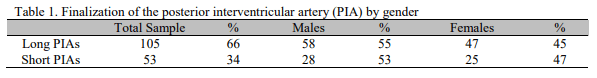
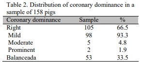
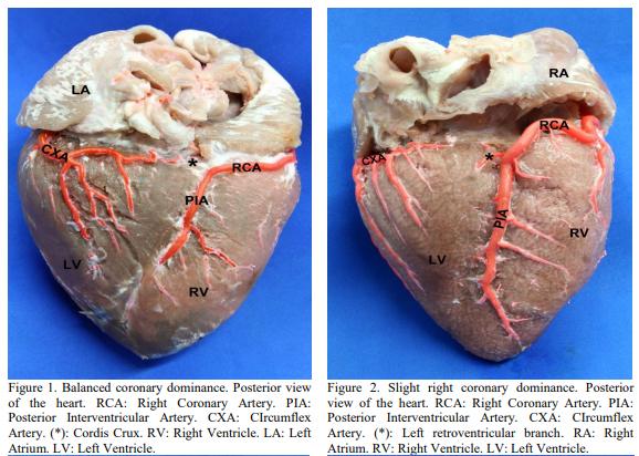
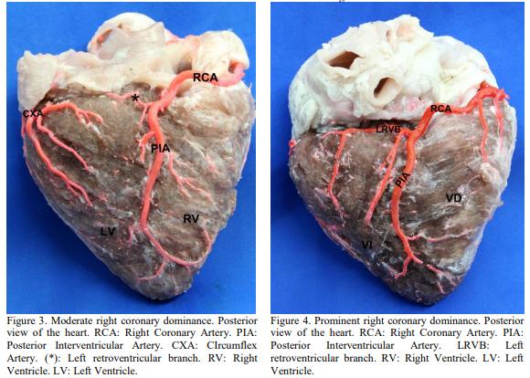
Coronary dominance in swine has been poorly evaluated. The frequencies of each type of dominance have been described, but few details have been given as to the different expressions of each one. The aim of this study was to characterize coronary dominance in commercial breed swine. One hundred and fifty eight pig hearts were evaluated. The coronary arteries (CA) were infused with synthetic resin (Palatal 85% and Styrene15%) through the ostia after channeling. The coronary artery that gives origin to the posterior interventricular artery (PIA), and the site of termination of both the circumflex arteries (CXA), and left retroventricular branch (LRVB) were determined in order to establish the coronary dominance pattern. Right coronary dominance was found in 105 hearts (66.5%), and a balanced circulation in 53 specimens (33.5%). No dominance was observed for the left coronary artery in the hearts studied. The CXA ended on the posterior aspect of the left ventricle in 101 samples (64%) and on the crux cordis in 55 specimens (34.8%). In two specimens (1.3%) it ended as a left marginal artery. In all cases the PIA was a branch of the RCA, and was long in 105 hearts (66%), 55% of which corresponded to males and 45% to females, but this difference was not statistically significant (p=0.77). The AIA ended on the apex in 126 specimens (80%), 71 of which (56%) corresponded to males and 55 (44%) to females (p=0.74). Regarding right coronary dominance, subtype I was observed in 98 specimens (93.3%), subtype II in 5 cases (4.8%), whereas subtype III was observed in 2 hearts (1.9%). Knowing coronary dominance patterns and their irrigation territories is useful for training purposes based on the use of experimental and hemodynamic models with this animal species.

Keywords: pig, coronary dominance, coronary arteries, left dominance, balanced circulation.

INTRODUCTION
Blood irrigation is supplied to the heart by the right coronary artery (RCA) and left coronary artery (LCA), which have their origin in the ascending aorta at the level of the respective right and left ostia. The high variation existing in the morphology of these vessels regarding their distribution in the irrigation of the posterior aspect of the heart serves as a basis for the coronary dominance concept (Cavalcanti et al., 1995; Ilia et al., 2001; Ballesteros et al., 2007). This anatomic trait has been poorly studied in swine, and the parameters to be followed to indicate whether one type of dominance or another prevails have not been elucidated (Weaver et al., 1986; Sahni et al., 2008)
Based on the above, we used a concept commonly utilized in humans to indicate that the dominance of the right or left coronary arteries depends on the coronary artery that gives origin to the posterior interventricular artery (PIA) or to the artery that supplies the largest portion of the posterior wall of the left ventricle (Cavalcanti et al., 1995; Ilia et al., 2001; Ballesteros et al., 2007). The diameter of the RCA is significantly smaller in the left dominance pattern, whereas the diameter of the circumflex artery (CXA) is smaller in the presence of a right dominance pattern. The cause of these relations is probably given by complex embryological processes that simultaneously determine the type of irrigation and the caliber of the coronary vessels to guarantee appropriate irrigation of all segments of the heart (Ballesteros et al., 2007).
Right coronary dominance occurs when the RCA irrigates the posterior aspect of the right ventricle, gives origin to the PIA, and extends beyond the crux cordis, through its left retroventricular branch, thus irrigating part of the posterior wall of the left ventricle. In the balanced coronary circulation pattern the RCA irrigates the right ventricle and the posterior portion of the interventricular septum through the PIA, whereas the LCA irrigates the left ventricle ending in the crux cordis. In the coronary left dominance pattern the LCA irrigates the posterior aspect of the left ventricle, the posterior segment of the interventricular septum that forms the PIA, and/or the posterior wall of the right ventricle (Schlesinger, 1940; James, 1965; Nerantzis et al., 1996).
Right dominance has been reported in swine with a frequency of 78-100%, balanced circulation 16-17%, and left dominance 4-5%. The latter pattern has not been found in most investigations carried out with this animal species (Weaver et al., 1986; Kato et al., 1987; Crick et al., 1998; Sahni et al., 2008). Left dominance has been reported in humans within a range of 2.5-36% (Schlesinger, 1940; Ortale et al., 2004; Pessa et al., 2004; Loukas et al., 2006).
In addition to a purely academic interest, the importance of knowing coronary dominance patterns in swine, given the great similarity existing between the human and the pig hearts, resides on the impact of this species as an experimental model for heart surgery and trauma, for hemodynamic procedures, and to determine the etiology of arrhythmias derived from occlusive coronary heart disease (Crick et al., 1998; López et al., 2004; Sahni et al., 2008).
Coronary dominance has been determined using classical dissection, injection and corrosion techniques, or radiographic studies (Kato et al., 1987; Crick et al., 1998; Ballesteros et al., 2007; Sahni et al., 2008). The sparse information on coronary dominance in swine gives relevance to the present study, developed in fresh cadaveric material with the purpose of establishing a differential evaluation of this anatomic feature between the hearts of swine and humans.
MATERIAL AND METHODS
This descriptive cross-sectional study assessed the coronary arteries (CA) of 158 hearts obtained from swine destined to the slaughterhouse and provided by Frigorífico Vijagual of Bucaramanga, Colombia. The samples were subjected to exsanguination for 6 hours. The ostia of the CAs were channeled and perfused with polyester resin (palatal GP41L 85% and styrene 15%), stained with mineral red. To facilitate casting, the anatomic pieces were kept immersed in water for 24 hours. The hearts were then impregnated with KOH for 5 minutes with the purpose of releasing the subepicardial fat, and affixed in a 5% formaldehyde solution.
The proximal and medium calibers of the RCA were measured and compared with the type of coronary dominance. The morphometric measurements were made with a digital caliper (Mitutoyo®). PIAs ending at the upper or medium thirds of the posterior interventricular sulcus (SIP) were rated as short, while those ending at the inferior segment of the said sulcus, the apex, or even the anterior aspect of the left ventricle were rated as long. The terminations of the right and left coronary arteries were assessed, and the type of dominance was established in the samples evaluated using Schlesinger’s criteria (1940). Similarly, the extension of the right coronary dominance was rated as mild when it reached the right third of the Left ventricle, moderate when it reached the middle third, and prominent when it reached the lateral third or the obtuse margin of the heart (Ballesteros et al., 2007). Photographs were taken of all anatomic pieces to support the findings. Continuous variables were expressed as means, medians, and standard deviations; discrete variables were expressed as frequencies and percentages. Continuous variables were analyzed using t-test, whereas discrete variables were analyzed using Pearson’s Chi-square test and Fisher’s exact test. The results were analyzed using the statistics “Epi – Info 3.5.3” software. The significance level used for this research was (p<0.005).
RESULTS
The mean weight of the 158 hearts evaluated was 360 ± 61.21 grams. The hearts were obtained from swine sacrificed weighing 90 kilograms. Right coronary dominance was found in 105 hearts in the evaluated sample. A right coronary dominance pattern was found in 105 hearts (66.5%), and balanced dominance pattern in 53 specimens (33.5%) (Figure 1). No left coronary artery dominance pattern was observed in the hearts studied.
The proximal and medium caliber of the RCA in the cases of right coronary dominance pattern was 3.84 ± 0.80 mm. However, this same caliber was found in the hearts with a balanced coronary dominance pattern (3.97 + 0.79 mm) (p=0.88). The CXAs ended as a left marginal branch (LMB) in 2 hearts (1.3%), at the posterior aspect of the left ventricle in 103 samples (65.2%), and at the crux cordis in 53 specimens (33.5%). In no case the CXA ended as a PIA.
In all cases the PIA emerged as the final branch of the RCA, being rated as long in 105 hearts (66%) and as short in 53 (34%). Of the long PIAs, 55% corresponded to males and the 45% to females, but this difference was not statistically significant (p=0.77) (Table 1). The AIA ended at the apex in 126 specimens (80%), 71 of which (56%) corresponded to males and 55 (44%) to females (p=0.74). In 20 cases (12.7%) the AIA ended at the posterior aspect of the left ventricle and in 12 samples (7.3%) this vessel reached the medium or lower thirds of the anterior interventricular sulcus.
Evaluation of coronary dominance in pigs; a comparative study with findings in human hearts - Image 1
Regarding the right coronary dominance pattern, subtype I (Figure 2) was observed in 98 specimens (93.3%), with a short left retroventricular branch (LRVB), subtype II was observed in 5 cases (4.8%), in which the LRVB reached the middle third of the left ventricle (Figure 3), whereas subtype III was observed in 2 cases (1.9%) (Figure 4). (Tab 2).
Evaluation of coronary dominance in pigs; a comparative study with findings in human hearts - Image 2
 
Evaluation of coronary dominance in pigs; a comparative study with findings in human hearts - Image 3
 
Evaluation of coronary dominance in pigs; a comparative study with findings in human hearts - Image 4
DISCUSSION
To define coronary dominance it is necessary to know the sites of termination of the CXA and the origin of the PIA, either from the RCA or the CXA. Prior studies in swine do not mention the termination of the PIA. In humans, the termination of this vessel has been reported in the proximal and medium thirds of the posterior interventricular sulcus in 25-35%, whereas its termination has been described at the distal third of the sulcus of the same name, the apex, and at the anterior aspect of the left ventricle within a range of 67.4-75% (James et al., 1965; Margaris et al., 1997; Ballesteros et al., 2011), whereas our report shows a slightly lower incidence. For short PIB, compensation phenomena are seen in the irrigation of the posteroinferior segments of the heart, given by the distal segment of the AIA that, after overcoming the apex, becomes distributed in the neighboring territory of the diaphragmatic aspect, irrigating the segment not reached by the PIA. In cases of short PIBs, compensation phenomena are observed in the irrigation of the lower posterior segments of the heart, given by the distal segment of the AIA that has passed the apex, and distributes itself in the neighboring territory of the diaphragmatic surface, thus irrigating the segment missed by the AIP.
No CXAs ended at the posterior interventricular sulcus. They ended more frequently at the posterior aspect of the left ventricle (64%) and in a smaller percentage at the crux cordis, in line with the majority of prior studies (Lumb and Singletary, 1962; Crick et al., 1998; Sahni et al., 2008). Shorter CXAs end as marginal branches, a fact observed in our study in 1.3% of the cases, with an incidence lower than that observed in humans (9.1-25%) (Baroldi and Scomazzoni, 1967; Baptista et al., 1991; Kalpana, 2003; Ballesteros et al, 2007). In these cases, the marginal branches, in addition to irrigating the obtuse aspect of the heart through their collateral branches, were seen to take part in the irrigation of the posterior aspect of the left ventricle together with the left ventricular branches of the RCA. The termination of the CXA at the posterior interventricular sulcus has been reported in human hearts with a frequency of 7– 23%, this being the trait of the left coronary artery dominance pattern (Baroldi and Scomazzoni, 1967; Baptista et al., 1991; Mouchet, 1993; Kalpana, 2003).
The frequency of right coronary dominance observed in the present study (66.5%), is lower than that reported in prior studies (78-100%) (Weaver et al., 1986; Kato et al., 1987; Crick et al., 1998; Sahni et al., 2008). These differences are possibly due to the number of samples evaluated and to the divergence of criteria used by the authors to perform these characterizations. Diverse studies conducted in human hearts similarly report a wide range of occurrence of the right coronary dominance pattern: Lower incidences within a range of the 48-70% (Schlesinger, 1940; Cavalcanti et al., 1995; Ortale et al., 2004; Kaimkhani et al., 2005; Loukas et al., 2006), medium incidences within a range of 70-84% (Blunk and DiDio, 1971; DiDio and Wakefield, 1975; Hadziselimovic, 1981; Sahni and Jit, 1990; Kalpana, 2003; Pessa et al., 2004; Ballesteros et al., 2007) and high incidences between 85 and 90% (James, 1965; Penthe et al., 1976; Nerantzis et al., 1996). No studies have been conducted in swine to typify right dominance in depth; our series found that mild right dominance (subtype I) was most common, unlike common findings in humans, where a higher incidence of moderate right dominance is described. Similarly, the low frequency of prominent coronary dominance found in our study is consistent with reports in humans (Baptista et al., 1989; Kalpana, 2003; Ortale et al., 2004; Ballesteros et al., 2007).
Our study failed to find any specimens with left coronary artery dominance, which is consistent with the reports by Kato et al. (1987) and Sahni et al. (2008). Some other authors (Weaver et al., 1986; Crick et al., 1998) have reported frequencies of between 4 and 5% for this feature. Left dominance has been reported in humans within a range of 5-20% (Schlesinger, 1940; Baroldi and Scomazzoni, 1967; Blunk and Didio, 1971; Sahni and Jit, 1990; Zbigniew and Mikusek, 2000; Kalpana, 2003; Ortale et al., 2004; Kaimkhani et al., 2005; Ballesteros et al., 2007). The low frequency of a left dominance pattern could be a trait of coronary circulation in swine, an issue that should be taken into account for compared anatomy purposes, and for the design of experimental hemodynamic procedures using this particular species.
Our series found a balanced circulation pattern in 33.5% of the hearts, a figure that is slightly higher than the one found in the studies conducted by Weaver et al. (1986); Kato et al. (1987) and Crick et al. (1998), who reported this morphologic trait within a range of 16-20%. Literature reports in humans present frequencies of between 7-26% (Polacek and Zechmeister, 1958; Zbigniew and Mikusek, 2000; Ortale et al., 2004; Loukas et al., 2006; Ballesteros et al., 2007). A balanced circulation expresses an equitable distribution of the irrigation of the diaphragmatic aspect of the heart by the coronary arteries and their branches.
One setback of the present work was the difficulty to obtain samples not damaged by the slaughter process because operators often cut through the heart cavities, so many hearts failed to meet the ideal conditions for a good perfusion of the vascular beds.
CONCLUSIONS
The absence of a left coronary artery dominance pattern in our study is consistent with the results of most prior studies and expresses a difference with coronary irrigation in humans characterized by presenting left dominances with considerable frequency. The incidence of a right dominance pattern found in our series is lower than that reported for swine in the literature, and is found in the lower range of what has been reported for humans. The findings in this study contribute to the knowledge of compared interspecies anatomy and human anatomy, which is useful for the design of hemodynamic procedures.
ACKNOWLEDGEMENTS
We thank Frigorífico Vijagual in Bucaramanga – Colombia, and Dr. Luz Stella Cortés, DMV for the donation of pieces for this investigation.
This article was originally published in Arquivo Brasileiro de Medicina Veterinária e Zootecnia., v.67, n.3, p.783-789, 2015. http://dx.doi.org/10.1590/1678-4162-6637. This is an Open Access article distributed under the terms of the Creative Commons Attribution Non-Commercial License.

BALLESTEROS, L.E.; CORZO, E.G.; SALDARRIAGA, B. Determinación de la dominancia coronaria en población mestiza colombiana. Un estudio anatómico directo. Int. J. Morphol., v.25, p.483-491, 2007.

BALLESTEROS, L.E.; RAMIREZ, L.M.; QUINTERO, I.D. Right coronary artery anatomy: anatomical and morphometric analysis. Rev. Bras. Cir Cardiovasc., v.26, p.230-237, 2011.

BAPTISTA, C.A.; DIDIO, L.J.; PRATES J.C. Types of division of the left coronary artery and the ramus diagonalis of the human heart. Jpn. Heart. J., v.32, p.323-335, 1991.

BAPTISTA, C.A.; DIDIO, L.J.; TEOFILOVSKIPARAPID, G. Variation in length and termination of the right coronary artery in man. Jpn. Heart. J., v.30, p.789-798, 1989.

BAROLDI, G.; SCOMAZZONI, G. Coronary circulation in the normal and the pathologic heart. Office of the Surgeon General. Washington, DC: Dept of the Army, 1967.

BLUNK, J.N.; DIDIO, L.J.A. Types of coronary circulation in the human hearts. Ohio St. Med. J., v.67, p.596-607, 1971.

CAVALCANTI, J.S.; DE LUCENA OLIVEIRA, M.; PAIS E MELO, A.V. et al. Anatomic variations of the coronary arteries. Arq. Bras. Cardiol., v.65, p.489- 492, 1995.

CRICK, S.; SHEPPARD, M.; YEN HO, S. et al. Anatomy of the pig heart: comparisons with normal human cardiac structure. J. Anat., v.193, p.105-119, 1998.

DIDIO, L.J.; WAKEFIELD, T.W. Coronary arterial predominance or balance on the surface of the human cardiac ventricles. Anat. Anz., v.137, p.147-158, 1975.

HADZISELIMOVIC, H. Age characteristics of blood vessels of the human heart. Acta. Anat. (Basel), v.109, p.231-237, 1981.

ILIA, R.; ROSENSHTEIN, G.; WEINSTEIN J. et al. Left anterior descending artery length in left and right coronary artery dominance. Coron. Artery. Dis., v.12, p.77-78, 2001.

JAMES, T.N. Anatomy of the coronary arteries in health and disease. Circulation., v.32, p.1020-1033, 1965.

KAIMKHANI, Z.A.; ALI, M.M.; FARUQI, A.M. Pattern of coronary arterial distribution and its relation to coronary artery diameter. J. Ayub. Med. Coll. Abbottabad., v.17, p.40-43, 2005.

KALPANA, R.A. Study on principal branches of coronary arteries in humans. J. Anat. Soc. India., v.52, p.137-140, 2003.

KATO, T.; YASUE, T.; SHOJI, Y. et al. Angiographic diference in coronary artery of man, dog, pig and monkey. Acta. Pathol. Jpn., v.37, p.361- 373, 1987.

LÓPEZ, M.; RUIZ, G.; RAMÍREZ, M. et al. Cambios fisiológicos en cerdo de cirugía experimental para trasplante cardíaco. Investigación en Salud, v.6, p.11- 13, 2004.

LOUKAS, M.; CURRY, B.; BOWERS, M. et al. The relationship of myocardial bridges to coronary artery dominance in the adult human. Heart. J. Anat., v.209, p.43-50, 2006.

LUMB, G.; SINGLETARY, H. Blood supply to the atrioventricular node and bundle of hiis: a comparative study in pig, dog, and man. Grant H-5o63 from the National Institutes of Health, United States Public Health Service, v.41, p.65-75, 1962.

MARGARIS, N.G.; KOSTOPOULOS, K.G.; NERANTZIS, C.E. et al. Posterior right diagonal artery. An angiographic study. Angiology, v.48, p.73- 77, 1997.

MOUCHET, A. Les arteres coronaires du coeur chez l’Homme. 2nd ed. Maloine, Paris. 1993.

NERANTZIS, C.E.; PAPACHRISTOS, J.C.; GRIBIZI, J.E. et al. Functional dominance of the right coronary artery: incidence in the human heart. Clin. Anat., v.9, p.10-13, 1996.

ORTALE, J.R.; KEIRALLA, L.C.; SACILOTTO, L. The posterior ventricular branches of the coronary arteries in the human heart. Arq. Bras. Cardiol., v.82, p.468-472, 2004.

PENTHE, P.; BARA, J.A.; BLANC, J.J. Etude anatomique descriptive des gros troncs coronariens et des principales collaterales epicardiques. Nouv. Presse. Med., v.5, p.71- 75, 1976.

PESSA, C.J.; GOMES, W.J.; CATANI, R. et al. Anatomical relationships between the posterior mitral valve annulus and the coronary arteries. Implications to operative treatment. Braz. J. Cardiovasc. Surg., v.19, p.372-377, 2004.

POLACEK, P.; ZECHMEISTER, A. The occurrence and significance of myocardical bridges and loops on coronary arteries. University. J. E. Purkyne, v.10, p.125-131, 1958.

SAHNI, D.; JIT, I. Blood supply of the human interventricular septum in north-west Indians. Indian. Heart. J., v.42, p.161-169, 1990.

SAHNI, D.; KAUR, G.D.; JIT, H. et al. Anatomy and distribution of coronary arteries in pig in comparison with man. Indian J. Med. Res., v.127, p.564-570, 2008.

SCHLESINGER M.J. Relation of anatomic pattern to phatologic conditions of the coronary arteries. Arch. Pathol., v.30, p.403-415, 1940.

WEAVER, M.E.; PANTELY, G.A.; BRISTOW, J.D. et al. A quantitative study of the anatomy distribution of coronary arteries in swine in comparison with other animals and man. Cardiovasc. Res., v.20, p.907-917, 1986.

ZBIGNIEW, K.; MIKUSEK, J. Clinical and morphological studies on varieties of coronary vascularisation of diaphragmatic surface of human heart. Med. Sci. Monit., v.6, p.253-257, 2000.

Related topics:
Authors:
Fabian Gomez
Universidad Industrial de Santander
Recommend
Comment
Share
Profile picture
Would you like to discuss another topic? Create a new post to engage with experts in the community.
Join Engormix and be part of the largest agribusiness social network in the world.