INTRODUCTION
It is well established that maize grain can be invaded by fungi, which generally cause losses in weight and quality. Moreover, some of the fungi associated with maize are known to contaminate the grain with mycotoxins posing serious hazard to consumer health. On the basis of adverse effects on human and animal health and widespread contamination, aflatoxins, deoxynivalenol (replaced in some areas by nivalenol), fumonisins, ochratoxin A and zearalenone are considered as the most important mycotoxins on a worldwide scale [1]. These mycotoxins are known to occur in maize [2], although ochratoxin A is more of a problem in small grain cereals such as barley and wheat [3].
Survey of the fungal biota and mycotoxins of maize continues to attract worldwide attention and has been the subject of investigations in many parts of the world. Comprehensive reviews on the global occurrence of mycotoxins [4, 5] cite few episodes from Sub-Saharan Africa, mainly pertaining to reports from South Africa. On the other hand, the health risk from mycotoxins is expected to be generally higher in developing countries because of the prevailing climate that favors fungal growth, the inadequate crop management and storage systems, and the existing gap between supply and demand which forces people to consume what they might have otherwise rejected, even when the food is moldy and organoleptically unacceptable [6]. There is an urgent need to monitor and manage the mycotoxin problem in staple food crops such as maize.
In recent years, data on mycotoxins of maize in Africa have begun to accumulate with reports, for instance, from Benin [7], Kenya [8, 9] and Nigeria [10]. In Ethiopia, there is limited information on the occurrence of aflatoxins in maize [11]. Worse still reports on fumonisins and the other Fusarium mycotoxins are so far not available. The prevalence of Aspergillus flavus [12] and the occurrence of toxigenic Fusaria [13] were also reported based on qualitative determination of fungi by plating surface disinfected maize kernels. Reportedly, there is occurrence of Aspergillus and Fusarium mycotoxins in Ethiopian barley, wheat, sorghum and teff [14]. There is a need to further extend the data base on the nature and extent of mycotoxin contamination and the associated fungi in the region. Such information is a prerequisite for assessing the risk to consumer health and for managing the mycotoxin problem.
This work reports the fungi occurring on the surface of and inside maize grain from Ethiopia and the incidence and extent of contamination of the grain by aflatoxins (AFT), fumonisins (FUM), deoxynivalenol (DON) and nivalenol (NIV).
MATERIALS AND METHODS
Samples
Seventeen samples (one kg each) of maize grain of the 2004/2005 harvest in Ethiopia were collected from Dire Dawa (east), Adama (central) and Ambo (west). Five samples each from Dire Dawa and Ambo, and seven samples from Adama were drawn. Samples were collected from farmers’ storage 7 – 9 months after harvest. The duration of grain storage by farmers in Ethiopia is on the average 8 – 10 months [6] and sampling was done close to the end of the storage period to capture the extent of fungal load that could develop. Systematic random sampling was carried out in which maize storages within 10 Km radius of the respective towns of Dire Dawa, Adama, and Ambo were sampled at 5 – 7 Km intervals. Each sample was composed of five portions of about 200 g each drawn into a plastic cup from different locations of the bagged grain by probing using sampling spear. The five incremental portions were mixed to get about 1 Kg composite sample. It is widely accepted in mycotoxicology that about 200 g incremental portion be taken for every 200 Kg product [6]. Based on information obtained from extension agents in the study areas, the maximum quantity of grain farmers store at the time of sampling was assumed to be about 1 ton; accordingly, the sample size was set at 1 kg. Samples were transported by air express to the analytical laboratory in Freising, Germany and were maintained at 4 ºC until analysis.
Determination of Fungi
For isolation of seed surface mycobiota, 10 g seed of each sample was washed in 90 ml saline solution (composed of 0.58 g NaH2PO4, 4 g NaCl, 2.6 g Na2HPO4 in 1 l distilled water) in screw cap bottles on a horizontal shaker (HS 501, IKA-Werke, Staufen, Germany) at 180 strokes minute-1 for 20 min and the wash solution was further diluted to 10-2, 10-3 and 10-4 with the saline solution. One ml of each dilution was pipetted separately into 9 cm diameter plastic petri plates (in duplicate). About 20 ml of autoclaved dichloran glycerine (DG-18) agar (Merck, Darmstadt, Germany) was then poured into each plate and mixed with the sample by gently swirling the plates. Plates were then incubated at room temperature for 1 – 2 weeks during which developing fungi were counted and identified; colony forming units (cfu) g-1 seed were determined. Frequency (out of the 17 samples) of occurrence and relative density (contribution to the total cfu g-1) of each species or genus was computed.
Isolation of internal mycobiota was done employing the dilution plate method involving the following procedures. A sub-sample of 10 g whole kernels was weighed in screw-capped baffled bottles (Kinematika GS 25K), surface sterilized by a quick rinse in 70% isopropanol followed by soaking for 1 – 2 min in 1% sodium hypochlorite solution. Seeds were ground in 90 ml of saline solution with the composition given above with an ultraturax (IKA-Werke, Staufen, Germany) for 30 seconds, and 2 ml of the resulting suspension were pipetted into 14.5 cm plastic petri plates. About 50 ml of autoclaved DG-18 agar (Merck, Darmstadt, Germany) cooled down to about 50 ºC was poured into each plate.
Fungi were identified based on colony and microscopic characteristics. Among the major groups, Aspergilli were identified according to Raper and Fennel [15]; Fusaria were identified according to Nelson et al. [16], with the exception that the recently accepted species name F. verticillioides (Sacc.) Nirenberg was used in place of F. monilliforme Sheldon.
Mycotoxin Analyses
A representative sub-sample of 100 g maize grain was ground to pass a 1-mm sieve and used for mycotoxin analysis.
Enzyme Linked Immunosorbent Assay of AFT and FUM
Commercially available ELISA kits, Ridascreenâ Aflatoxin Total and Ridascreenâ Fast Fumonisin (R-Biopharm, Darmstadt, Germany) were used for the analysis of AFT and FUM, respectively. After a competitive enzyme immunoassay [17], the A450 was measured using a plate reader (Multiscan Plus, Titertek).
Aflatoxin analysis was done as follows. Ten grammes of ground grain sample was extracted with 50 ml of 70% methanol by stirring for 3 min on a magnetic stirrer. After filtration, 100 µl of the extract was mixed with 600 µl buffer (provided with the ELISA kit) and assayed for AFT content in the ELISA. A calibration curve using A450 values was constructed from five standard concentrations between 50 and 4050 ng kg-1. All the standard and sample values were previously adjusted as percentages of the zero standard; 0 µg kg-1 was equal to 100% absorbance.
Fumonisin analysis involved the following procedures. Ten grammes of ground grain sample was extracted with 50 ml of 70% methanol by stirring for 3 min on a magnetic stirrer. After filtration, 100 µl of the extract was mixed with 1.3 ml distilled water and assayed for FUM content. A calibration curve using A450 values was constructed from four standard concentrations between 0.1 and 6.4 mg kg-1.
High Performance Liquid Chromatography Analysis of DON and NIV
The HPLC conditions were as below. The HPLC equipment for DON and NIV analysis consisted of a Merck L-6200 HPLC pump, a Merck 655A-40 auto sampler, a reversed phase Lichrospher 100 RP-18 column, 125 × 4 mm, 5 µm particle size (Merck), a post column system of Besta E-100 (2×) pumps and reaction loops of 0.5 mm × 8 m and 0.5 mm × 4 m (Omnifit), and a Merck F-1050 fluorescence detector.
A ground sub-sample (12.5 g) was extracted in 50 ml of acetonitrile : water (84:16) by stirring for 2 h. Initial clean-up of portions of extracts through a column of 0.75 g charcoal-celite-neutral alumina, 25:15:5 (wt/wt) was followed by a further purification through a column packed with 0.5 g florisil [18]. The final eluate was evaporated and the residue was taken up in 1 ml of the HPLC mobile phase. One hundred microliters were injected. Acetonitrile : water (1:9) was used as eluent at a flow rate of 1 ml min-1. Post-column derivatization using 0.1 N NaOH (Merck, Darmstadt, Germany) at 0.4 ml min-1 was followed by reaction with a solution of acetyl acetone (5 ml) and ammonium acetate (62.5 g) in 1 l of water at a flow rate of 0.75 ml min-1 and 115°C in oil bath (MGW Lauda C6) and detection of DON and NIV was at 503 nm (397 nm excitation). The chromatography data system Chromeleon Version 6.0 (Dionex) was used for recording chromatograms.
RESULTS
Fungi Associated with Maize Grain in Ethiopia
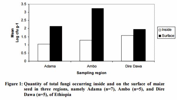
The total fungal density ranged from 2.8 × 10² to 9.4 × 10³ cfu g-1 in samples from Dire Dawa, 2.4 × 10² to 3.5 × 10⁴ cfu g-1 in those from Adama, and 2.2 × 10⁴ to 1.3 × 10⁶ for samples from Ambo (Figure 1). There were substantial variations in cfu g-1 among the samples regardless of their geographic origin. In general, higher fungal counts were encountered on the surface of than inside the seeds (Figure 1).
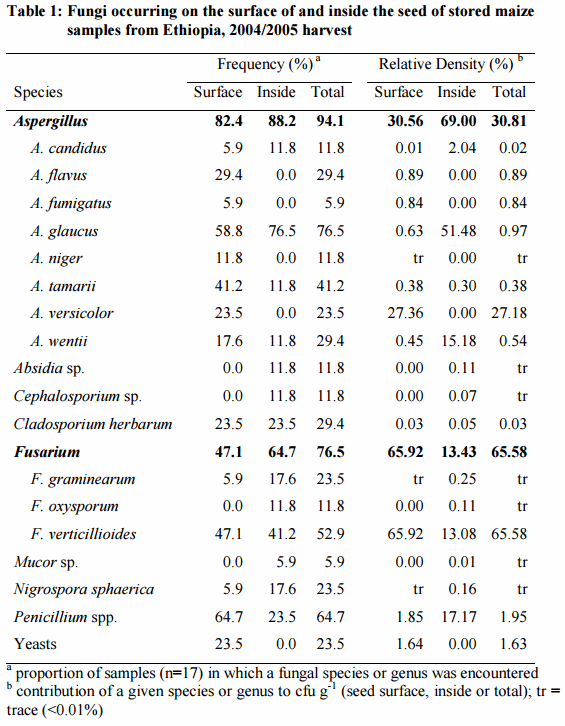
Fifteen species of fungi were identified from the maize samples (Table 1). Aspergilli were the most frequent fungi, occurring in 94% of the samples. Fusarium spp. occurred in 76.5% of the samples while Penicillium spp. were found in 64% of the samples. In terms of cfu g-1, Fusarium was the dominant genus representing about 66% of the seed surface fungi and that of the total fungi isolated. Fusarium verticillioides comprised more than 99% of the Fusaria isolates. The eight Aspergillus spp. isolated in the study constituted 69% of the internal fungi, but only 30.8% of the total cfu g-1 (Table 1). Aspergillus versicolor and A. glaucus showed high levels of cfu g -1, on the surface and inside the seed, respectively (Table 2). Aspergillus glaucus and A. tamari followed by A. flavus and A. wentii were more frequently encountered among the Aspergilli. Aspergillus flavus occurred only on the seed surface of 29% of samples at a maximum of 2500 cfu g-1 (Table 1); it represented 0.89% of the total fungi isolated (Table 1). Species of Absidia, Cephalosporium, Cladosporium, Mucor and Nigrospora were isolated generally at trace levels. Yeasts occurred on the seed surface in 23.5% of the samples.
Occurrence of Aflatoxins and Fusariotoxins
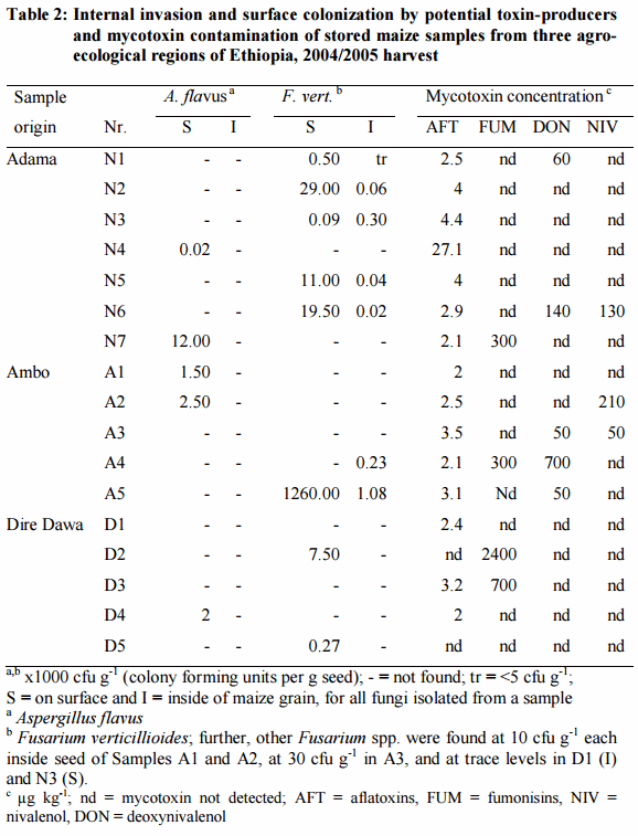
Aflatoxins were detected in 15 (88%) of the 17 maize samples; the concentrations were below 5 µg kg-1, except in one sample from Adama which had 27 µg kg-1. FUM occurred in two samples from Dire Dawa at the concentrations of 700 and 2400 µg kg-1 and at 300 µg kg-1 in one sample each from Adama and Ambo. Five samples contained DON at 50 – 700 µg kg-1. NIV was detected in three samples at 50, 130 and 210 µg kg-1.
DISCUSSION
The most prevalent fungi were Aspergilli similar to other tropical countries although the species and their levels of occurrence were not always comparable. The frequency (29%) of A. flavus, which is the main aflatoxin producer in maize, sharply contrasts reports from South East Asia. For instance, in a recent report on maize from Vietnam [19], 92% of 13 samples analyzed in the study contained the fungus, with density reaching 70,000 cfu g-1 in surface disinfected seed. In the present study, A. flavus was detected in all sampling regions showing its ubiquitous distribution but it occurred solely as surface contaminant of the seed, at lower cfu g-1 and frequency. It is noteworthy that xerophilic species such as A. glaucus and A. versicolor occurred at higher densities than A. flavus. The high frequency of these two species in cereals and sorghum from Ethiopia was reported earlier [6]. Aspergillus wentii was reported in maize from Vietnam [19], while A. tamarii was found in maize fields in Nigeria [20]. Among storage fungi, Penicillium was the other most frequently encountered genus. Isolates of Penicillium were not identified on species level. In general Aspergilli occur more commonly in warmer regions while Penicillia are more frequent in colder climates [21]. In Ethiopia, the altitude-mediated ecological diversity might expose the grain to a range of fungi.
Fusaria were frequent contaminants of the samples next to Aspergilli. Fusarium verticillioides, which is known to produce FUM, was the dominant species of the genus; F. proliferatum, a polyphialidic and toxigenic member of Section Liseola, was not found in the samples. This agrees with earlier reports on maize from Ethiopia [13] and neighboring Kenya [9]. Fusaria declined only slightly in maize kept at safe storage moisture levels in Benin [7]. The high average relative density of Fusaria, however, was related to the exceptionally high density (nearly 1.3 x 10⁶ cfu g-1) of F. verticillioides in sample Nr. A5 in which the fungus had apparently sporulated on the seed surface.
The samples were taken from grain stored for 7 – 9 months in bags by small holder farmers. Hot drought conditions have been one of the most frequent factors reported to favor preharvest infection of maize by A. flavus [22] and F. verticillioides [23]. During storage, seed moisture content higher than safe levels appears to be the most important single factor influencing fungal invasion of grain [21]. The three sampling regions represent different agro-ecological zones [24]. Ambo represents tropical highland maize growing areas with altitude exceeding 2000 m while Adama is described as tropical mid-altitude. Dire Dawa is representative of tropical lowland areas with predominantly dry and hot climate. The marked difference between sampling regions in their fungal load, with the lowest cfu g-1 recorded in samples from Dire Dawa might thus be explained by differences in weather factors during storage. It is well established that preharvest weather factors influence the mycobiota of freshly harvested grain but growth and ecological succession of species of fungi in response to prevailing storage conditions dictate the kind of fungi and the extent of their invasion in stored grain.
Regardless of the sampling regions, the levels of cfu g-1 of the fungi isolated in the present study were generally lower than those reported from other countries such as Burundi [25] and Vietnam [19], which is in agreement to Ayalew-Mamed [6], who also reported that cfu g-1 levels of fungi in cereals from Ethiopia were much lower than other tropical countries.
Although AFT occurred in most maize samples, the concentrations (generally less than 5 µg Kg-1 with a maximum of 27 µg Kg-1 in one sample) were comparatively lower than earlier reports from Nigeria [10], Kenya [8] and Vietnam [19]. Comparatively low aflatoxin levels were also found in barley, wheat, tef and sorghum from Ethiopia [14]. Maize is one of the high risk crops for aflatoxin contamination worldwide. Higher levels of AFT in maize leading to outbreaks of acute aflatoxicosis in humans are common in neighboring Kenya [8]. Maize in east Africa is reported to be heavily contaminated by AFT while in southern Africa (Botswana, South Africa and Swaziland), AFT are not a major contaminant of maize and levels are usually <20 µg kg-1 [26]; the results of the present study contrast that conclusion as the AFT levels were comparable to that of the southern Africa.
The occurrence of FUM (with a mean of 925 µg kg-1 in 23% of the samples) was somewhat comparable to reports from Eastern Africa. In Zambia FUM occurred in maize with 35% frequency and a mean level of 410 µg kg-1 [27] while in Kenya it occurred at 47% frequency and 672 µg kg-1 [9] were recorded. In Benin, FUM were detected at an average level of 640 µg kg-1 in higher proportion (64%) of the maize samples [27]. Higher levels of FUM were also reported for maize from Europe mostly southern parts [28], Argentina and China [5].
The low frequency and levels of DON and NIV agree with a previous report for cereals and sorghum from Ethiopia [14]. On the other hand, 60% of European cereals was contaminated with DON at 4 – 67,000 µg kg-1 while NIV occurred in 16% of samples at 3 – 7800 µg kg-1 [28].
Many countries have set tolerance limits for AFT in food and feeds at 20 µg kg-1 although some countries have more stringent regulations [29]. Nearly 40 countries have enforced limits for DON in wheat and other cereals generally at 750 µg kg-1, reaching up to 2000 µg kg-1 in some countries. Six countries have set limits for FUM in maize at 1000 to 3000 µg kg-1 [29]. By comparison to such tolerance limits, the levels of AFT and fusariotoxins in maize grain of 2004/05 harvest in Ethiopia were too low to be of major concern. Identifying low mycotoxin risk areas may contribute to the management of mycotoxin problems in poorer parts of Africa where as indicated by Shephard [30] regulation is less practical under the prevailing food scarcity and lack of capacity to enact or enforce legislation on mycotoxin levels. Specific ELISA methods were used for analysis of AFT and FUM with quantification limits of 1.75 and 222 µg kg-1, respectively. The detection limit for the HPLC procedure was 30 µg kg-1 for DON.
The occurrence of AFT in 88% of the samples while A. flavus occurred in only 29% of the samples of the present study is in agreement with earlier observations that the mycotoxins are highly stable and could be detected long after the producing fungi have died out or replaced by other fungi due to ecological succession [6]; A. flavus has relatively high moisture requirements among storage fungi [21]. Although both samples containing higher levels of FUM were from Dire Dawa, no clear trend was observed on the incidence of mycotoxins across the three sampling regions. The major FUM-producer, F. verticillioides, is a field fungus with higher seed moisture requirement for growth, i.e. above 19% moisture content in starchy cereal grain such as maize [6]. Thus, so as long as seed moisture levels allow growth of storage fungi, the extent of invasion by F. verticillioides declines in stored grain. On the other hand, FUM are stable compounds, and they could be detected in grain after considerable storage period. This might explain the comparatively higher concentrations of FUM despite the low levels of F. verticillioides detected in maize samples from Dire Dawa. Poor correlation between the incidence of F. verticillioides and fumonisin contamination has been reported [9].
CONCLUSION
The fungi isolated in the present study were from the same genera that are common in maize and cereals. A. flavus was less frequent. Fusarium verticillioides was the predominant member of Fusarium while other toxin-producing species such as F. graminearum occurred at trace levels. There was no clear relationship between the occurrence of mycotoxins and potential producer fungi in the samples. Aflatoxins, FUM, DON, and NIV played a minor role in the contamination of maize of the 2004/2005 harvest in Ethiopia. Had there been widespread, heavy mycotoxin contamination of maize in the study areas, it would have been possible to detect it, given the robust analytical systems used. However, in view of the ubiquitous occurrence of potential toxin producers in all sampling regions, and the frequent occurrence of aflatoxicosis outbreaks in neighboring Kenya with comparable highland maize production regions, further monitoring of mycotoxins in maize from Ethiopia, including the relatively hot, humid western and southwestern maize-producing regions of the country, coupled with detailed analysis of preharvest and storage factors is justified. Identification of low risk areas which is an established pest mitigation measure in mainstream crop protection deserves to be explored in mycotoxin management.
ACKNOWLEDGEMENTS
I am indebted to Dr. Robert Beck and Dr. J. Lepschy for reviewing the manuscript, Ms. Sabine Topor and Ms. Edith Böck for technical assistance in mycological analysis and Ms. Gitta Clasen in DON and NIV analysis, and Dr. P. Büttner for help in identification of fungi. The German Academic Exchange Service (DAAD) sponsored the study which is gratefully acknowledged.
REFERENCES
1. Miller JD Mycotoxins in small grains and maize: old problems, new challenges. Food Addit. Contam. 2008; 25: 219–230.
2. Scudamore KA, Nawaz S and MT Hetmanski Mycotoxins in ingredients of animal feed stuffs: II. Determination of mycotoxins in maize and maize products. Food Addit. Contam. 1998; 15: 30–55.
3. Kuiper-Goodman T and PM Scott Risk assessment of the mycotoxin ochratoxin A. Biomed. Environ. Sci. 1989; 2: 179–248.
4. Jelinek CF, Pohland AE and GE Wood Worldwide occurrence of mycotoxins in foods and feeds – an update. J. Assoc. Off. Anal. Chem. 1989; 72: 223–230.
5. Placinta CM, D’Mello JPF and AMC Macdonald A review of worldwide contamination of cereal grains and animal feed with Fusarium mycotoxins. Anim. Feed Sci. Technol. 1999; 78: 21–37.
6. Ayalew-Mamed A Mycoflora and Mycotoxins of Major Cereal Grains and Antifungal Effects of Selected Medicinal Plants from Ethiopia, Ph.D. Dissertation. Goettingen, Germany: Georg August University of Goettingen, 2002.
7. Hell K, Cardwell KF and HM Poehling Relationship between management practices, fungal infection and aflatoxin for stored maize. J. Phytopathol. 2003; 151: 690–698.
8. Probst C, Njapau H and PJ Cotty Outbreak of an acute aflatoxicosis in Kenya in 2004: identification of the causal agent. Appl. Env. Microbiol. 2007; 73: 2762– 2764.
9. Kedera CJ, Plattner RD and AE Desjardins Incidence of Fusarium spp. and levels of fumonisin B1 in maize in Western Kenya. Appl. Environ. Microbiol. 1999; 65: 41–44.
10. Bankole SA and OO Mabekoje Occurrence of aflatoxins and fumonisins in preharvest maize from southwestern Nigeria. Food Addit. Contam. 2004; 21: 251–255.
11. Geyid A and A Maru A survey of aflatoxin contents in maize, sorghum and teff samples. Ethiop. J. Health Dev. 1987; 2: 59–70.
12. Abate D and BA Gashe Prevalence of Aspergillus flavus in Ethiopian cereal grains: a preliminary survey. Ethiop. Med. J. 1985; 23: 143–148.
13. Wubet T and D Abate Common toxigenic Fusarium species in maize grain in Ethiopia. SINET: Ethiop. J. Sci. 2000; 23: 73–87.
14. Ayalew A, Fehrmann H, Lepschy J, Beck R and D Abate Natural occurrence of mycotoxins in staple cereals from Ethiopia. Mycopathologia 2006; 162: 57– 63.
15. Raper KB and DJ Fennell The Genus Aspergillus. Baltimore: William and Wilknis, 1965.
16. Nelson PE, Tousson TA and WFO Marasas Fusarium Species: an Illustrated Manual for Identification. University Park: Pennsylvania State University, 1983.
17. Biermann A and G Terplan Erfahrung mit einem Mikro-ELISA zur Aflatoxin B1-Bestimmung. Arch. Lebensmittelhyg. 33: 1–32.
18. Lepschy J, Dietrich R, Märtlbauer E, Schuster M, Süβ A and G Terplan A survey on the occurrence of Fusarium mycotoxins in Bavarian cereals from the 1987 harvest. Z. Lebensm. Unter. Forsch. 1989; 188: 521–526.
19. Trung TS, Tabuc C, Bailly S, Querin A, Guerre P and JD Bailly Fungal mycoflora and contamination of maize from Vietnam with aflatoxin B1 and fumonisin B1. World Mycotox. J. 2008; 1: 87–94.
20. Donner M, Atehnkeng J, Sikora RA, Bandyopadhyay R and PJ Cotty Distribution of Aspergillus section Flavi in soils of maize fields in three agroecological zones of Nigeria. Soil Biol. Biochem. 2008; 41: 37–44.
21. Northolt MD, Frisvad JC and RA Samson Occurrence of food-borne fungi and factors for growth. In: RA Samson (Ed). Introduction to Food-Borne Fungi. Baarn: Centraalbureau voor Schimmelcultures, 1995: 243–250.
22. Pyne GA and NW Widstrom Aflatoxin in maize. Cr. Rev. Plant Sci. 1992; 10: 423–440.
23. Miller JD Factors that affect the occurrence of fumonisin. Environ. Health Persp. 2001; 109: 321–324.
24. Hartkamp AD, White JW, Rodríguez Aguilar A, Bänziger M, Srinivasan G, Granados G and J Crossa Maize Production Environments Revisited: a GISBased Approach. Mexico, D.F.: CIMMYT, 2000.
25. Munimbazi C and LB Bullerman Molds and mycotoxins in foods from Brundi. J. Food Prot. 1996; 59: 869–875.
26. Siame BA and IN Nawa Mycotoxin contamination of food systems in eastern and southern Africa. In: JF Leslie, R Bandyopadhyay and A Visconti (Eds). Mycotoxins: Detection Methods, Management, Public Health and Agricultural Trade. Wallingford: CAB International, 2008: 117–125.
27. Doko MB, Rapior S, Visconti A and JE Schjoth Incidence and levels of fumonisin contamination in maize genotypes grown in Europe and Africa. J. Agr. Food Chem. 1995; 43: 429–434.
28. Bottalico A Fusarium diseases of cereals: species complex and related mycotoxin profiles in Europe. J. Plant Pathol. 1998; 80: 85–103.
29. FAO. United Nations Food and Agriculture Organization. Worldwide Regulations for Mycotoxins in Food and Feed in 2003. Food and Nutrition Paper 81. FAO, Rome, 2004.
30. Shephard GS Impact of mycotoxins on human health in developing countries. Food Addit. Contam. 2008; 25: 146–151.







.jpg&w=3840&q=75)