Nanoparticles as a Solution for Eliminating the Risk of Mycotoxins
Mycotoxins are toxic secondary metabolites produced by certain filamentous fungi. The occurrence of mycotoxins in food and feed causes negative health impacts on both humans and animals. Clay binders, yeast cell walls, or antioxidant additives are the most widely used products for mycotoxin elimination to reduce their impact. Although conventional methods are constantly improving, current research trends are looking for innovative solutions. Nanotechnology approaches seem to be a promising, effective, and low-cost way to minimize the health effects of mycotoxins. This review aims to shed light on the critical knowledge gap in mycotoxin elimination by nanotechnology. There are three main strategies: mold inhibition, mycotoxin adsorption, and reducing the toxic effect via nanoparticles. One of the most promising methods is the use of carbon-based nanomaterials. Graphene has been shown to have a huge surface and high binding capacity for mycotoxins. Attention has also been drawn to polymeric nanoparticles; they could substitute adsorbents or enclose any substance, which would improve the health status of the organism. In light of these findings, this review gives new insights into possible future research that might overcome challenges associated with nanotechnology utilization for mycotoxin elimination from agricultural products.
Keywords: mycotoxin; nanotechnology; agriculture; toxicity; nanoparticles.
1. Edwards, S.G. In?uence of agricultural practices on fusarium infection of cereals and subsequent contamination of grain by trichothecene mycotoxins. Toxicol. Lett. 2004, 153, 29–35.
2. Simpson, D.R.; Weston, G.E.; Turner, J.A.; Jennings, P.; Nicholson, P. Differential control of head blight pathogensofwheatbyfungicidesandconsequencesformycotoxincontaminationofgrain. Eur. J.PlantPathol. 2001, 107, 421–431.
3. Cunha, S.C.; Sa, S.V.M.; Fernandes, J.O. Multiple mycotoxin analysis in nut products: Occurrence and risk characterization. Food Chem. Toxicol. 2018, 114, 260–269.
4. Santini, A.; Meca, G.; Uhlig, S.; Ritieni, A. Fusaproliferin, beauvericin and enniatins: Occurrence in food—A review. World Mycotoxin J. 2012, 5, 71–81.
5. Jestoi, M. Emerging fusarium-mycotoxins fusaproliferin, beauvericin, enniatins, and moniliformin—A review. Crit. Rev. Food Sci. Nutr. 2008, 48, 21–49.
6. Marin, S.; Ramos, A.J.; Cano-Sancho, G.; Sanchis, V. Mycotoxins: Occurrence, toxicology, and exposure assessment. Food Chem. Toxicol. 2013, 60, 218–237.
7. Madalena,M.;Sobral,C.;Faria,M.A.;Cunha,S.C.;Ferreira,I.Toxicologicalinteractionsbetweenmycotoxins from ubiquitous fungi: Impact on hepatic and intestinal human epithelial cells. Chemosphere 2018, 202, 538–548.
8. Della?ora, L.; Dall’Asta, C.; Galaverna, G. Toxicodynamics of mycotoxins in the framework of food risk assessmentan in silico perspective. Toxins 2018, 10, 52.
9. Freire,L.;Sant’Ana,A.S.Modi?edmycotoxins: Anupdatedreviewontheirformation,detection,occurrence, and toxic effects. Food Chem. Toxicol. 2018, 111, 189–205.
10. Berthiller, F.; Maragos, C.M.; Dall’Asta, C. Introduction to masked mycotoxins. In MaskedMycotoxins inFood: Formation, Occurrence and Toxicological Relevance; DallAsta, C., Berthiller, F., Eds.; Royal Society of Chemistry: London, UK, 2016; Volume 24, pp. 1–13.
11. Osweiler, G.D. Mycotoxins—Contemporary issues of food animal health and productivity. Vet. Clin. N. Am. Food Anim. Pract. 2000, 16, 511–530.
12. Dell’Orto, V.; Baldi, G.; Cheli, F. Mycotoxins in silage: Checkpoints for effective management and control. World Mycotoxin J. 2015, 8, 603–617.
13. Cheat,S.;Oswald,I.P.;Kolf-Clauw,M.MycotoxinOutbreakinAnimalFeed;CRCPress-Taylor&FrancisGroup: Boca Raton, FL, USA, 2016; pp. 257–286.
14. Rodrigues, I. A review on the effects of mycotoxins in dairy ruminants. Anim. Prod. Sci. 2014, 54, 1155–1165. Nanomaterials 2018, 8, 727 13 of 21
15. Krstanovic, V.; Sarkanj, B.; Velic, N.; Mastanjevic, K.; Santek, B. Mycotoxins in malting and brewing by-products used for animal feed. J. Biotechnol. 2017, 256, S68–S69.
16. Aslam, N.; Rodrigues, I.; McGill, D.M.; Warriach, H.M.; Cowling, A.; Haque, A.; Wynn, P.C. Transfer of a?atoxins from naturally contaminated feed to milk of Nili-Ravi buffaloes fed a mycotoxin binder. Anim. Prod. Sci. 2016, 56, 1637–1642.
17. Dal Pozzo, M.; Viegas, J.; Kozloski, G.V.; Stefanello, C.M.; da Silveira, A.M.; Bayer, C.; Santurio, J.M. The effect of mycotoxins adsorbents beta glucans or montmorillonite on bovine ruminal fermentation in vitro. Acta Sci. Vet. 2016, 44, 6.
18. Bhatti, S.A.; Khan, M.Z.; Ul Hassan, Z.; Saleemi, M.K.; Saqib, M.; Khatoon, A.; Akhter, M. Comparative ef?cacy of bentonite clay, activated charcoal and trichosporon mycotoxinivorans in regulating the feed-to-tissue transfer of mycotoxins. J. Sci. Food Agric. 2018, 98, 884–890.
19. Brown, K.A.; Mays, T.; Romoser, A.; Marroquin-Cardona, A.; Mitchell, N.J.; Elmore, S.E.; Phillips, T.D. Modi?ed hydra bioassay to evaluate the toxicity of multiple mycotoxins and predict the detoxi?cation ef?cacy of a clay-based sorbent. J. Appl. Toxicol. 2014, 34, 40–48.
20. Campagnollo, F.B.; Franco, L.T.; Rottinghaus, G.E.; Kobashigawa, E.; Ledoux, D.R.; Dakovic, A.; Oliveira, C.A.F. In vitro evaluation of the ability of beer fermentation residue containing saccharomyces cerevisiae to bind mycotoxins. Food Res. Int. 2015, 77, 643–648.
21. Mendieta, C.R.; Gomez, G.V.; Del Rio, J.C.G.; Cuevas, A.C.; Arce, J.M.; Avila, E.G. Effect of the addition of saccharomyces cerevisiae yeast cell walls to diets with mycotoxins on the performance and immune responses of broilers. J. Poult. Sci. 2018, 55, 38–46.
22. Nathanail, A.V.; Gibson, B.; Han, L.; Peltonen, K.; Ollilainen, V.; Jestoi, M.; Laitila, A. The lager yeast saccharomycespastorianusremovesandtransformsfusariumtrichothecenemycotoxinsduringfermentation of brewer’s wort. Food Chem. 2016, 203, 448–455.
23. Singh, A.; Prasad, S.M. Nanotechnology and its role in agro-ecosystem: A strategic perspective. Int. J. Environ. Sci. Technol. 2017, 14, 2277–2300.
24. Chen, H.D.; Seiber, J.N.; Hotze, M. Acs select on nanotechnology in food and agriculture: A perspective on implications and applications. J. Agric. Food Chem. 2014, 62, 1209–1212.
25. Pfeiffer, C.; Rehbock, C.; Huhn, D.; Carrillo-Carrion, C.; de Aberasturi, D.J.; Merk, V.; Barcikowski, S.; Parak, W.J. Interaction of colloidal nanoparticles with their local environment: The (ionic) nanoenvironment around nanoparticles is different from bulk and determines the physico-chemical properties of the nanoparticles. J. R. Soc. Interface 2014, 11, 20130931.
26. Kaushik,A.;Solanki,P.R.;Ansari,A.A.;Ahmad,S.;Malhotra,B.D.Ananostructuredceriumoxide?lm-based immunosensor for mycotoxin detection. Nanotechnology 2009, 20, 055105.
27. Anfossi, L.; Giovannoli, C.; Baggiani, C. Mycotoxin detection. Curr. Opin. Biotechnol. 2016, 37, 120–126.
28. Chauhan, R.; Singh, J.; Sachdev, T.; Basu, T.; Malhotra, B.D. Recent advances in mycotoxins detection. Biosens. Bioelectron. 2016, 81, 532–545.
29. Berthiller, F.; Brera, C.; Iha, M.H.; Krska, R.; Lattanzio, V.M.T.; MacDonald, S.; Malone, R.J.; Maragos, C.; Solfrizzo, M.; Stranska-Zachariasova, M.; et al. Developments in mycotoxin analysis: An update for 2015–2016. World Mycotoxin J. 2017, 10, 5–29.
30. Guo, L.J.; Feng, J.S.; Fang, Z.C.; Xu, J.; Lu, X.N. Application of micro?uidic “lab-on-a-chip” for the detection of mycotoxins in foods. Trends Food Sci. Technol. 2015, 46, 252–263.
31. Selvaraj, J.N.; Zhou, L.; Wang, Y.; Zhao, Y.J.; Xing, F.G.; Dai, X.F.; Liu, Y. Mycotoxin detection—Recent trends at global level. J. Integr. Agric. 2015, 14, 2265–2281.
32. Sadhasivam, S.; Britzi, M.; Zakin, V.; Kostyukovsky, M.; Trostanetsky, A.; Quinn, E.; Sionov, E. Rapid detection and identi?cation of mycotoxigenic fungi and mycotoxins in stored wheat grain. Toxins 2017, 9, 302.
33. Rai, M.; Jogee, P.S.; Ingle, A.P. Emerging nanotechnology for detection of mycotoxins in food and feed. Int. J. Food Sci. Nutr. 2015, 66, 363–370.
34. Rhouati, A.; Bulbul, G.; Latif, U.; Hayat, A.; Li, Z.H.; Marty, J.L. Nano-aptasensing in mycotoxin analysis: Recent updates and progress. Toxins 2017, 9, 349. Nanomaterials 2018, 8, 727 14 of 21
35. Gontero, D.; Lessard-Viger, M.; Brouard, D.; Bracamonte, A.G.; Boudreau, D.; Veglia, A.V. Smart multifunctional nanoparticles design as sensors and drug delivery systems based on supramolecular chemistry. Microchem. J. 2017, 130, 316–328.
36. Xie, Y.J.; Yang, Y.; Kong, W.J.; Yang, S.H.; Yang, M.H. Application of nanoparticle probe-based lateral ?ow immunochromatographic assay in mycotoxins detection. Chin. J.Anal. Chem. 2015, 43, 617–628.
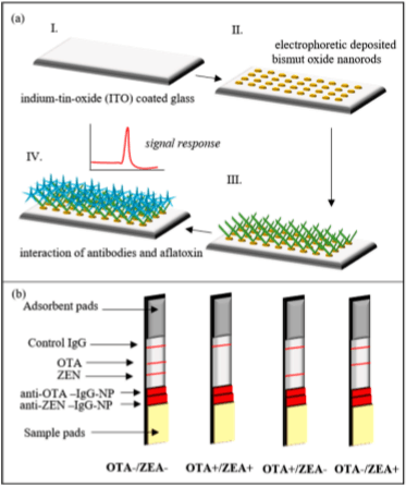
37. Solanki, P.R.; Singh, J.; Rupavali, B.; Tiwari, S.; Malhotra, B.D. Bismuth oxide nanorods based immunosensor for mycotoxin detection. Mater. Sci. Eng. C Mater. Biol. Appl. 2017, 70, 564–571.
38. Zhang, W.J.; Li, G.X. Third-generation biosensors based on the direct electron transfer of proteins. Anal. Sci. 2004, 20, 603–609.
39. Petrakova, A.V.; Urusov, A.E.; Zherdev, A.V.; Liu, L.; Xu, C.; Dzantiev, B.B. Application of magnetite nanoparticles for the development of highly sensitive immunochromatographic test systems for mycotoxin detection. Appl. Biochem. Microbiol. 2017, 53, 470–475.
40. Lv,X.;Li,Y.;Yan,T.;Pang,X.;Cao,W.;Du,B.;Wu,D.;Wei,Q.Electrochemiluminescencemodi?edelectrodes basedonRuSi@Ru(bpy)3 2+ loadedwithgoldfunctionednanoporousCO/Co3O4 fordetectionofmycotoxin deoxynivalenol. Biosens. Bioelectron. 2015, 70, 28–33.
41. Zhao, Y.; Yang, Y.; Luo, Y.; Yang, X.; Li, M.; Song, Q. Double detection of mycotoxins based on sers labels embedded Ag@Au core-shell nanoparticles. Acs Appl. Mater. Interfaces 2015, 7, 21780–21786.
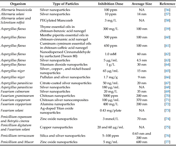
42. Sun, Y.; Xing, G.; Yang, J.; Wang, F.; Deng, R.; Zhang, G.; Hu, X.; Zhang, Y. Development of an immunochromatographic test strip for simultaneous qualitative and quantitative detection of ochratoxin a and zearalenone in cereal. J. Sci. Food Agric. 2016, 96, 3673–3678.
43. Kong, D.; Liu, L.; Song, S.; Suryoprabowo, S.; Li, A.; Kuang, H.; Wang, L.; Xu, C. A gold nanoparticle-based semi-quantitative and quantitative ultrasensitive paper sensor for the detection of twenty mycotoxins. Nanoscale 2016, 8, 5245–5253.
44. Berthiller, F.; Crews, C.; Dall’Asta, C.; De Saeger, S.; Haesaert, G.; Karlovsky, P.; Oswald, I.P.; Seefelder, W.; Speijers, G.; Stroka, J. Masked mycotoxins: A review. Mol. Nutr. Food Res. 2013, 57, 165–186.
45. Goryacheva, I.Y.; De Saeger, S. Immunochemical detection of masked mycotoxins: A short review. World Mycotoxin J. 2012, 5, 281–287.
46. Huybrechts, B.; Martins, J.C.; Debongnie, P.; Uhlig, S.; Callebaut, A. Fast and sensitive LC-MS/MS method measuring human mycotoxin exposure using biomarkers in urine. Arch. Toxicol. 2015, 89, 1993–2005.
47. Nakagawa, H.; Ohmichi, K.; Sakamoto, S.; Sago, Y.; Kushiro, M.; Nagashima, H.; Yoshida, M.; Nakajima, T. Detection of a new fusarium masked mycotoxin in wheat grain by high-resolution LC-OrbitrapTM MS. Food Addit. Contam. Part A Chem. Anal. Control Expo. Risk Assess. 2011, 28, 1447–1456.
48. Aqai, P.; Peters, J.; Gerssen, A.; Haasnoot, W.; Nielen, M.W.F. Immunomagnetic microbeads for screening with ?ow cytometry and identi?cation with nano-liquid chromatography mass spectrometry of ochratoxins in wheat and cereal. Anal. Bioanal. Chem. 2011, 400, 3085–3096.
49. Sureka,S.;Chalcravorty,A.;Holmes,E.C.;Spassibojko,O.;Bhatt,N.;Wu,D.L.;Turgeon,B.G.Standardization of functional reporter and antibiotic resistance cassettes to facilitate the genetic engineering of ?lamentous fungi. ACS Synth. Biol. 2014, 3, 960–962.
50. Niemirowicz, K.; Durnas, B.; Piktel, E.; Bucki, R. Development of antifungal therapies using nanomaterials. Nanomedicine 2017, 12, 1891–1905.
51. Roque, L.; Molpeceres, J.; Reis, C.; Rijo, P.; Reis, C.P. Past, recent progresses and future perspectives of nanotechnology applied to antifungal agents. Curr. Drug Metab. 2017, 18, 280–290.
52. Niemirowicz, K.; Bucki, R. Enhancing the fungicidal activity of antibiotics: Are magnetic nanoparticles the key? Nanomedicine 2017, 12, 1747–1749.
53. Soliman, G.M. Nanoparticles as safe and effective delivery systems of antifungal agents: Achievements and challenges. Int. J. Pharm. 2017, 523, 15–32.
54. Voltan, A.R.; Quindos, G.; Alarcon, K.P.M.; Fusco-Almeida, A.M.; Mendes-Giannini, M.J.S.; Chorilli, M. Fungal diseases: Could nanostructured drug delivery systems be a novel paradigm for therapy? Int. J. Nanomed. 2016, 11, 3715–3730. Nanomaterials 2018, 8, 727 15 of 21
55. Adelere, I.A.; Lateef, A. A novel approach to the green synthesis of metallic nanoparticles: The use of agro-wastes, enzymes, and pigments. Nanotechnol. Rev. 2016, 5, 567–587.
56. Gupta, D.; Chauhan, P. Fungicidal activity of silver nanoparticles against Alternaria brassicicola. In 2nd InternationalConferenceonEmergingTechnologies: MicrotoNano2015;Sharma,N.N.,Gaol,F.L.,Akhtar,J.,Eds.; AIP Publishing: Melville, NY, USA, 2016; Volume 1724.
57. Abdel-Hafez,S.I.I.;Nafady,N.A.;Abdel-Rahim,I.R.;Shaltout,A.M.;Daros,J.A.;Mohamed,M.A.Assessment of protein silver nanoparticles toxicity against pathogenic Alternariasolani. 3Biotech 2016, 6, 199.
58. Majumder, S.; Shakil, N.A.; Kumar, J.; Banerjee, T.; Sinha, P.; Singh, B.B.; Garg, P. Eco-friendly peg-based controlled release nano-formulations of mancozeb: Synthesis and bioef?cacy evaluation against phytopathogenic fungi alternaria solani and sclerotium rolfsii. J. Environ. Sci. Health Part B Pest. Contam. Agric. Wastes 2016, 51, 873–880.
59. Khalili, S.T.; Mohsenifar, A.; Beyki, M.; Zhaveh, S.; Rahmani-Cherati, T.; Abdollahi, A.; Bayat, M.; Tabatabaei, M. Encapsulation of thyme essential oils in chitosan-benzoic acid nanogel with enhanced antimicrobial activity against aspergillus ?avus. LWT-Food Sci. Technol. 2015, 60, 502–508.
60. Beyki, M.; Zhaveh, S.; Khalili, S.T.; Rahmani-Cherati, T.; Abollahi, A.; Bayat, M.; Tabatabaei, M.; Mohsenifar, A. Encapsulation of mentha piperita essential oils in chitosan-cinnamic acid nanogel with enhanced antimicrobial activity against aspergillus ?avus. Ind. Crops Prod. 2014, 54, 310–319.
61. Zhaveh, S.; Mohsenifar, A.; Beiki, M.; Khalili, S.T.; Abdollahi, A.; Rahmani-Cherati, T.; Tabatabaei, M. Encapsulation of cuminum cyminum essential oils in chitosan-caffeic acid nanogel with enhanced antimicrobial activity against aspergillus ?avus. Ind. Crops Prod. 2015, 69, 251–256.
62. Li,H.B.; Shen,Q.S.;Zhou,W.; Mo,H.Z.;Pan,D.D.;Hu,L.B.Nanocapsulardispersionofcinnamaldehydefor enhancedinhibitoryactivityagainsta?atoxinproductionbyaspergillus?avus. Molecules2015,20,6022–6032.
63. Zhao, J.; Wang, L.; Xu, D.; Lu, Z.S. Involvement of ros in nanosilver-caused suppression of a?atoxin production from aspergillus ?avus. RSC Adv. 2017, 7, 23021–23026.
64. Babaei, E.; Dehnad, A.; Hajizadeh, N.; Valizadeh, H.; Reihani, S.F.S. A study on inhibitory effects of titanium dioxide nanoparticles and its photocatalytic type on Staphylococcus aureus, Escherichia coli and Aspergillus ?avus. Appl. Food Biotechnol. 2016, 3, 115–123.
65. Yu, K.P.; Huang, Y.T.; Yang, S.C. The antifungal ef?cacy of nano-metals supported TiO2 and ozone on the resistant aspergillus niger spore. J. Hazard. Mater. 2013, 261, 155–162.
66. Pinto, R.J.B.; Almeida, A.; Fernandes, S.C.M.; Freire, C.S.R.; Silvestre, A.J.D.; Neto, C.P.; Trindade, T. Antifungal activity of transparent nanocomposite thin ?lms of pullulan and silver against aspergillus niger. Colloid Surf. B Biointerfaces 2013, 103, 143–148.
67. Mitra, C.; Gummadidala, P.M.; Afshinnia, K.; Merri?eld, R.C.; Baalousha, M.; Lead, J.R.; Chanda, A. Citrate-coated silver nanoparticles growth-independently inhibit a?atoxin synthesis in aspergillus parasiticus. Environ. Sci. Technol. 2017, 51, 8085–8093.
68. Mousavi, S.A.A.; Pourtalebi, S. Inhibitory effects of silver nanoparticles on growth and a?atoxin B-1 production by aspergillus parasiticus. Iran. J. Med. Sci. 2015, 40, 501–506.
69. Rashed,A.-O.M.;Mohamed,A.-E.-A.A.R.;Abobakr,M.M.Wheatprotectionfromrootrotcausedbyfusarium culmorum using silver nanoparticles. J. Chem. Soc. Pak. 2016, 38, 898–903.
70. Kheiri, A.; Moosawi Jorf, S.A.; Malihipour, A.; Saremi, H.; Nikkhah, M. Synthesis and characterization of chitosan nanoparticles and their effect on fusarium head blight and oxidative activity in wheat. Int. J. Biol. Macromol. 2017, 102, 526–538.
71. Dananjaya, S.H.S.; Erandani, W.; Kim, C.H.; Nikapitiya, C.; Lee, J.; De Zoysa, M. Comparative study on antifungal activities of chitosan nanoparticles and chitosan silver nano composites against fusarium oxysporum species complex. Int. J. Biol. Macromol. 2017, 105, 478–488.
72. Shenashen, M.; Derbalah, A.; Hamza, A.; Mohamed, A.; El Safty, S. Antifungal activity of fabricated mesoporous alumina nanoparticles against root rot disease of tomato caused by fusarium oxysporium. Pest Manag. Sci. 2017, 73, 1121–1126.
73. Boxi, S.S.; Mukherjee, K.; Paria, S. Ag doped hollow TiO2 nanoparticles as an effective green fungicide againstfusariumsolaniandventuriainaequalisphytopathogens. Nanotechnology2016,27,085103. Nanomaterials 2018, 8, 727 16 of 21
74. He, L.L.; Liu, Y.; Mustapha, A.; Lin, M.S. Antifungal activity of zinc oxide nanoparticles against botrytis cinerea and penicillium expansum. Microbiol. Res. 2011, 166, 207–215.
75. Khamis, Y.; Hashim, A.F.; Margarita, R.; Alghuthaymi, M.A.; Abd-Elsalam, K.A. Fungicidal ef?cacy of chemically-produced copper nanoparticles against penicillium digitatum and fusarium solani on citrus fruit. Philipp. Agric. Sci. 2017, 100, 69–78.
76. Kotzybik, K.; Graf, V.; Kugler, L.; Stoll, D.A.; Greiner, R.; Geisen, R.; Schmidt-Heydt, M. In?uence of different nanomaterials on growth and mycotoxin production of penicillium verrucosum. PLoS ONE 2016, 11, e0150855.
77. Zeng, X.L.; Zhang, F.Q.; He, N.Y.; Zhang, B.Y.; Liu, X.Y.; Li, X.L. Zno nanoparticles of different shapes and their antimycotic property against penicillium and mucor. Nanosci. Nanotechnol. Lett. 2016, 8, 688–694.
78. Medda, S.; Hajra, A.; Dey, U.; Bose, P.; Mondal, N.K. Biosynthesis of silver nanoparticles from Aloe vera leaf extract and antifungal activity against Rhizopus sp. and Aspergillus sp. Appl. Nanosci. 2015, 5, 875–880.
79. Ammar, H.A.M.; El-Desouky, T.A. Green synthesis of nanosilver particles by aspergillus terreus HA1N and penicilliumexpansumHA2Nanditsantifungalactivityagainstmycotoxigenicfungi. J.Appl. Microbiol. 2016, 121, 89–100
80. Devipriya, D.; Roopan, S.M. Cissus quadrangularis mediated ecofriendly synthesis of copper oxide nanoparticles and its antifungal studies against aspergillus niger, aspergillus ?avus. Mater. Sci. Eng. C Mater. Biol. Appl. 2017, 80, 38–44.
81. Yassin, M.A.; El-Samawaty, A.M.A.; Dawoud, T.M.; Abd-Elkader, O.H.; Al Maary, K.S.; Hatamleh, A.A.; Elgorban, A.M. Characterization and anti-aspergillus ?avus impact of nanoparticles synthesized by penicillium citrinum. Saudi J. Biol. Sci. 2017, 24, 1243–1248.
82. Shakibaie, M.; Mohazab, N.S.; Mousavi, S.A.A. Antifungal activity of selenium nanoparticles synthesized by bacillus species Msh-1 against aspergillus fumigatus and candida albicans. Jundishapur J. Microbiol. 2015, 8, e26381.
83. Xue, B.J.; He, D.; Gao, S.; Wang, D.Y.; Yokoyama, K.; Wang, L. Biosynthesis of silver nanoparticles by the fungus arthroderma fulvum and its antifungal activity against genera of candida, aspergillus and fusarium. Int. J. Nanomed. 2016, 11, 1899–1906.
84. Madbouly, A.K.; Abdel-Aziz, M.S.; Abdel-Wahhab, M.A. Biosynthesis of nanosilver using chaetomium globosum and its application to control fusarium wilt of tomato in the greenhouse. IETNanobiotechnol. 2017, 11, 702–708.
85. Pietrzak, K.; Twaruzek, M.; Czyzowska, A.; Kosicki, R.; Gutarowska, B. In?uence of silver nanoparticles on metabolism and toxicity of moulds. Acta Biochim. Pol. 2015, 62, 851–857.
86. Lara, H.H.; Romero-Urbina, D.G.; Pierce, C.; Lopez-Ribot, J.L.; Arellano-Jimenez, M.J.; Jose-Yacaman, M. Effect of silver nanoparticles on candida albicans bio?lms: An ultrastructural study. J.Nanobiotechnol. 2015, 13, 91.
87. Tang, Z.Y.; Kotov, N.A. One-dimensional assemblies of nanoparticles: Preparation, properties, and promise. Adv. Mater. 2005, 17, 951–962. 88. Stroka, J.; Maragos, C.M. Challenges in the analysis of multiple mycotoxins. World Mycotoxin J. 2016, 9, 847–861.
89. Chemical and physical characteristics of the principal mycotoxins. IARC Sci. Public 2012, 158, 31–38.
90. Gibson, N.; Shenderova, O.; Luo, T.J.M.; Moseenkov, S.; Bondar, V.; Puzyr, A.; Purtov, K.; Fitzgerald, Z.; Brenner, D.W. Colloidal stability of modi?ed nanodiamond particles. Diam. Relat. Mater. 2009, 18, 620–626.
91. Chen, W.; Duan, L.; Zhu, D.Q. Adsorption of polar and nonpolar organic chemicals to carbon nanotubes. Environ. Sci. Technol. 2007, 41, 8295–8300.
92. Kovac, T.; Sarkanj, B.; Klapec, T.; Borisev, I.; Kovac, M.; Nevistic, A.; Strelec, I. Fullerol C60(OH)24 nanoparticles and mycotoxigenic fungi: A preliminary investigation into modulation of mycotoxin production. Environ. Sci. Pollut. Res. 2017, 24, 16673–16681.
93. Aoshima, H.; Kokubo, K.; Shirakawa, S.; Ito, M.; Yamana, S.; Oshima, T. Antimicrobial activity of fullerenes and their hydroxylated derivatives. Biocontrol Sci. 2009, 14, 69–72 Nanomaterials 2018, 8, 727 17 of 21
94. Puzyr, A.P.; Purtov, K.V.; Shenderova, O.A.; Luo, M.; Brenner, D.W.; Bondar, V.S. The adsorption of a?atoxin B1 by detonation-synthesis nanodiamonds. Dokl. Biochem. Biophys. 2007, 417, 299–301.
95. Gibson, N.M.; Luo, T.J.M.; Brenner, D.W.; Shenderova, O. Immobilization of mycotoxins on modi?ed nanodiamond substrates. Biointerphases 2011, 6, 210–217.
96. Golokhvast, K.S.; Chaika, V.V.; Kuznetsov, L.V.; Elumeeva, K.V.; Kusaikin, M.I.; Zakharenko, A.M.; Kiselev, N.N.; Panichev, A.M.; Reva, G.V.; Usov, V.V.; et al. Effects of multiwalled carbon nanotubes receivedorallyduring6daysonthegastrointestinaltract. Bull. Exp. Biol. Med. 2013,155,788–792.
97. Boczkowski, J.; Lanone, S. Potential uses of carbon nanotubes in the medical ?eld: How worried should patients be? Nanomedicine 2007, 2, 407–410.
98. Han, Z.; Jiang, K.; Fan, Z.; Diana Di Mavungu, J.; Dong, M.; Guo, W.; Fan, K.; Campbell, K.; Zhao, Z.; Wu, Y. Multi-walled carbon nanotubes-based magnetic solid-phase extraction for the determination of zearalenone and its derivatives in maize by ultra-high performance liquid chromatography-tandem mass spectrometry. Food Control 2017, 79, 177–184.
99. Ying, Y.-F.; Wu, Y.-L.; Wen, Y.; Yang, T.; Xu, X.-Q.; Wang, Y.-Z. Simultaneous determination of six resorcylic acid lactones in feed using liquid chromatography-tandem mass spectrometry and multi-walled carbon nanotubes as a dispersive solid phase extraction sorbent. J. Chromatogr. A 2013, 1307, 41–48.
100. Moreno, V.; Zougagh, M.; Rios, A. Hybrid nanoparticles based on magnetic multiwalled carbon nanotube-nanoC18SiO2 composites for solid phase extraction of mycotoxins prior to their determination by LC-MS. Microchim. Acta 2016, 183, 871–880.
101. Dong, M.; Si, W.; Wang, W.; Bai, B.; Nie, D.; Song, W.; Zhao, Z.; Guo, Y.; Han, Z. Determination of type a trichothecenes in coix seed by magnetic solid-phase extraction based on magnetic multi-walled carbon nanotubes coupled with ultra-high performance liquid chromatography-tandem mass spectrometry. Anal. Bioanal. Chem. 2016, 408, 6823–6831.
102. Jiang, K.; Huang, P.; Luan, L.; Fan, K.; Guo, W.; Zhao, Z.; Wu, Y.; Han, Z. Iron (II, III) oxide/multi-walled carbon nanotube composite as solid-phase extraction sorbent followed by ultra-high performance liquid chromatography tandem mass spectrometry for simultaneous determination of zearalenone and type a trichothecenes in salviae miltiorrhizae radix et rhizoma (danshen). J. Chromatogr. A 2017, 1482, 1–10.
103. Dong,M.; Si, W.; Jiang,K.; Nie,D.; Wu,Y.; Zhao,Z.; DeSaeger,S.; Han,Z.Multi-walled carbonnanotubesas solid-phaseextractionsorbentsforsimultaneousdeterminationoftypeatrichothecenesinmaize,wheatand rice by ultra-high performance liquid chromatography-tandem mass spectrometry. J. Chromatogr. A 2015, 1423, 177–182.
104. Singh, C.; Srivastava, S.; Ali, M.A.; Gupta, T.K.; Sumana, G.; Srivastava, A.; Mathur, R.B.; Malhotra, B.D. Carboxylated multiwalled carbon nanotubes based biosensor for a?atoxin detection. Sens. Actuators B Chem. 2013, 185, 258–264.
105. Pirouz, A.A.; Selamat, J.; Iqbal, S.Z.; Mirhosseini, H.; Karjiban, R.A.; Bakar, F.A. The use of innovative and ef?cient nanocomposite (magnetic graphene oxide) for the reduction on of fusarium mycotoxins in palm kernel cake. Sci. Rep. 2017, 7, 12453.
106. Magro, M.; Moritz, D.E.; Bonaiuto, E.; Baratella, D.; Terzo, M.; Jakubec, P.; Malina, O.; Cepe, K.; Falcao de Aragao,G.M.;Zboril,R.;etal. Citrininmycotoxinrecognitionandremovalbynakedmagneticnanoparticles. Food Chem. 2016, 203, 505–512.
107. Khajarern, J.M.; Khajarern, S.; Moon, T.H.; Lee, J.H. Effects of dietary supplementation of fermented chitin-chitosan (fermkit) on toxicity of mycotoxin in ducks. Asian-Australas. J. Anim. Sci. 2003, 16, 706–713.
108. Huang, J.; Huang, Z.; Kang, Y.; Wang, A.; Zong, L. Mycotoxin Adsorbent Preparation Used for Removing Feed Mycotoxin Zearalenone and Reducing Diarrhea, and Used as Antibacterial, Involves Dissolving Chitosan in Organic Acid Solution, and then Adding Rectorite is Added to Solution. CN Patent CN103831088-A, 4 June 2014; CN103831088-B, 27 January 2016.
109. Desai, K.G.H. Chitosan nanoparticles prepared by ionotropic gelation: An overview of recent advances. Crit. Rev. Ther. Drug Carrier Syst. 2016, 33, 107–158. Nanomaterials 2018, 8, 727 18 of 21
110. Huang, Y.; Lapitsky, Y. On the kinetics of chitosan/tripolyphosphate micro- and nanogel aggregation and their effects on particle polydispersity. J. Colloid Interface Sci. 2017, 486, 27–37.
111. Sacco,P.;Paoletti,S.;Cok,M.;Asaro,F.;Abrami,M.;Grassi,M.;Donati,I.Insightintotheionotropicgelation of chitosan using tripolyphosphate and pyrophosphate as cross-linkers. Int. J. Biol. Macromol. 2016, 92, 476–483.
112. Graham, L.M.; Nguyen, T.M.; Lee, S.B. Nanodetoxi?cation: Emerging role of nanomaterials in drug intoxication treatment. Nanomedicine 2011, 6, 921–928.
113. Zhao, Z.; Liu, N.; Yang, L.; Wang, J.; Song, S.; Nie, D.; Yang, X.; Hou, J.; Wu, A. Cross-linked chitosan polymers as generic adsorbents for simultaneous adsorption of multiple mycotoxins. Food Control 2015, 57, 362–369.
114. Luo, Y.; Zhou, Z.; Yue, T. Synthesis and characterization of nontoxic chitosan-coated Fe3O4 particles for patulin adsorption in a juice-pH simulation aqueous. Food Chem. 2017, 221, 317–323.
115. Swain, P.S.; Rao, S.B.N.; Rajendran, D.; Dominic, G.; Selvaraju, S. Nano zinc, an alternative to conventional zinc as animal feed supplement: A review. Anim. Nutr. 2016, 2, 134–141.
116. Shi, Y.H.; Xu, Z.R.; Feng, J.L.; Wang, C.Z. Ef?cacy of modi?ed montmorillonite nanocomposite to reduce the toxicity of a?atoxin in broiler chicks. Anim. Feed Sci. Technol. 2006, 129, 138–148.
117. Al-Shemmari,F.H.J.;Rabah,A.A.;Al-Mulla,E.A.J.;AbdAlrahman,N.O.M.Preparationandcharacterization of natural rubber latex/modi?ed montmorillonite clay nano-composite. Res. Chem. Intermed. 2013, 39, 4293–4301.
118. Yuan, P.; Tan, D.Y.; Annabi-Bergaya, F. Properties and applications of halloysite nanotubes: Recent research advances and future prospects. Appl. Clay Sci. 2015, 112, 75–93.
119. Lvov,Y.;Wang,W.C.;Zhang,L.Q.;Fakhrullin,R.Halloysiteclaynanotubesforloadingandsustainedrelease of functional compounds. Adv. Mater. 2016, 28, 1227–1250.
120. Lun, H.L.; Ouyang, J.; Yang, H.M. Natural halloysite nanotubes modi?ed as an aspirin carrier. RSC Adv. 2014, 4, 44197–44202.
121. Patel, S.; Jammalamadaka, U.; Sun, L.; Tappa, K.; Mills, D.K. Sustained release of antibacterial agents from doped halloysite nanotubes. Bioengineering 2016, 3, 1.
122. Hanif, M.; Jabbar, F.; Sharif, S.; Abbas, G.; Farooq, A.; Aziz, M. Halloysite nanotubes as a new drug-delivery system: A review. Clay Miner. 2016, 51, 469–477.
123. Sun, L.; Mills, D.K.; IEEE. Halloysite nanotube-based drug delivery system for treating osteosarcoma. InProceedingsofthe36thAnnualInternationalConferenceoftheIEEEEngineeringinMedicineandBiology Society, Chicago, IL, USA, 26–30 August 2014; pp. 2920–2923.
124. Long, Z.R.; Zhang, J.; Shen, Y.; Zhou, C.R.; Liu, M.X. Polyethyleneimine grafted short halloysite nanotubes for gene delivery. Mater. Sci. Eng. C Mater. Biol. Appl. 2017, 81, 224–235.
125. Jana, S.; Kondakova, A.V.; Shevchenko, S.N.; Sheval, E.V.; Gonchar, K.A.; Timoshenko, V.Y.; Vasiliev, A.N. Halloysite nanotubes with immobilized silver nanoparticles for anti-bacterial application. Colloid Surf. B Biointerfaces 2017, 151, 249–254.
126. Tham, W.L.; Poh, B.T.; Mohd Ishak, Z.A.; Chow, W.S. Transparent poly(lactic acid)/halloysite nanotube nanocomposites with improved oxygen barrier and antioxidant properties. J. Ther. Anal. Calorim. 2016, 126, 1331–1337.
127. Vergaro, V.; Lvov, Y.M.; Leporatti, S. Halloysite clay nanotubes for resveratrol delivery to cancer cells. Macromol. Biosci. 2012, 12, 1265–1271.
128. Hari, J.; Gyurki, A.; Sarkozi, M.; Foldes, E.; Pukanszky, B. Competitive interactions and controlled release of a natural antioxidant from halloysite nanotubes. J. Colloid Interface Sci. 2016, 462, 123–129.
129. Zhong, B.C.; Lin, J.; Liu, M.L.; Jia, Z.X.; Luo, Y.F.; Jia, D.M.; Liu, F. Preparation of halloysite nanotubes loaded antioxidant and its antioxidative behaviour in natural rubber. Polym. Degrad. Stab. 2017, 141, 19–25.
130. Fu, Y.; Zhao, D.T.; Yao, P.J.; Wang, W.C.; Zhang, L.Q.; Lvov, Y. Highly aging-resistant elastomers doped with antioxidant-loaded clay nanotubes. ACS Appl. Mater. Interfaces 2015, 7, 8156–8165.
131. Zhang, Y.Y.; Gao, R.; Liu, M.; Shi, B.M.; Shan, A.S.; Cheng, B.J. Use of modi?ed halloysite nanotubes in the feed reduces the toxic effects of zearalenone on sow reproduction and piglet development. Theriogenology 2015, 83, 932–941.
Nanomaterials 2018, 8, 727 19 of 21
132. Gao, R.; Meng, Q.W.; Li, J.N.; Zhang, Y.Y.; Bi, C.P.; Shan, A.S. Modi?ed halloysite nanotubes reduce the toxic effects of zearalenone in gestating sows on growth and muscle development of their offsprings. J. Anim. Sci. Biotechnol. 2016, 7, 14.
133. Zhang, Y.Y.; Gao, R.; Liu, M.; Yan, C.J.; Shan, A.S. Adsorption of modi?ed halloysite nanotubes in vitro and the protective effect in rats exposed to zearalenone. Arch. Anim. Nutr. 2014, 68, 320–335.
134. Jia, Z.Q.; Yin, S.T.; Liu, M.; Zhang, Y.Y.; Gao, R.; Shi, B.M.; Shan, A.S.; Chen, Z.H. Modi?ed halloysite nanotubes and the alleviation of kidney damage induced by dietary zearalenone in swine. Food Addit. Contam. Part A Chem. Anal. Control Expo. Risk Assess. 2015, 32, 1312–1321.
135. Fakhrullina, G.I.; Akhatova, F.S.; Lvov, Y.M.; Fakhrullin, R.F. Toxicity of halloysite clay nanotubes in vivo: A caenorhabditis elegans study. Environ. Sci. Nano 2015, 2, 54–59.
136. Fastov, S.A.; Terent’ev, A.O.; Fastov, I.S.; Firstova, V.V.; Kalmantaeva, O.V. Study of halloysite clay nanotube toxicityasrelatedtotheirstructure. InAbstractsofPapersoftheAmericanChemicalSociety;AmericanChemical Society: Washington, DC, USA, 2013; Volume 245.
137. Cardoso, M.M.; Peca, I.N.; Roque, A.C.A. Antibody-conjugated nanoparticles for therapeutic applications. Curr. Med. Chem. 2012, 19, 3103–3127.
138. Carter, T.; Mulholland, P.; Chester, K. Antibody-targeted nanoparticles for cancer treatment. Immunotherapy 2016, 8, 941–958.
139. Welch,N.G.;Scoble,J.A.;Muir,B.W.;Pigram,P.J.Orientationandcharacterizationofimmobilizedantibodies for improved immunoassays. Biointerphases 2017, 12, 02D301.
140. Kumada,Y.Site-specificimmobilizationofrecombinantantibodyfragmentsthroughmaterial-bindingpeptides forthesensitivedetectionofantigensinenzymeimmunoassays.Biochim. Biophys. Acta-ProteinsProteom.2014, 1844,1960–1969.[CrossRef]
141. Rusmini, F.; Zhong, Z.; Feijen, J. Protein immobilization strategies for protein biochips. Biomacromolecules 2007, 8, 1775–1789.
142. Duval, F.; van Beek, T.A.; Zuilhof, H. Key steps towards the oriented immobilization of antibodies using boronic acids. Analyst 2015, 140, 6467–6472.
143. Kim, H.-J.; Kim, S.-H.; Lee, J.-K.; Choi, C.-U.; Lee, H.-S.; Kang, H.-G.; Cha, S.-H. A novel mycotoxin puri?cation system using magnetic nanoparticles for the recovery of a?atoxin B1 and zearalenone from feed. J. Vet. Sci. 2012, 13, 363–369.
144. Xiong, Y.; Tu, Z.; Huang, X.; Xie, B.; Xiong, Y.; Xu, Y. Magnetic beads carrying poly(acrylic acid) brushes as “nanobody containers” for immunoaf?nity puri?cation of a?atoxin B1 from corn samples. RSC Adv. 2015, 5, 77380–77387.
145. Ali, I.; Peng, C.S.; Khan, Z.M.; Naz, I. Yield cultivation of magnetotactic bacteria and magnetosomes: A review. J. Basic Microbiol. 2017, 57, 643–652.
146. Lin,W.;Pan,Y.X.;Bazylinski,D.A.Diversityandecologyofandbiomineralizationbymagnetotacticbacteria. Environ. Microbiol. Rep. 2017, 9, 345–356.
147. Pi, F.; Sun, J.; Liu, W.; Sun, X.; Zhang, Y. Elimination of a?atoxin B1 in vegetable oil based on immunomagnetosomes probes from a novel magnetotactic bacterium. Food Control 2017, 80, 319–326.
148. Awad, W.A.; Ghareeb, K.; Dadak, A.; Hess, M.; Bohm, J. Single and combined effects of deoxynivalenol mycotoxin and a microbial feed additive on lymphocyte DNA damage and oxidative stress in broiler chickens. PLoS ONE 2014, 9, e88028.
149. Hou, Y.J.; Zhao, Y.Y.; Xiong, B.; Cui, X.S.; Kim, N.H.; Xu, Y.X.; Sun, S.C. Mycotoxin-containing diet causes oxidative stress in the mouse. PLoS ONE 2013, 8, e60374.
150. Horky, P.; Skladanka, J.; Nevrkla, P.; Slama, P. Effect of diet supplemented with antioxidants (selenium, copper, vitamins E and C) on antioxidant status and ejaculate quality of breeding boars. Ann. Anim. Sci. 2016, 16, 521–532.
151. Horky, P.; Ruttkay-Nedecky, B.; Kremplova, M.; Krystofova, O.; Kensova, R.; Hynek, D.; Babula, P.; Zitka, O.; Zeman, L.; Adam, V.; et al. Effect of different doses of organically bound selenium on antioxidant status and levels of metal ions in postpartum sows. Int. J. Electrochem. Sci. 2013, 8, 6162–6179.
152. Skalickova, S.; Milosavljevic, V.; Cihalova, K.; Horky, P.; Richtera, L.; Adam, V. Selenium nanoparticles as a nutritional supplement. Nutrition 2017, 33, 83–90. Nanomaterials 2018, 8, 727 20 of 21
153. Kovacic, P.; Somanathan, R. Biomechanisms of nanoparticles (toxicants, antioxidants and therapeutics): Electron transfer and reactive oxygen species. J. Nanosci. Nanotechnol. 2010, 10, 7919–7930.
154. Nelson, B.C.; Johnson, M.E.; Walker, M.L.; Riley, K.R.; Sims, C.M. Antioxidant cerium oxide nanoparticles in biology and medicine. Antioxidants 2016, 5, 15.
155. Raveendran, P.; Fu, J.; Wallen, S.L. Completely “green” synthesis and stabilization of metal nanoparticles. J. Am. Chem. Soc. 2003, 125, 13940–13941.
156. Kohli, E.; Raj, H.G.; Kumari, R.; Rohil, V.; Kaushik, N.K.; Prasad, A.K.; Parmar, V.S. Comparison of the prevention of a?atoxin B1-induced genotoxicity by quercetin and quercetin pentaacetate. Bioorg. Med. Chem. Lett. 2002, 12, 2579–2582.
157. Choi, K.C.; Chung, W.T.; Kwon, J.K.; Yu, J.Y.; Jang, Y.S.; Park, S.M.; Lee, S.Y.; Lee, J.C. Inhibitory effects of quercetin on a?atoxin B1-induced hepatic damage in mice. Food Chem. Toxicol. 2010, 48, 2747–2753.
158. Periasamy, R.; Kalal, I.G.; Krishnaswamy, R.; Viswanadha, V. Quercetin protects human peripheral blood mononuclear cells from OTA-induced oxidative stress, genotoxicity, and in?ammation. Environ. Toxicol. 2016, 31, 855–865.
159. Abbas, Z.; Blank, R.; Wein, S.; Wolffram, S. Effect of quercetin on the toxicokinetics of ochratoxin A in rats. Food Addit. Contam. Part A Chem. Anal. Control Expo. Risk Assess. 2013, 30, 861–866.
160. Orsolic, N.; Jazvinscak Jembrek, M.; Terzic, S. Honey and quercetin reduce ochratoxin A-induced DNA damage in the liver and the kidney through the modulation of intestinal micro?ora. Food Agric. Immunol. 2017, 28, 812–833.
161. Lesniak-Walentyn, A.; Kolesarova, A.; Medvedova, M.; Maruniakova, N.; Capcarova, M.; Kalafova, A.; Hrabia, A.; Sirotkin, A.V. Proliferation and apoptosis in the rabbit ovary after administration of T-2 toxin and quercetin. J. Anim. Feed Sci. 2013, 22, 264–271.
162. Ben Salem, I.; Prola, A.; Boussabbeh, M.; Guilbert, A.; Bacha, H.; Lemaire, C.; Abid-Esse?, S. Activation of ER stress and apoptosis by alpha- and beta-zearalenol in HCT116 cells, protective role of Quercetin. Neurotoxicology 2016, 53, 334–342.
163. Ben Salem, I.; Prola, A.; Boussabbeh, M.; Guilbert, A.; Bacha, H.; Abid-Esse?, S.; Lemaire, C. Crocin and quercetin protect HCT116 and HEK293 cells from Zearalenone-induced apoptosis by reducing endoplasmic reticulum stress. Cell Stress Chaperones 2015, 20, 927–938.
164. Fernandez-Blanco, C.; Font, G.; Ruiz, M.-J. Role of quercetin on Caco-2 cells against cytotoxic effects of alternariol and alternariol monomethyl ether. Food Chem. Toxicol. 2016, 89, 60–66.
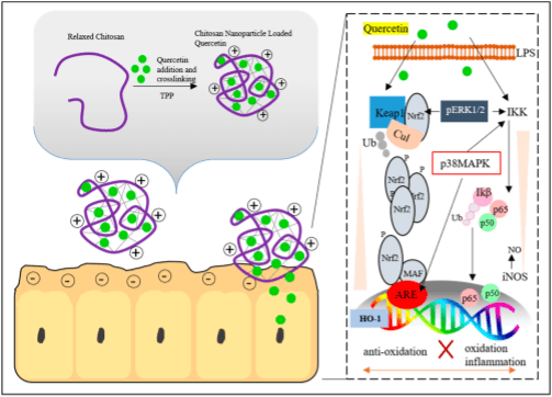
165. Abdel-Wahhab, M.A.; Aljawish, A.; El-Nekeety, A.A.; Abdel-Aziem, S.H.; Hassan, N.S. Chitosan nanoparticles plus quercetin suppress the oxidative stress, modulate DNA fragmentation and gene expression in the kidney of rats fed ochratoxin A-contaminated diet. Food Chem. Toxicol. 2017, 99, 209–221.
166. Sun, G.Y.; Chen, Z.H.; Jasmer, K.J.; Chuang, D.Y.; Gu, Z.Z.; Hannink, M.; Simonyi, A. Quercetin attenuates in?ammatoryresponsesinBV-2microglialcells: RoleofMAPKsonthenrf2pathwayandinductionofheme oxygenase-1. PLoS ONE 2015, 10, e0141509.
167. Oberdorster, G.; Oberdorster, E.; Oberdorster, J. Nanotoxicology: An emerging discipline evolving from studies of ultra?ne particles. Environ. Health Perspect. 2005, 113, 823–839.
168. Kaundal,B.;Dalai,S.;Choudhury,S.R.Nanomaterialtoxicityinmicrobes,plantsandanimals. InNanoscience inFoodandAgriculture5; Ranjan, S., Dasgupta, N., Lichtfouse, E., Eds.; Springer International Publishing: Cham, Switzerland, 2017; pp. 243–266.
169. Suresh,A.K.;Pelletier,D.A.;Doktycz,M.J.Relatingnanomaterialpropertiesandmicrobialtoxicity. Nanoscale 2013, 5, 463–474.
170. Chatterjee, S.; Kumari, R.M.; Nimesh, S. Nanotoxicology: Evaluation of toxicity potential of nanoparticles. In Advances in Nanomedicine for the Delivery of Therapeutic Nucleic Acids; Nimesh, S., Chandra, R., Gupta, N., Eds.; Elsevier: New York City, NY, USA, 2017; pp. 187–201.
171. Jo,H.Y.;Kim,Y.;Park,H.W.;Moon,H.E.;Bae,S.;Kim,J.;Kim,D.G.;Paek,S.H.TheunreliabilityofMTTassay in the cytotoxic test of primary cultured glioblastoma cells. Exp. Neurobiol. 2015, 24, 235–245. Nanomaterials 2018, 8, 727 21 of 21
172. Bondarenko,O.;Juganson,K.;Ivask,A.;Kasemets,K.;Mortimer,M.;Kahru,A.ToxicityofAg,CuOandZnO nanoparticles to selected environmentally relevant test organisms and mammalian cells in vitro: A critical review. Arch. Toxicol. 2013, 87, 1181–1200.
173. Wang, Z.Y.; Zhang, L.; Zhao, J.; Xing, B.S. Environmental processes and toxicity of metallic nanoparticles in aquatic systems as affected by natural organic matter. Environ. Sci. Nano 2016, 3, 240–255.
174. Goyal, P.; Basniwal, R.K. Toxicity of nanoparticles and their impact on environment. In Nanoscience and Plant-Soil Systems; Ghorbanpour, M., Khanuja, M., Varma, A., Eds.; Springer: Berlin, Germany, 2017; Volume 48, pp. 531–543.
175. Alshannaq, A.; Yu, J.H. Occurrence, toxicity, and analysis of major mycotoxins in food. Int. J. Environ. Res. Public Health 2017, 14, 632.
176. Kharisov, B.I.; Kharissova, O.V.; Chavez-Guerrero, L. Synthesis techniques, properties, and applications of nanodiamonds. Synth. React. Inorg. Met.-Org. Nano-Met. Chem. 2010, 40, 84–101.
177. Moore, L.; Yang, J.Y.; Lang, T.T.H.; Osawa, E.; Lee, D.K.; Johnson, W.D.; Xi, J.Z.; Chow, E.K.H.; Ho, D. Biocompatibilityassessmentofdetonationnanodiamondinnon-humanprimatesandratsusinghistological, hematologic, and urine analysis. ACS Nano 2016, 10, 7385–7400.
178. Sherlala, A.I.A.; Raman, A.A.A.; Bello, M.M.; Asghar, A. A review of the applications of organo-functionalized magnetic graphene oxide nanocomposites for heavy metal adsorption. Chemosphere 2018, 193, 1004–1017.
179. Liu, J.H.; Wang, T.C.; Wang, H.F.; Gu, Y.G.; Xu, Y.Y.; Tang, H.; Jia, G.; Liu, Y.F. Biocompatibility of graphene oxide intravenously administrated in mice-effects of dose, size and exposure protocols. Toxicol. Res. 2015, 4, 83–91.
180. Cervini-Silva, J.; Palacios, E.; Gomez-Vidales, V. Nontronite as natural source and growth template for (nano)maghemite gamma-Fe2O3 and (nano)wustite Fe1−xO. Appl. Clay Sci. 2018, 156, 178–186.
181. Shen, J.M.; Huang, G.; Zhou, X.; Zou, J.; Yang, Y.; Chen, Y.F.; Men, S.K. Safety evaluation of graphene oxide-based magnetic nanocomposites as mri contrast agents and drug delivery vehicles. RSCAdv. 2014, 4, 50464–50477.
182. Divya, K.; Jisha, M.S. Chitosan nanoparticles preparation and applications. Environ. Chem. Lett. 2018, 16, 101–112.
183. Bowman, K.; Leong, K.W. Chitosan nanoparticles for oral drug and gene delivery. Int. J. Nanomed. 2006, 1, 117–128. 184. Wang, X.Y.; Du, Y.M.; Luo, J.W. Biopolymer/montmorillonite nanocomposite: Preparation, drug-controlled release property and cytotoxicity. Nanotechnology 2008, 19, 065707.
185. Baek, M.; Lee, J.A.; Choi, S.J. Toxicological effects of a cationic clay, montmorillonite in vitro and in vivo. Mol. Cell. Toxicol. 2012, 8, 95–101.


.jpg&w=3840&q=75)