Effect of selenium source on production, reproduction and immunity of lactating dairy cows in Florida and California
A nutraceutical is defined as a product isolated or purified from feeds that is demonstrated to have a physiological benefit or provide protection against chronic disease. Indeed selenium (Se) is an essential nutrient in that selenium deficiency is associated with increased incidences of retained fetal membranes, clinical mastitis, calf mortality and increased milk somatic cell counts (SCC).
Selenium supplementation in Se-deficient diets reduces the incidence of these clinical problems. Consequently, selenium should be considered a nutraceutical that has an array of biological responses.
Cows fed Se-deficient diets have reduced blood glutathione peroxidase (GSH-Px) activity compared with those fed Se-supplemented diets (Grasso et al., 1990; Gunter et al., 2003). Likewise, cows receiving injections containing sodium selenite have greater concentrations of selenium in plasma and greater GSH-Px activity than cows not injected with sodium selenite (Hogan et al., 1990). Glutathione peroxidase is one of the selenoenzymes (Se-GSH-Px) capable of protecting the cell against oxidative injury.
The Se-GSH-Px catalyzes the reduction of hydrogen peroxide (H2O2) to water and organic hydroperoxides to alcohols while utilizing the peptide glutathione as a cofactor (Mezzetti et al., 1990). It is important, for example, that neutrophils provide a high oxidizing intracellular environment to kill phagocytized bacteria, but it is essential that neutrophils regulate the balance between reactive oxygen metabolites, superoxide (O2 -) and H2O2, in order not to damage cells, leading to cell death.
Iodothyronine deiodinase enzyme is a selenoenzyme that catalyzes the activation and inactivation of thyroid hormones that regulate metabolic processes and may contribute to production responses.
Basal feed ingredients and selenium yeast provide mostly selenoamino acids (i.e., selenomethionine (SeMet) and selenocysteine (SeCys)), whereas inorganic selenium supplements provide selenate and selenite. The basic pathways for selenium metabolism have been summarized by Weiss (2003).
Inorganic selenium sources undergo reduction to form selenide, which leads to the formation of SeCys (i.e., the hydroxyl group of a serine molecule linked to a specific tRNA [UGA codon] is replaced with a selenol moiety to form SeCys-tRNA, which is inserted into selenoproteins). Thus, various selenium sources (both inorganic and organic) must first be converted to inorganic selenide before synthesis of SeCys, which contributes to the bioactive components of selenoproteins. After absorption of SeMet from the intestinal tract, SeMet can be found in blood proteins and in the plasma methionine pool as it is transported to body tissues.
For example, the mammary gland extracts large quantities of methionine to synthesize milk proteins. This would account for the large amounts of selenium found in milk, which may benefit the neonate or serve as a selenium source for human consumption.
During the immediate postpartum period, the cow’s immune system is challenged severely (Goff, 2006) and the innate and humoral defense systems are reduced. The incidence of diseases and disorders can be high during this time period and have a negative impact on reproductive performance. For example, the ‘risk’ of pregnancy (odds ratio) is reduced if cows have retained fetal membranes (RFM) or lose one body condition score (BCS) (Loeffler et al., 1999). Reduction in adaptive and innate immunity at parturition increases the risk of health disorders such as RFM, metritis, and mastitis.
Selenium has long been associated with immunity. In some studies, cattle supplemented with selenium yeast had an 18% increase in plasma selenium compared with those fed sodium selenite (Weiss, 2003).
We have conducted joint experiments between the laboratories of W.W. Thatcher at the University of Florida (Silvestre et al., 2006a,b) and J.E.P. Santos at the University of California, Davis (Rutigliano et al., 2006) to evaluate a supplemental source of organic selenium on immune, health, reproductive, and lactation responses by dairy cows.
Objectives were to evaluate effects of organic selenium on pregnancy rate (PR) at the first and second postpartum artificial insemination (AI) services, uterine health, milk yield and periparturient immune responses during the summer heat stress period. The concept of replicating the experiment at two sites was to have sufficient numbers of lactating dairy cows to test the effects of selenium supplements on pregnancy rates to first and second services. The state of Florida is a selenium-deficient area, and lactating dairy cows are exposed to a seasonal period of heat stress that impairs reproductive performance and health.
Materials and methods
EXPERIMENTAL DESIGN
Florida. Cows were assigned (23 ± 8 days prepartum) to diets containing organic selenium (Sel-Plex®, Alltech Inc.; n = 289) or inorganic selenium (sodium selenite [SS; n = 285]) fed at 0.3 mg/kg (dry matter (DM) basis) for more than 81 days postpartum (dpp).
Rectal temperature was recorded each morning for 10 days postpartum. Vaginoscopies were performed at 5 and 10 days postpartum. Cows within diet were assigned randomly to two reproductive management programs (Presynch-Ovsynch vs CIDR-Ovsynch).
Cows enrolled in the Presynch-Ovsynch received two injections of 25 mg of PGF2α (dinoprost tromethamine; Lutalyse sterile solution, Pfizer Animal Health, New York, NY) on study days 45 + 3 and 59. Cows in the CIDR-Ovsynch received an intravaginal insert containing 1.38 g of progesterone (EAZI-BREED CIDR; Pfizer Animal Health, New York, NY) from study days 53 to 60. At removal of the intravaginal insert, cows received an injection of PGF2α.
All cows were artificially inseminated (AI) at fixed time on study day 81 (i.e., 81 dpp) following the Ovsynch protocol starting at 12 or 3 days after Presynch- and CIDR-presynchronization protocols, respectively. For the Ovsynch protocol, cows received an injection of 100 μg of GnRH (gonadorelin diacetate tetrahydrate, Cystorelin; Merial, Ltd, Iselin, NJ), followed 7 days later by an injection of 25 mg of PGF2α, and a second injection of 100 μg of GnRH 48 hrs later, and AI 12 hrs after the final injection of GnRH.
All cows were resynchronized for a second service with Ovsynch 20 to 23 days after first service. An ultrasound pregnancy diagnosis was conducted 27 to 30 days after first timed AI (TAI) and re-confirmed 55 days after TAI. Cows in estrus following Presynchs were AI up to the second TAI service. The pregnancy rate at second service was determined by rectal palpation ~42 days after AI. Strategic blood sampling determined anovulatory status at Ovsynch and ovulatory response after TAI to first service. Lactation performance was monitored daily through 81 days of lactation and monthly via DHIA testing throughout lactation.
California. Cows were assigned to treatments as a randomized block design in a 2 x 2 factorial arrangement of treatments at approximately 25 ± 3 days before the expected date of calving (study day 0 = day of calving). Animals were blocked by parity (201 primiparous, and 365 multiparous) and expected calving date and, within each block, randomly assigned to one of the four treatments. Treatments were the two sources of dietary selenium and the two methods of presynchronization (see Florida, above).
Selenium was supplemented in the pre- and postpartum diets at 0.3 mg/kg of diet DM either as sodium selenite or Sel-Plex®. Cows were also assigned to one of the two presynchronization methods. Cows enrolled in the Presynch-Ovsynch received two injections of 25 mg of PGF2α on study days 37 and 51.
Cows in the CIDR-Ovsynch received the intravaginal progesterone insert from study days 53 to 60. At removal of the intravaginal insert, cows received an injection of PGF2α. All cows were artificially inseminated at fixed time on study day 72 following the Ovsynch protocol starting at 12 or 3 days after the presynchronization with Presynch and CIDR-PS protocols, respectively. Body condition was scored at study enrollment. Lactation performance was followed for the first 80 days postpartum. Cows were diagnosed for pregnancy at 28, 42 and 56 days after AI.
DIETS
Florida and California: Diets were formulated to meet or exceed nutrient requirements for pre-partum and lactating cows (NRC, 2001). Table 1 depicts the respective diets for pre-partum and lactating experimental cows in Florida and California.
Results and discussion
PLASMA SELENIUM AND IMMUNE RESPONSES
Florida. Blood was sampled on (n=20 cows/diet) -25, 0, 7, 14, 21, and 37 days postpartum to determine selenium and immune status. Plasma selenium increased in Sel-Plex® -fed cows (0.087 vs 0.069 ± 0.004 μg/mL; P<0.01) and the patterns throughout the sampling period are depicted in Figure 1. It is clear that concentration of plasma selenium was lower in cows given selenite, and a concentration of 0.069 ug/mL is low even with selenite supplementation. In contrast, feeding Sel-Plex® caused a 1.26-fold increase in plasma selenium concentrations.
Table 1. Dietary composition for experimental cows in the Florida and California studies.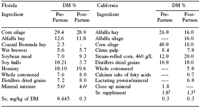
18 mg/kg
219 mg/kg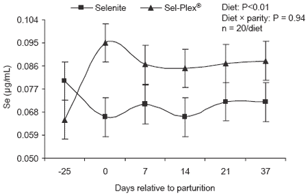
Figure 1. Plasma selenium concentrations in lactating cows fed diets containing either Sel-Plex® or sodium selenite.
Innate immunity (i.e., neutrophil function) was determined by phagocytic and oxidative burst capacity of neutrophils in whole blood using a dual color flow cytometric method.
Samples were collected from a subsample of 36 cows at -26 days and from 40 cows at 0, 7, 14, 21 and 37 days postpartum and analyzed for neutrophil function. Adaptive immunity (ability to induce an antibody response) was monitored by measuring immunoglobulin G (IgG) concentrations in blood in response to ovalbumin (Ovalb) injections. Ovalb antigen (1 mg [i.m.]) was dissolved in an E. coli J5 endotoxemia preventive vaccine and injected at -60 and -22 ± 6 days postpartum (day of initiating Sel-Plex® [n = 38] and selenite [n = 47] diets). Ovalb was dissolved in PBS with Quil- A adjuvant and injected again at parturition (day 0). Serum samples were collected on days of immunization and at 21 and 42 days postpartum.
Percentage of gated neutrophils that phagocytized E. coli and underwent oxidative burst activity did not differ between dietary groups at -26 days postpartum (44.6 ± 4.6%).
For subsequent samples, Sel-Plex® improved neutrophil function in which phagocytosis and killing activity were increased. This improvement is reflected in Figure 2; however, the average response to Sel-Plex® was greater for primiparous animals than multiparous cows (Figure 2). This difference was due to a diet × parity × day interaction (P<0.05); namely, Sel-Plex® improved neutrophil function at parturition in multiparous cows (42 ±6.14% vs 24.3 ±7.2%) and at 7, 14 and 37 days postpartum in primiparous cows (53.9 vs 30.7%, 58.6 vs 41.9%, and 53.4 vs 34.8%, respectively; pooled SE = 6.8%).
Overall neutrophil function was suppressed in primiparous cows at the time of parturition and was not restored until 7 to 14 days postpartum. In contrast, multiparous cows did not have restoration of neutrophil function until 14 to 21 days postpartum.
Organic selenium improved phagocytosis and killing activity of neutrophils in both multiparous and primiparous cows. However, the primiparous cows seemed to be more responsive in that Sel-Plex® stimulated neutrophil function throughout 0 to 21 days postpartum, whereas Sel-Plex® stimulation in multiparous cows was evident on the day of parturition only. In most of our postpartum experiments, we detect distinct differences between primiparous and multiparous cows for a multiplicity of physiological and biochemical responses.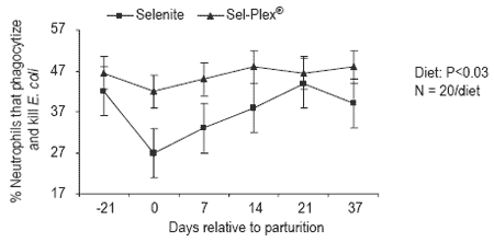
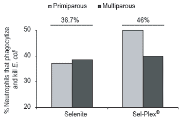
Figure 2. Phagocytosis and killing activity of neutrophils in whole blood from periparturient lactating dairy cows fed diets containing inorganic selenium or organic selenium (Sel-Plex®) supplements.
Anti-IgG to Ovalb did not differ between dietary groups at -60 and -22 days postpartum (0.18 ±0.01 and 0.97 ±0.04 optical density [OD]). Concentrations of anti-IgG to Ovalb were higher (P<0.01) in Sel-Plex® multiparous cows at 21 and 42 days postpartum (Figure 3). However, anti-IgG to Ovalb did not differ between selenium supplements for primiparous cows (1.40 ±0.08 OD). Thus, measurement of adaptive immunity was improved in multiparous dairy cows in response to Sel-Plex® but not in primiparous cows.
Our findings indicated that feeding selenium as organic selenium (Sel-Plex®), beginning at 26 days prepartum, elevated plasma selenium concentrations, increased neutrophil function at the time of parturition, and improved immuno-responsiveness (i.e., multiparous cows).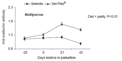
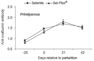
Figure 3. Abundance (optical density) of serum anti-ovalbumin antibody in multiparous and primiparous cows immunized with ovalbumin and fed either Sel-Plex® or sodium selenite.
California. A subset of 15 primiparous and 24 multiparous cows in the selenite group and 10 primiparous and 26 multiparous cows in the Sel-Plex® group was used to evaluate innate (cellular) and adaptive (humoral) immune responses. Concentrations of selenium in plasma were determined at -45, 0, 21, 42 and 60 days relative to calving. GSH-Px activity in plasma, neutrophil phagocytic activity and its oxidative metabolism were determined on days 0 and 42 postpartum. Each animal received an i.m. injection of 1 mg of ovalbumin at -45, -25 and 0 days relative to calving. Anti-ovalbumin IgG concentrations in serum were analyzed at every injection and at 21 and 42 days postpartum.
Concentration of selenium in plasma was similar (P=0.38) for cows given Sel-Plex® and selenite throughout the study (0.107 vs 0.101 μg/mL), but concentrations increased with days postpartum. GSH-Px activity in plasma was not affected (P=0.70) by source of selenium.
Source of supplemental selenium had no effect on phagocytic and bactericidal activities of neutrophils harvested from cows at calving and 42 days postpartum (Table 2). Cows at calving had a smaller (P<0.05) percentage of neutrophils phagocytizing bacteria, mean number of phagocytized bacteria per neutrophil, percentage of phagocytized bacteria killed, number of phagocytized bacteria, and percentage of neutrophils killing at least one bacterium compared with cows at 42 days postpartum.
Multiparous cows tended (P=0.08) to kill a greater percentage of intracellular bacteria compared with primiparous cows (46.9 vs 40.5%).
Oxidative burst of neutrophils was evaluated in vitro by reduction of nitroblue tetrazolium (NBT). Source of selenium and parity did not affect oxidative burst in nonstimulated and stimulated neutrophils. A greater (P=0.04) proportion of nonstimulated neutrophils collected from cows at study day 42 reduced NBT compared with those from cows at calving, but days postpartum did not affect oxidative burst in stimulated neutrophils.
Table 2. Effect of selenium source on neutrophil function (LS means) in experimental cows of California.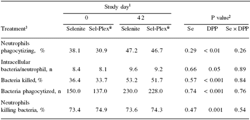
1 Study day 0 = calving.
2 Se = source of Se; Study day; and Se × DPP = interaction between Se and DPP.
3 Percentage of neutrophils containing at least one intracellular bacterium (live or dead)
4 Number of bacteria phagocytized / number of neutrophils phagocytizing at least one bacterium.
5 Number of dead, phagocytized bacteria / (number of live + dead phagocytized bacteria) × 100.
6 Number of neutrophils containing at least one dead bacterium / number of neutrophils phagocytizing at least one bacterium.
Source of selenium did not influence serum titers for anti-ovalbumin IgG of cows in the first 42 days postpartum. Concentrations of anti-ovalbumin IgG in serum increased (P=0.002) over the study period as expected, but no interaction was observed between source of selenium and study day on serum anti-ovalbumin IgG titers. Primiparous and multiparous cows had similar responses to immunization with ovalbumin, and no treatment × parity interaction on serum titers for anti-ovalbumin IgG was observed.
UTERINE HEALTH AND PREGNANCY RATES
Florida and California. Both in Florida and California, frequencies of retained placenta, mastitis, ketosis, displacement of abomasum, and subclinical endometritis were not affected by diets (Table 3). Likewise, frequencies of cows cycling were not affected by diets or reproductive program (Table 3).
Sel-Plex® reduced the frequency of multiparous cows detected with more than one event of fever in Florida (rectal temperature >39.5 ºC; Sel-Plex®, 13.3% [25/188] vs selenite, 25.5% [46/181]; P<0.05), but the Sel-Plex® effect was not observed in primiparous cows, which had a much higher frequency of fever (40.5%). In California, there was no effect of dietary selenium on occurrence of a fever during the first 10 days postpartum as indicated by a rectal temperature >39.5 ºC. As in Florida, multiparous cows were less likely (P<0.001) to develop postpartum fever (38.6% [141/365]) than primiparous cows (59.5% [119/200].
Table 3. Effect of selenium source on incidence of postpartum disorders.
1Selenite (n=285 FL, n=285 CA; Sel-Plex®, n=289 FL, n=281 CA).
In Florida cervical discharge scores measured at 5 and 10 days postpartum were better for the Sel-Plex® group (Table 4). The frequency of cows with a purulent-fetid discharge was reduced and proportion of cows with a clean discharge was increased. These findings lend additional support to the conclusion that feeding Sel-Plex® in the Florida experiment improved immunocompetence and was associated with improved uterine health measured at 5 and 10 days postpartum.
In Florida, diet failed to alter first service pregnancy rate at ~30 days post AI or pregnancy losses between ~30 and ~55 days post AI (Table 5). These low pregnancy rates and high embryonic losses are typical of cows managed during the summer heat stress period of Florida. Diet did indeed alter second service pregnancy rate (Table 5).
The benefit of Sel-Plex® on second service pregnancy rate is very interesting. We hypothesize that cows of the Sel-Plex® group were better able to reestablish an embryotrophic environment at second service after either early or late embryonic losses. For example, cows presented for second service may not have been pregnant to the first service by 30 days at the ultrasound diagnosis or were pregnant and underwent embryonic loss and required a second service. Indeed, pregnancy rate to the second service for cows that had lost an embryo was 22.7% for the Sel-Plex® group vs 4.2% in the selenite group.
Table 4. Overall frequencies of cervical discharge scores measured at 5 and 10 days postpartum.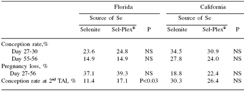
Parity (P<0.01); Day (P<0.02); Calving difficulty (P<0.01); RFM (P<0.01)
Table 5. Effect of selenium source on reproductive outcomes of lactating dairy cows in Florida and California experiments.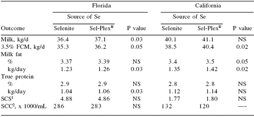
The product Mu-Se® (Schering-Plough Animal Health Corp, NJ), which contains inorganic selenium (50 mg) and vitamin E (500 mg), increased pregnancy rate to second service (i.e., no effect at first service), reduced services per conception, and decreased the interval from calving to conception (Arechiga et al., 1998).
These results are somewhat similar to the Florida results (Table 5) in which Sel-Plex® increased pregnancy rate to the second service in cows that did not become pregnant at first service. In California, dietary selenium supplements failed to alter conception rates at day 28 and day 56, pregnancy loss from 28 to 56 days of gestation, or conception rate at the second timed insemination (Table 5).
MILK PRODUCTION AND SOMATIC CELLS
Florida. Mean milk yield and milk composition determined from monthly samples for cows fed supplemental inorganic and organic selenium are presented in Table 6. Both monthly milk yield and fat-corrected milk (FCM) were greater for cows fed Sel-Plex®.
An additional series of analyses were conducted to determine when the differences in milk production occurred. Milk production was monitored on a daily basis for the first 81 days of lactation and mean daily milk yield did not differ between treatments (35.6 kg/d).
Additional analyses were conducted with the monthly estimates of milk production from monthly DHIA measurements and are presented in Figure 4. Monthly milk yields were elevated in the Sel-Plex® group in later stages of lactation between 6-8 months of lactation. The increase in milk production in later stages of lactation occurred primarily in primiparous cows but not in multiparous cows (Figure 4).
Table 6. Effect of selenium source on performance of lactating dairy cows in Florida and California experiments.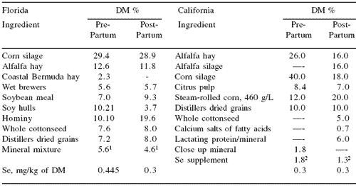
1SCS = somatic cell score.
2SCC = somatic cell count.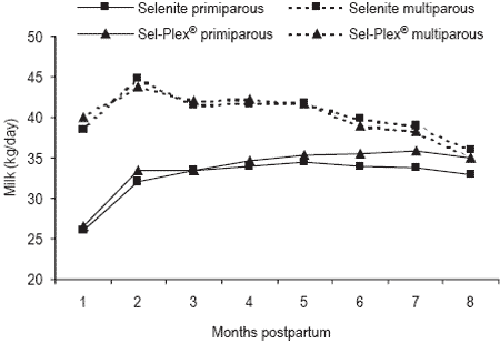
Figure 4. Mean monthly milk yield (kg/day) for primiparous and multiparous cows fed Sel-Plex® or sodium selenite. Diet × parity × month interaction, P<0.05.
The slight increase in milk production due to Sel-Plex® may be due to a carry-over effect of Sel-Plex® since the minimal period of supplementation for all cows was 81 days of lactation. The monthly profiles of somatic cells in milk (Figure 5) indicated that concentrations of somatic cells were lower in Sel-Plex® -treated cows during the later stages of lactation (i.e., months 6-8). Perhaps this improvement represented a lower loss of alveolar epithelial cells which possibly accounted for the subtle increase in milk yield. Milk somatic cells were lower in primiparous cows than multiparous cows between 3 to 8 months of lactation.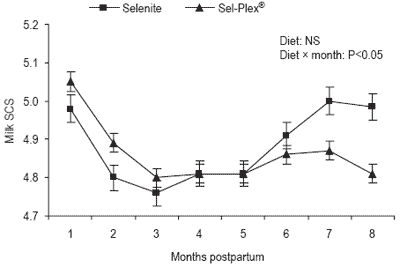
Figure 5. Mean monthly concentrations of milk somatic cells, Somatic Cell Score (SCS), for lactating cows fed Sel-Plex® or sodium selenite. Parity × month interaction P<0.05.
California: Cows fed Sel-Plex® produced more (P=0.02) 3.5% FCM and milk fat than those fed selenite (Table 6). Estimates of daily production of 3.5% FCM, determined from monthly estimates of milk production for the first 3-month period of lactation, indicated that cows fed Sel-Plex® had a 5% increase in daily production of 3.5% FCM.
Replacing selenite with Sel-Plex® did not improve health or immunological status of periparturient dairy cows in California, but increased yields of 3.5% fat-corrected milk and fat, and concentration of fat in milk. The lack of treatment effects, other than on milk yield, during the first 90 days of lactation is perplexing. One of the potential selenoproteins induced by organic supplementation of selenium is iodothyronine deiodinase, which may increase the availability of triiodothyronine to enhance milk production.
Further research is warranted to investigate the effects of feeding Sel-Plex® on increasing milk production. The results in California appeared to be an immediate response in early lactation in which Sel-Plex® was fed to cows that were not Se-deficient.
In contrast, the increase in milk production of the Florida experiment occurred well after the supplementation period, suggesting that prior feeding of Sel-Plex® appeared to possibly increase persistency in first calf heifers (Figure 4). Whether shedding of mammary alveolar cells was reduced leading to enhanced milk production of primiparous cows fed Sel-Plex® earlier in lactation warrants consideration.
Discussion and summary of site differences
Weiss (2003) pointed out that in many of the clinical and experimental efforts, most control diets would be considered deficient in selenium and the companion diets contained either 0.1 or 0.3 mg/kg of supplemental selenium.
Supplementation of selenium to deficient diets often elicits a positive response whereas additional supplementation of Se-adequate diets would not be expected to produce additional clinical benefits. This appears to be the case when comparing the efficacy of Sel-Plex® in the present studies in which supplemental Sel-Plex® had distinctive effects on immune responses, uterine health, plasma selenium concentrations, and second service pregnancy rates in Se-deficient Florida. In contrast, there were virtually no Sel-Plex® effects within the California study other than a significant increase in milk production.
Both studies were conducted during the summer heat stress period. However, the cows in Florida were exposed to a longer period of heat stress based on the Temperature Humidity Index (THI) exceeding 72 (threshold value above which cows reduce milk production as a regulatory response to hyperthermia). In Florida, the THI exceeded 72 in early May versus early July in California. Average THI in the plateau phase of summer was ~82 for a 4-month period in Florida, whereas it was ~78 for a 1-month period in California. The more stressful environment in Florida likely contributed to the lower pregnancy rates and increased pregnancy losses compared with those in California (Table 5).
Analyses of the composite feed samples for both experiments identified clear differences in dietary selenium concentrations. Selenium concentrations in the prepartum diets differed substantially between the Florida (selenite of 0.44 and Sel-Plex® of 0.49 mg/kg of DM) and California (selenite of 0.60 and Sel-Plex® of 0.74 mg/kg of DM) experiments.
Differences in selenium concentrations (mg/kg of DM) for the postpartum diets were even greater; that is, Florida with 0.36 for control and 0.36 for Sel-Plex® and California with 0.71 for control and 0.60 for Sel-Plex®. Based upon the higher overall selenium concentrations (i.e., supplemental + background Se levels) in the California experiment, it was not surprising that clinical and immuno-responses did not differ.
In contrast, Florida was essentially a Se-deficient environment in which the virtually all dietary selenium was derived via the two supplements. Under these conditions, Sel-Plex® (0.33 mg Se/kg) fed in the ration beginning at 26 days prepartum, elevated plasma selenium concentrations, increased neutrophil function at the time of parturition, improved immuno-responsiveness in multiparous cows, improved uterine health, and increased second service pregnancy rate during summer in an environment that is selenium deficient.
Such effects were not observed in the California study. It is noteworthy that in the California study, Sel-Plex® supplementation during early lactation did result in a substantial increase in milk production. In contrast, this effect on milk production in early lactation was not evident in the Florida experiment although Sel-Plex® had numerous effects on immunological, uterine health, and pregnancy rate to second service outcomes with a population of cows managed in a selenium deficient environment.
References
Authors: FLAVIO T. SILVESTRE1, HELOISA M. RUTIGLIANO2, WILLIAM W. THATCHER1, JOSÉ E.P. SANTOS2 and CHARLES R. STAPLES1Arechiga, C.F., S. Vazquez-Flores, O. Ortz, J. Hernandez-Ceron, A. Porras, L.R. McDowell and P. J. Hansen. 1998. Effect of injection of ß-carotene or vitamin E and selenium on fertility of lactating dairy cows. Theriogenology 50:65-76.
Goff, J.P. 2006. Major advances in our understanding of nutritional influences on bovine health. J. Dairy Sci. 89: 1292–1301.
Grasso, P.J., R.W. Scholz, R.J. Erskine, and R.J. Eberhart. 1990. Phagocytosis, bactericidal activity, and oxidative metabolism of mammary neutrophils from dairy cows fed selenium-adequate and selenium-deficient diets. Am. J. Vet. Res. 51:269– 274.
Gunter, S.A., P.A. Beck and J.M. Phillips. 2003. Effects of supplementary selenium source on the performance and blood measurements in beef cows and their calves. J. Anim. Sci. 81: 856–864.
Hogan, J.S., K.L. Smith, W.P. Weiss, D.A. Todhunter and W.L. Shockey. 1990. Relationships among vitamin E, selenium, and bovine blood neutrophils. J. Dairy Sci. 73: 2372–2378.
Loeffler, S.H., M.J. de Vries and Y.H. Schukken. 1999. The effects of time of disease occurrence, milk yield, and body condition on fertility of dairy cows. J. Dairy Sci.82: 2589-2604.
Mezzetti, A., C. Di Ilio, A.M. Calafiore, A. Aceto, L. Marzio, G. Frederici, and F. Cuccurullo. 1990. Glutathione peroxidase, glutathione reductase and glutathione transferase activities in the human artery, vein and heart. J. Mol. Cardiol. 22: 935- 938.
National Research Council. 2001. Nutrient Requirements of Dairy Cattle. Natl. Acad. Press, Washington DC.
Rutigliano, H.M., F.S. Lima, R.L.A. Cerri, L.F. Greco, J.M. Vilela, V. Magalhães, J. Hillegass, W.W. Thatcher and J.E.P. Santos. 2006. Effects of source of supplemental selenium and method of presynchronization on reproduction and lactation of dairy cows. J. Dairy Sci. 89 (Suppl. 1):207 (Abstr.).
Silvestre, F.T., D.T. Silvestre, C. Crawford, J.E.P. Santos, C.R. Staples and W.W. Thatcher. 2006a. Effect of selenium (Se) source on innate and adaptive immunity of periparturient dairy cows. Biol. Repro. 39th Annual Meeting. Special Issue. p. 132.
Silvestre, F.T., D.T. Silvestre, J.E.P. Santos, C. Risco, C.R. Staples and W.W. Thatcher. 2006b. Effects of selenium (Se) sources on dairy cows. J. Anim. Sci. 89: (Suppl 1.) 52.
Weiss, W.P. 2003. Selenium nutrition of dairy cows: comparing responses to organic and inorganic selenium forms. In: Nutritional Biotechnology in the Feed and Food Industries: Proceedings of Alltech’s 19th Annual Symposium (T.P. Lyons and K.A. Jacques, eds). Nottingham University Press, UK, pp. 333-343.
1 University of Florida, Gainesville, Florida, USA
2 University of California-Davis, Tulare, California, USA


