Demonstration of Listeria monocytogenes by Immunohistochemistry in Formalin-Fixed Brain Tissues from Natural Cases of Ovine and Bovine Encephalitis
In the present work, evidence of Listeria monocytogenes antigens based on the avidin–biotin complex (ABC) immunoperoxidase technique was performed on formalin-fixed central nervous system tissues (CNS) from a total of 23 natural cases of encephalitis (four ovine and 19 bovine). Listeria monocytogenes serotype 4 was isolated from 10 of 17 cultured specimens.
Meningoencephalitis characterized by focal necrosis, microabscesses, perivascular cuffing, and gliosis with presence of macrophages and/or neutrophils was observed at histological examination. Positive L. monocytogenes antigens were successfully identified by immunohistochemistry (IHC) in the CNS of all 23 cases.
Paraffin-embedded tissues assayed were stored up for 17 years. Morbidity of the outbreaks was between 0.3–3% and 0.1–1% for ovine and bovine cases, respectively. In all the ovine cases, flocks involved were under extensive grazing conditions. In nine of the 19 bovine cases (47.3%), supplementation with corn silage was used. The ABC test can help as a practical tool for the diagnosis of natural cases of L. monocytogenes encephalitis on formalin-fixed specimens from ovine and bovine.
Introduction
Listeriosis caused by Listeria monocytogenes, is an important infectious disease of ruminants, and a highly fatal opportunistic foodborne infection in human beings. Listeria monocytogenes is a facultative intracellular gram-positive small rod, aerobic and facultatively anaerobic, found in soil, vegetation and faeces (Jubb and Huxtable, 1985, 1993; Rebhun, 1987).
The bacteria have a group of genes, which allow invasion, survival, multiplication and mobility in intracellular environments. The organism resists freezing and thawing and is able to survive for several years in faeces, straw, silage and in some soils (Blood and Radostis, 1989; Smith and Sherman, 1994) Listeria monocytogenes can replicate at low environmental temperatures.
Listeriosis is a disease that is most frequently encountered in cattle, sheep and goats, and is also occasionally found in wild species. Clinical manifestations of invasive listeriosis in ruminants are usually severe and include abortion, neonatal septicaemia, and meningoencephalitis (Ladds et al., 1974).
Bovine listerial encephalitis has a high fatality rate with a clinical course of about 1 week but with low morbidity (Rebhun, 1987). In addition, listerial encephalitis in small ruminants is more commonly found in sheep than in goats (Wilesmith and Gitter, 1986) with higher mortality rate than that observed in cattle (Lippman, 1969). Microabscesses, focal gliosis and perivascular cuffing characterize listerial encephalitis.
Severe lesions are confined to the brainstem, especially the pons and medulla oblongata (Ladds et al., 1974; Jubb and Huxtable, 1993).
Isolation of L. monocytogenes may be unsuccessful even when appropriate samples are submitted. Culture-negative cases can be associated with few or no bacteria in the lesions.
Therefore, a rapid and sensitive test such as immunohistochemistry (IHC) is a helpful tool for diagnosis found by different authors not only in natural cases but also in experimental ones (Barlow and Mcgorum, 1985; Domingo et al., 1986; Johnson et al., 1995; Krueger et al., 1995; Marco et al., 1988; Weinstock et al.,1995).
However, no mention of the use of this diagnostic tool in natural cases of bovine and ovine encephalitic listeriosis was performed in Argentina. The successful demonstration of L. monocytogenes antigens with the IHC technique performed on formalin-fixed central nervous system (CNS) tissues from natural cases of encephalitis in ovine and bovine, from different outbreaks that occurred in Buenos Aires province between 1984 to 2001, is described in this report.
Materials and Methods
Case retrieval
A retrospective evaluation of confirmed and suspected cases (four adult ovine: three ewes and one ram; 19 bovine: 11 steers, three calves, three bulls, one cow and one heifer) of listeriosis was performed on CNS specimens from commercial flocks and herds with clinical signs of nervous dysfunction which were submitted and processed by the Diagnostic Laboratories (INTA, Balcarce, Argentina) from 1984 to 2001. Information regarding grazing, feedlot conditions, corn silage supplementation and other epidemiological data was collected when it was possible.
Isolation and characterization of L. monocytogenes
Isolation of bacteria was performed, when it was possible, from CNS tissue which was inoculated directly onto 7% Columbia bovine blood agar and incubated aerobically at 37ºC. Simultaneously, cold enrichment incubation (4ºC for 15 days) from suspensions of CNS in brain–heart infusion broth was performed. Listeria monocytogenes isolation was identified by standard microbiological and serotyping techniques (Seeliger and Hohne, 1979; Albritton et al., 1980; Cipolla et al., 1998).
Histopathological examination
Carcass for a complete postmortem examination or formalinfixed brain was available for histological evaluation. Diagnosis of listeriosis was based on gross and microscopic assessment of fixed tissues, and bacterial isolation. CNS tissue samples from the neocortex, hippocampus, thalamus, corpora quadrigemina, pons cerebri, cerebellum, medulla oblongata and upper cervical spinal cord were selected and fixed in 10% formalin saline, dehydrated and embedded in paraffin. Sections 4–5 lm thick were cut and stained with haematoxylin and eosin (H&E).
Suspected cases were those with histological lesions suggestive of listeriosis. Routine neuropathological examination of those brains demonstrated perivascular cuffing, glial foci and microabscesses (Ladds et al., 1974; Charlton and Garcia, 1977; Jubb and Huxtable, 1993).
Immunohistochemistry
Central nervous system specimens with histopathological lesions compatible with those produced by L. monocytogenes were processed by immunoperoxidase technique (Hsu et al., 1981). Paraffin-embedded sections of CNS tissues 3–4 lm thick were mounted on histoslides (Probe-On, Fisher Scientific, Pittsburgh, PA, USA) and stained on a capillary gap system (Micro-Probe Staining System, Fisher Scientific, Pittsburgh, PA, USA).
Sections were routinely deparaffinized in three sequential steps: 10 min in xylene, and for 2 min in 100% alcohol for two times. After that, endogenous peroxidase was inhibited with methanol and 3% H2O2 (Sigma, St. Louis, MO, USA) for 10 min, and sections were then treated with enzyme Pepsin 0.4% (Sigma, St. Louis, MO, USA) in phosphate buffered saline pH 7.4 (PBS; Sigma, St. Louis, MO, USA) for 15 min at 37ºC in water bath. Slides were rehydrated in graded ethanol and treated with blocking solution.
A commercially available kit system based on avidin–biotin complex immunoperoxidase (ABC) method was used (Vectastain ABC Elite Rabbit IgG, Vector Laboratories, Burlingame, CA, USA). Commercially available rabbit polyclonal antiserum against L. monocytogenes (Listeria O antiserum poly, serotypes 1 and 4, Difco, Detroit, MI, USA) was used as the primary antibody at 1 : 1500 for 45 min at 37ºC. Secondary antiserum and ABC were incubated at 37ºC for 20 min.
Subsequently, slides were washed and the final reaction was developed in 3-amino-9-ethylcarbazole (Aminoethylcarbazole Substrate Chromogen System, Dako, Carpinteria, CA, USA).
Sections were rinsed and counterstained with Mayer’s haematoxylin (Sigma, St. Louis, MO, USA), rinsed and mounted.
Formalin-fixed brain tissue with histological lesions of listeriosis from a sheep from which L. monocytogenes had been isolated in pure culture was used as positive control and to titre the primary antibody. Negative controls consisted of a nonimmune normal rabbit serum applied to Listeria-infected brain-tissue sample, and a positive antiserum applied to normal sheep and bovine brain tissue negative by culture to L. monocytogenes. Beyond that, positive L. monocytogenes control slides were used omitting the secondary antibodies or the ABC solution.
Results
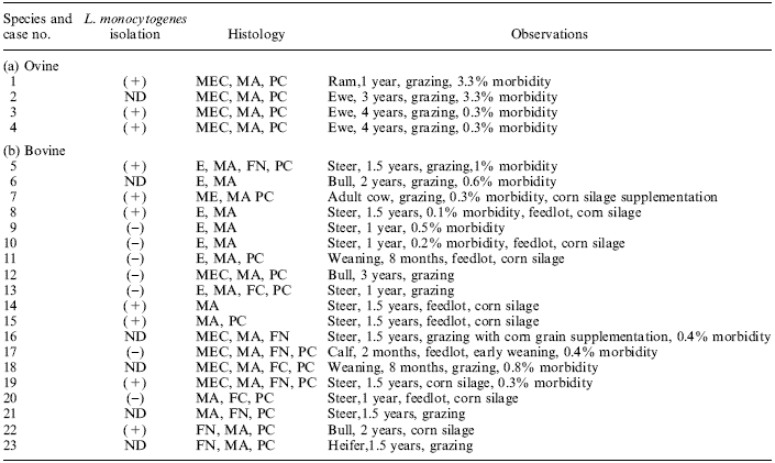
Morbidity of the outbreaks were between 0.3–3% and 0.1%–1% for ovine and bovine cases, respectively. The ovine cases involved flocks that had been grazing under extensive conditions. In nine of the 19 bovine cases (47.3%), supplementation with corn silage had been used (Table 1).
Bacterial isolation of L. monocytogenes was accomplished in 10 (three cases from ovine and seven cases from bovine) of 17 cultured specimens (Tables 1 and 2). All isolates were of serotype 4. Additionally, L. monocytogenes antigens were detected by IHC in seven bovine specimens negative by bacteriological culture and in six cases (one from ovine and five from bovine specimens) in which bacterial cultures were not attempted (Table 2).
On histological examination, all the cases showed multiple microabscesses in the brain stem between the midbrain and upper cervical spinal cord, being more severe and extensive in the medulla and/or pons in all specimens (Table 1). Microscopic findings consisted of single or multiple microabscesses (Figure 1) with necrotic and liquefactive changes with infiltration by neutrophils and mononuclear cells located either in the gray and/or white matter. The proportion of glial cells to neutrophils in inflammatory foci varied greatly within the same brain and also between brain tissues that were examined.
Meningeal lesions showed a mild to moderate degree of mononuclear infiltration of leptomeninges in all cases. Besides, perivascular mononuclear cell accumulation, occasionally with prominent perivascular lymphoid cuffing (Figure 2), and diffuse infiltration of microglia in the neuroparenchyma were observed. There were no significant differences between the histopathological changes seen in sheep and cattle.
In all of the 23 specimens investigated by ABC technique (Table 1), L. monocytogenes antigens could be identified.
Immunostaining of L. monocytogenes was best visualized in the areas of CNS where microabscesses and necrotic foci were present. Listeria monocytogenes antigens appeared as small rods, circular or ovoid granules, localized in microabscesses and in large foci of necrosis, mainly in the cytoplasm of phagocytes and outside the cells. Occasionally, small numbers of phagocytes containing L. monocytogenes antigens were detected in the perivascular cuff, glial foci and areas of neutrophil-free of inflammation. No reaction was seen in the negative control sections of IHC.
Discussion
To the knowledge of the authors, this is the first report on successful identification of L. monocytogenes antigens by IHC in the brain of spontaneous infected sheep and cattle after retrospective evaluation of confirmed and suspected cases of listeriosis in Argentina. The morbidity of the outbreaks found in this work fits within the range mentioned by other authors (Ladds et al., 1974; Rebhun, 1987; Jubb and Huxtable, 1993).
Similarly to other reports, in the present study, 47.3% of the cases of bovine listeriosis were associated with animals that had been fed with corn silage (Ladds et al., 1974; Rebhun, 1987; Jubb and Huxtable, 1993; Low and Donachie, 1997; Moisan et al., 1998).
An increased susceptibility of sheep, in comparison with cattle, was mentioned when they were fed with contaminated silage (Rebhun, 1987). The trend towards trench and other make-shift corn silos is frequent for beef and dairy cattle supplementation under intensive and/or feedlot conditions in Argentina. In the present work, despite the fact that silage was not used in ovine, the morbidity rate of sheep outbreaks was higher than that found in bovine cases.
Unfortunately, silage conditions were not established in this work. In consequence, the causes of this high incidence in sheep for these particular cases remain undetermined.
| Table 1. Ovine and bovine cases with encephalitis caused by L. monocytogenes detected by immunohistochemistry |
 To enlarge the image, click here |
| ND, not done; MEC, meningoencephalitis; MA, microabscesses; PC, perivascular cuffing; E, encephalitis; FN, focal necrosis. |
| Table 2.Detection of L. monocytogenes in four ovine and 19 bovine cases |
 |
| aPercentage of total cases (n = 23). |
 |
| Fig. 1. Photomicrograph of bovine brain stem with typical microabscess in which L. monocytogenes was isolated, (H&E) ·200. |
 |
| Fig. 2. Photomicrograph of a perivascular lymphoid cuff in bovine brain with encephalitis caused by L. monocytogenes, (H&E) ·200. |
Animals that develop meningoencephalitis caused by L. monocytogenes are frequently under individual stress conditions that could affect the cellular immune response.
This pathological condition facilitates the invasiveness of the bacteria. Poor management conditions and stress associated with listeriosis are mentioned in the literature (Meredith and Schneider, 1984; Rebhun, 1987; Jubb and Huxtable, 1993). In the present work, some of these facts, like wet, muddy conditions and poor management were noted (data not shown). For example, in the case no. 17 (Table 1) in which early weaning was performed, these severe stress conditions could have played a deciding role in precipitating the disease.
Unfortunately, other detailed management information was not always available in our work.
The neurological form of listeriosis associated with menigoencephalitis most likely follows an ascending infection along trigeminal nerve to the brain stem. This infection of the trigeminal nerve could arise from the nasal cavity, oral cavity, or conjunctiva resulting in progressive encephalitis and meningitis (Jubb and Huxtable, 1993; Rebhun, 1987). In the present work, the affected animals were under 5 years old. Young animals are under increased risk of the disease because of the dental changes as was suggested (Barlow and Mcgorum, 1985).
In the present work, in all the cases L. monocytogenes isolated was of serotype 4, which is the target of the primary antiserum used in the ABC system. Listeria ivanovii, a second pathogenic species of the genus Listeria, which is specific for ruminants and formerly classified as serotype 5 of L. monocytogenes, has been mentioned as sporadic cause of bovine and ovine abortion (Seeliger et al., 1984; Alexander et al., 1992; Gill et al., 1997) and eventually involved with central nervous and reproductive symptoms associated with the ingestion of silages (Alexander et al., 1992). Therefore, L. ivanovii was not detected by the primary antiserum used in the ABC system used in this work.
The histological findings in the CNS of the Listeria cases in the present work were in agreement with those described in literature (Ladds et al., 1974; Charlton and Garcia, 1977; Rebhun 1987; Jubb and Huxtable, 1993; Johnson et al., 1995; Krueger et al., 1995).
Characteristic microscopic lesions showed microabscesses with necrosis and liquefactive changes which were different from those described in other bovine neurological diseases, like malignant catharral fever, infectious bovine rhinotracheitis, thromboembolic meningoencephalitis caused by Haemophilus sommnus, and sporadic bovine encephalomyelitis found in generalized chlamydial infection (Jubb and Huxtable, 1993).
In the present work, L. monocytogenes was detected in formalin-fixed, paraffin-embedded tissues from ovine and bovine. The ABC technique described here represents a significant improvement for diagnosis that can be used in routine field cases. IHC is currently being used to demonstrate a variety of infectious agents in processed material (Haines and Chelack, 1991).
In the present study, L. monocytogenes antigens were more frequently found by IHC in necrotic areas and microabscesses of the CNS as was reported previously (Weinstock et al., 1995). Our results indicate that the sensitivity of the IHC used here was good enough to detect positive cases maintained for up to 17 years (ovine case no. 1) stored in formalin. This finding correlates with data published by Weinstock et al. (1995) who mentioned positive IHC cases up to 10 years old.
On the other hand, it is probable that antibiotic treatment and/or the low number of organisms present in the CNS cultured was mainly responsible for the failure to obtain L. monocytogenes isolation in the other cases which were detected by IHC. In this work, the presence of typical microabscesses and necrotic foci where L. monocytogenes antigens detected by ABC was characteristic in all cases included those where no bacteria were isolated and/or culture was not performed.
In spite of the isolation of L. monocytogenes remains as an important criteria for the diagnosis of listeriosis, the use of combined histopathology and IHC enhanced the overall sensitivity of the diagnosis. Furthermore, the recovery of Listeria in the absence of lesions is not diagnostic for listerial encephalitis (Grønstøl, 1980). The findings of the present work suggest that ABC test for L. monocytogenes in formalin-fixed, paraffin-embedded specimens from CNS of ovine and bovine cases is a rapid and specific technique which can be used as a useful tool in confirming the diagnosis of encephalitic listeriosis in ovine and bovine.
References
Albritton, W. L., G. L. Wiggins, W. E. Dewitt, and J. C. Feeley, 1980: Listeria monocytogenes. In: Lennette, E. H., A. Balows, W. J. Hausler, and J. P. Truant (eds), Manual of Clinical Microbiology, Chapter 11, 3rd edn, pp. 139–142. American Society for Microbiology, Washington, D.C., USA.
Alexander, A. V., R. L. Walker, B. J. Johnson, B. R. Charlton, and L. W. Woods, 1992: Bovine abortion attributable to Listeria ivanovii: four cases (1988–1990). J. Am. Vet. Med. Assoc. 200, 711–714.
Barlow, R. M. and B. Mcgorum, 1985: Ovine listerial encephalitis: analysis, hypothesis and synthesis. Vet. Rec. 116, 233–236.
Blood, D. C., and O. M. Radostis, 1989: Diseases caused by bacteria. I. In: Blood D. C. O. M. Radostis (eds) Veterinary Medicine. A Textbook of the Diseases of Cattle, Sheep, Pigs, Goats, and Horses, 7th edn, pp. 582–587. Bailliere Tindall, London, UK.
Charlton, K. M., and M. M. Garcia, 1977: Spontaneous listeric encephalitis and neuritis in sheep. Light microscopic studies. Vet. Pathol. 14, 250–253.
Cipolla, A. L., F. Paolicchi, N. Leardini, M. Gomez, H. Terzolo, A. Moreira, C. M. Campero, E. Odriozola, E. R. Cobo, and L. Vagnoni, 1998: Caracterizacion bioquımica y serologica de cepas de Listeria monocytogenes aisladas de bovinos y ovinos con sindrome encefalıtico. Resumenes del XIV Congreso Latinoamericano de Microbiologıa, II Congreso de Microbiologıa del Mercosur y II Congreso Paraguayo de Microbiologıa. p. 88.
Domingo, M., J. A. Ramos, L. Dominguez, L. Ferrer, and A. Marco, 1986: Demonstration of Listeria monocytogenes with the PAP technique in formalin fixed and paraffin embedded tissues of experimental infected mice. J. Vet. Med. B. 33, 537–542.
Gill, P. A., J. G. Boulton, G. C. Fraser, A. E. Stevenson, and L. A. Reddacliff, 1997: Bovine abortion caused by of Listeria ivanovii. Aust. Vet. J. 75, 214.
Grønstøl, H., 1980. Listeriosis in sheep. Isolation of Listeria monocytogenes from organs of slaughtered animals and dead animals submitted for post-mortem examination. Acta Vet. Scand. 21, 11–17.
Haines, D. M., and B. J. Chelack, 1991: Technical considerations for developing enzyme immunohistochemical staining procedures on formalin-fixed paraffin-embedded tissues for diagnostic pathology. J. Vet. Diagn. Invest. 3, 101–112.
Hsu, S. M., L. Raine, and H. Fanger, 1981: The use of avidin biotin peroxidase complex (ABC) in immunoperoxidase techniques: a comparison between ABC and unlabeled antibody (PAP) procedures. J. Histochem. Cytochem. 29, 577–580.
Johnson, G. C., W. H. Fales, C. W. Maddox, and J. A. Ramos-vara, 1995: Evaluation of laboratory tests for confirming the diagnosis of encephalitic listeriosis in ruminants. J. Vet. Diagn. Invest. 7, 223– 228.
Jubb, K. V. F., C. R. Huxtable, 1993: The nervous system. In: Jubb K. V.F., P. C. Kennedy, N. Palmer (eds), Pathology of Domestic Animals, 4th edn, pp. 393–397. Academic Press, San Diego, CA, USA.
Krueger, N., C. Low, and W. Donachie, 1995: Phenotypic characterization of the cells of the inflammatory response in ovine encephalitic listeriosis. J. Comp. Pathol. 113, 263–275.
Ladds, P. W., S. M. Dennis, and C. O. Njoku, 1974: Pathology of listeric infection in domestic animals. Vet. Bull. 44, 67–74.
Lippman, R., 1969: Clinical, diagnostic and therapeutic studies on spontaneous nervous system listeriosis of sheep. Acta Vet. Sci. Hung. 19, 161–169.
Low, J. C., and W. Donachie, 1997: A review of Listeria monocytogenes and listeriosis. Vet. J. 153, 9–29.
Marco, A., J. A. Ramos, L. Dominguez, M. Domingo, and L. Gonzalez, 1988: Immunocytochemical detection of Listeria monocytogenes in tissue with the peroxidase–antiperoxidase technique. Vet. Pathol. 25, 385–387.
Meredith, C. D., and D. J. Schneider, 1984: An outbreak of ovine listeriosis associated with poor flock management practices. J. S. Afric. Vet. Assoc. 55, 55–56.
Moisan, P. G., G. A. Andrews, B. M. Debey, J. C. Nietfeld, D. J. Bush, and E. Woods-lavole, 1998: Listeriosis associated with silage feeding in six midwestern cattle herds. Large Anim. Pract. 19, 40–46.
Rebhun, W. C., 1987: Listeriosis. Vet. Clin. North Am: Food Anim. Pract. 3, 75–83.
Seeliger, H. P. R., and K. Hohne, 1979: Serotyping of Listeria monocytogenes and related species. In: Bergen T. and J.R. Norris (eds), Methods in Microbiology, pp. 31–49. Academic Press, London, UK.
Seeliger, H. P. R., J. Rocourt, A. Schrettenbrunner, P. A. DGrimont, and D. Jones, 1984: Listeria ivanovii sp. nov. Int. J. Sys. Bacteriol. 34, 336–337.
Smith, M. C., and D. M. Sherman, 1994: Listeriosis. Goat Medicine, Lea & Febiger, Philadelphia, PA, USA. pp. 141–144.
Weinstock, D., S. B. Horton, and P. H Rowland, 1995: Rapid diagnosis of Listeria monocytogenes by immunohistochemistry in formalin-fixed brain tissue. Vet. Pathol. 32, 193–195.
Wilesmith, J. W., and M. Gitter, 1986: Epidemiology of ovine listeriosis in Great Britain. Vet. Rec. 119, 467–470.
Authors: C. M. Campero1, A. C. Odeón1, A. L. Cipolla1, D . P. Moore2, M. A. Poso1 and E. Odriozola1
1 Instituto Nacional de Tecnología Agropecuaria (INTA), C.C. 276, (7620) Balcarce.
2 Consejo Nacional de Investigaciones Científicas y Técnicas (CONICET), (1033) Buenos Aires, Argentina.


United States