Introduction
Chlamydia abortus is the causative agent of ovine enzootic abortion (OEA), a disease with a high economic impact on ruminants and endemic in Mexico since 2016 [1]. This pathogen causes abortions during the last third of gestation or the birth of weak offspring that often die within 48 hours [2]. Moreover, C. abortus is zoonotic, generating severe complications such as spontaneous abortions or premature births [3].
Animals acquire this microorganism mainly through contact with contaminated materials from aborted fetuses or by ingesting infected food. This bacterium has been found in colostrum, milk, vaginal secretions, urine, and feces [4,5]. Furthermore, compared with other abortive diseases, females affected by OEA may give birth to apparently healthy offspring, complicating the detection of the disease based on clinical signs [2].
The conventional diagnostic techniques for C. abortus detection include bacterial culture, serology, and polymerase chain reaction [6]. While bacterial culture is precise, it is a laborious technique that requires facilities with high levels of biosecurity [2,7]. The most sensitive and specific technique for the molecular detection of this pathogen is PCR. Nonetheless, it requires a laboratory and highly trained personnel, which limits its field application, and the cost of equipment and reagents can be considerable [8].
In recent years, the loop-mediated isothermal amplification (LAMP) technique has gained popularity as an alternative diagnostic method capable of overcoming the aforementioned limitations. The LAMP technique is fast, efficient, and does not require a thermocycler; it is performed at a constant temperature, facilitating pathogen detection under field conditions [9].
In Mexico, a LAMP assay was previously implemented for the molecular detection of C. abortus (Chla-LAMP), and the results demonstrated accuracy and great potential as a simpler technique compared with conventional methods. However, the visualization of results was based on agarose gels that required a laboratory [10]. The versatility of LAMP could allow the incorporation of colorimetric detection assays, enabling the visual interpretation of results without the need for additional equipment such as electrophoresis [11].
Therefore, the objective of this study was to validate a new LAMP assay using colorimetry for the visual detection of Chlamydia abortus in domestic ruminants.
Materials and methods
Ethical considerations
The present study did not use animals directly, only biological samples donated from a previous experiment [12]. However, according to report No. 2024-04, the Animal Welfare and Research Ethics Committee of the Instituto Tecnológico de Sonora approved the procedures of the project.
Study location
The study was conducted in the Laboratorio de Desarrollo e Innovación en Biotecnología Veterinaria of the Departamento de Ciencias Agronómicas y Veterinarias at the Instituto Tecnológico de Sonora (ITSON), Sonora, México.
Biological material
Fifty-six DNA samples from a previous study [12] were used as biological controls. The samples were provided by the Centro de Investigación Nacional Interdisciplinaria en Salud Animal e Inocuidad (CENID) of the Instituto Nacional de Investigaciones Forestales, Agrícolas y Pecuarias (INIFAP) in México. The DNA extractions were obtained from vaginal swab samples collected from ovine and bovine females with a history of abortion. The positive and negative biological controls were previously identified by real-time PCR (qPCR) by [12]. They were also tested by LAMP in agarose gels (Chla-LAMP) under the conditions previously reported by [10].
Design of LAMP primers and synthetic positive
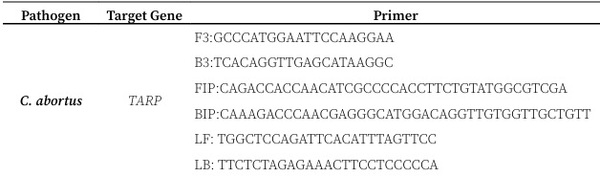
To design the LAMP primers for C. abortus, the Translocated Actin Recruiting Phosphoprotein (TARP) gene was selected as the target. The accession numbers of the reference sequences used for primer design were CP158097.1, CP070224.1, and LS974600.1. LAMP primers were designed using the NEB LAMP Primer Design Tool platform, version 1.4.1 (https:// lamp.neb.com/#!/), generating six primers [F3, B3, FIP (F1c+F2), BIP (B1c+B2), LF, LB] capable of recognizing a total of eight regions of the TARP gene (Table 1). The specificity of the oligonucleotides was confirmed by in silico analysis using the Primer-BLAST alignment tool of the NCBI GenBank® database (https://www. ncbi.nlm.nih.gov/).
The BLAST tool of the NCBI database was used to synthesize the positive control (DNAs+). The CP158097.1 sequence was obtained from the alignment using the primers downloaded in FASTA format, including a total of 50 bp upstream from F3 and 50 bp downstream from the first B3, where the oligonucleotides were aligned.
Table1. Sequences of primer sets designed for the detection of C. abortus.
Optimization of colorimetric LAMP assay conditions (crLAMP-Chla)
To optimize colorimetric LAMP conditions for C. abortus (crLAMP-Chla), primer-specific amplification was initially confirmed by a conventional PCR assay (PCR-Chla) using external primers Cb-B3 and Cb-F3, which amplify a ∼219 bp fragment of the target gene. PCR amplification was performed with GoTaq® Green Master Mix polymerase (Promega, Madison, WI, USA) and 1 ng/µL of DNAs+, following the manufacturer’s concentrations adapted to a final volume of 25 µL. Cycling conditions were: one step of 95 °C for 2 min, followed by 35 cycles of 95 °C for 30 s, 60 °C for 30 s, and 72 °C for 30 s. The final extension cycle was 72 °C for 5 min.
The crLAMP-Chla reaction was performed using DNAs+ of the target gene. To optimize reaction times, the test was conducted in a final volume of 20 µL containing the Cb-LAMP oligonucleotides in a 10X mixture with the WarmStart® Colorimetric LAMP 2X Master Mix kit with UDG (New England Biolabs, USA) and 1 ng of DNAs+, at a temperature of 65 °C for up to 40 min. Within this time, the reaction was visually inspected to determine color changes of the phenol red dye. Negative reactions remained pink, while positive reactions changed to yellow. To confirm the reaction process, 10 µL of the PCR product, 5 µL of the crLAMP-Chla product, and 1 µL of 6X loading buffer (Invitrogen™, USA) were subjected to electrophoresis on a 1.5% agarose gel stained with ethidium bromide.
Analytical sensitivity of the crLAMP-Chla assay
According to the optimized protocol, DNAs+ was used as a template to determine the sensitivity of the crLAMP-Chla system based on the lower limit of detection. Ten serial dilutions were performed in a concentration range of 1 ng/µL to 1 ag/µL to corroborate the sensitivity for each dilution.
Comparison of crLAMP-Chla with other molecular tests using biological samples
According to the established conditions of the optimized crLAMP-Chla protocol, the fifty six DNA isolates from the biological controls were used to validate the crLAMP-Chla test, including DNAs+. Finally, the results were visualized colorimetrically as described in the interpretation section and compared with the results of PCR-Chla analyses and the LAMP test protocol for C. abortus based on agarose gels (Chla-LAMP) described by [10].
Statistical analysis for concordance, sensitivity, and specificity of crLAMP-Chla
To measure the capability of crLAMP-Chla for detecting the biological control samples previously identified by real-time qPCR [12], the degree of concordance was calculated using Cohen's Kappa coefficient (K) as follows:
K = (Po – Pa) / (1 – Pa)
Where:
Po: Proportion of observed relative agreements between tests.
Pa: Hypothetical proportion of random agreements.
Additionally, the sensitivity (Se) and specificity (Sp) were calculated for crLAMP-Chla. The K, Se, and Sp analyses were performed with 95% confidence using the WinEpi online platform (http://www.winepi.net/sp/index. htm), employing a table of positive and negative frequencies for qPCR and crLAMP-Chla tests according to [13]. The K value was interpreted according to [14].
Results
Design of LAMP primers and DNAs+
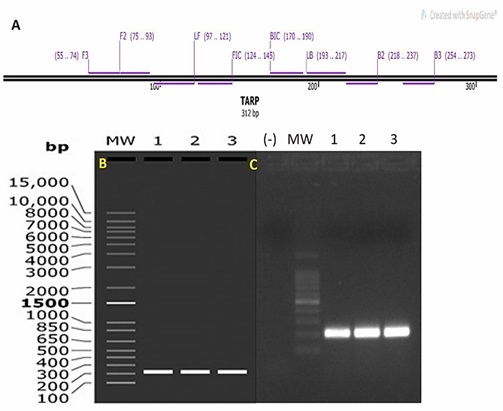
The in silico analysis and the PCR-Chla using primers Cb-F3 and Cb-B3 with the DNAs+ showed a band of approximately 219 bp in both electrophoresis methods (in silico and agarose gel, Figure 1A–1C), confirming the specific binding of the primers to the target gene.
Figure 1. A) In silico primer alignment. B) 1.5% agarose gel simulation. C) 1.5% agarose gel electrophoresis. (-) = Negative control, MW = Molecular weight, 1, 2 and 3 positive bands (~219 bp).
Optimization of colorimetric LAMP assay conditions (crLAMP-Chla)
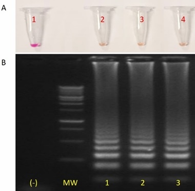
The colorimetric crLAMP-Chla assay conditions were optimized with 1 ng of DNAs+ from the TARP gene, showing a color change from pink (negative) to yellow (positive) at a temperature of 65 °C. The time required for the amplification of 1 ng of C. abortus DNAs+ was 15 minutes, although the sample was removed after 40 minutes as indicated previously in the protocol. Subsequently, ladder-shaped bands (characteristic of LAMP) were observed by electrophoresis in the same samples that changed to yellow, matching the colorimetric results. For confirmation purposes, the comparison was performed in triplicate (Figure 2).
Figure 2. Colorimetry and agarose gel electrophoresis with positive results with DNAs+ for the detection of C. abortus. A) In crLAMP-Chla, tube 1: negative (pink and no band pattern); tubes 2, 3, and 4: positives (yellow and band pattern). B) crLAMP-Chla in agarose gel electrophoresis. Bands in track (-) = Negative control; MW = Molecular weight; 1, 2, and 3: positive bands.
Analytical sensitivity of crLAMP-Chla assay
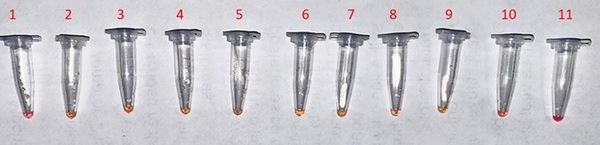
The sensitivity of the crLAMP-Chla assay was analyzed by colorimetry after incubation with different concentrations of DNAs+ or different copy numbers in a nuclease-free solution. In the crLAMP-Chla colorimetric reactions, the color change from pink to yellow was visible directly in the first tube at a concentration of 1 ng in 15 minutes, and the colorimetric change was completed at 0.01 fg in 45 minutes, corresponding to approximately 10 copies (Figure 3).
Comparison of crLAMP-Chla with other molecular tests using biological samples
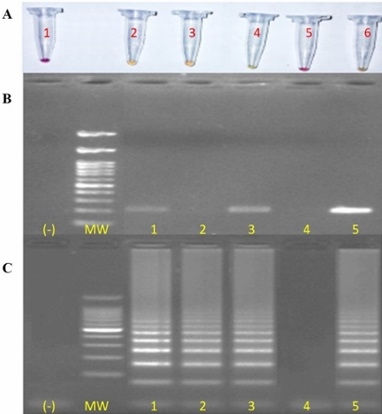
From the four negative and twenty-six positive samples analyzed by qPCR, the crLAMP-Chla and Chla-LAMP detected 100% of them, respectively. Conventional PCR only detected 60% and 40% of the true positive and negative biological controls of qPCR. A visual comparison of the crLAMP Chla, Chla-LAMP, and PCR is shown in Figure 4.
Concordance, sensitivity, and specificity analysis
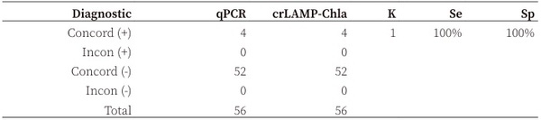
The concordance between qPCR and crLAMP Chla was K = 1, and Se and Sp were 100% and 100%, respectively (P = 0.05).
Discussion
The BLAST alignment of the sequence amplified by the external primers (F3-B3) showed 100% homology to the C. abortus genomic
Figure 3. Sensitivity analysis of crLAMP-Chla. Tubes 1: negative; 2: 1 ng (+); 3: 0.1 ng (+); 4: 0.01 ng (+); 5: 1 pg (+); 6: 0.1 pg (+); 7: 0.01 pg (+); 8: 1 fg (+); 9: 0.1 fg (+); 10: 0.01 fg (+); 11: 1 ag negative result.
Figure 4. Comparison of crLAMP-Chla with various molecular tests. A) Colorimetry of crLAMP Chla with biological samples. Tubes 1: Negative control; 2: Positive; 3: Positive; 4: Positive; 5: Negative; 6: DNAs+. B) Agarose gel electrophoresis of PCR-Chla with primers F3 and B3 in biological samples. Bands in track (-) = Negative control; MW = Molecular weight; 1, 3: positive samples; 2, 4: negative samples; 5: DNAs+. C) Agarose gel electrophoresis of Chla-LAMP with biological samples. Bands in track (-) = Negative control; MW = Molecular weight; 1, 2, 3: positive samples; 4: negative sample; 5: DNAs+
sequence and allowed the synthesis of DNAs+, which reduces the standardization time for other diagnostic tests. Furthermore, it avoids direct contact with infectious pathogens for personnel. Additionally, it has been reported to provide high sensitivity in molecular diagnostics [15,10]. It has also been demonstrated that synthetic fragments can be used as positive controls for
Table 2. Concordance (K), sensitivity (Se), and specificity (Sp) of crLAMP in relation to qPCR as the reference test.
Con = Concordance; Incon = Inconcordance; K = concordance; Se = test sensitivity; Sp = test specificity. Confidence level at 95%. Interpretation of K value: < 0.00 = no agreement; > 0.00–0.20 = negligible; 0.21–0.40 = discrete; > 0.41–0.60 = moderate; 0.61–0.80 = substantial; 0.81–1.00 = almost perfect.
the standardization of tests designed to identify microorganisms that are difficult to culture and require long processing time. Moreover, the synthesized sequences used as positive controls ensure the standardization of bioassays when the biological isolate is not available.
Comparing the sensitivity of the crLAMP-Chla assay with other LAMPs for C. abortus, important differences were observed in detection capacity, as well as in the applicability of the methods and detection time. For instance, [16] reported that a LAMP assay targeting the MOMP gene (a different gene from this study) had a sensitivity equivalent to nested PCR and superior to isolation in chicken embryos. Nonetheless, the exact amount of detectable DNA was not specified, as was determined for the crLAMP Chla assay in this study (10 DNA copies). [10] described an isothermal test for the detection of C. abortus and also a LAMP technique, reporting an analytical sensitivity of 30,893 DNA copies. This indicates that previous tests have adequate analytical sensitivity but may not reach the same level of accuracy as the crLAMP-Chla assay. Another important difference in the crLAMP Chla assay is the use of colorimetry to interpret the results with the naked eye, a suitable feature for application under field conditions without sophisticated infrastructure or additional equipment.
Before 2016, C. abortus was considered an exotic disease in Mexico by the country's health authority. Due to several reports from research centers and universities, it was possible to change the sanitary status to an endemic disease [1]. In this sense, a diagnostic tool for the accurate identification of clinical cases is required. Considering the positive and negative controls from the reference test (qPCR), the concordance between qPCR and crLAMP-Chla, according to [14], was almost perfect (K=1). Furthermore, the Se and Sp values for crLAMP-Chla were 100% and 100%, respectively, demonstrating a high capability to identify genetic material of C. abortus. The LAMP technique is therefore an emerging and alternative tool for the molecular detection of pathogens with relevance in veterinary and public health. It is fast and portable and has high specificity and sensitivity, comparable to qPCR and higher than conventional PCR. The incorporation of colorimetry into the LAMP technique has increased its popularity in veterinary diagnostics, since it allows a simple visual interpretation based on a color change, eliminating the need for sophisticated equipment. Studies performed with different microorganisms (such as Bovine alphaherpesvirus or Mycoplasma bovis) using colorimetry-based LAMP assays have demonstrated the effectiveness of this technique with a variety of pathogens that affect various animal species [17,18].
Conclusion
The crLAMP-Chla assay demonstrated the same capability as qPCR to identify the presence or absence of C. abortus genetic material by colorimetry with the naked eye. This positions the crLAMP-Chla assay as an effective and practical diagnostic tool for the rapid and accurate detection of C. abortus in resource-limited settings.
This article was originally published in Revista Colombiana de Ciencias Pecuarias; 39(Jan-Dec):e358631. https://doi.org/10.17533/udea.rccp.e358631. This is an Open Access article distributed under the terms and conditions of the Creative Commons Attribution (CC BY) license.