Vitrification of Equine Embryos
Vitrification of Equine Embryos: Effect of polyvinyl alcohol (PVA) in vitrification of equine embryos
Published: January 3, 2008
By: Jason Hudson, Brandi Hudson, James Bailey, Shelly Williams, Chris Seagle, Travis Meredith (Courtesy of Dr Jorge Murga - AMMVE)
Techniques for cryopreserving equine embryos have advanced in recent history allowing many practitioners to reliably collect and vitrify embryos on-site or ship embryos to referral centers where equine embryos are routinely vitrified. In recent studies, it has been demonstrated that equine embryos can be vitrified, then warmed and transferred achieving acceptable pregnancy rates [1,4].
There are endless possibilities when considering the management of vitrified embryos. Many owners elect to simply cryopreserve valuable genetics from a particular stallion and mare combination. Others sell and purchase cryopreserved embryos and have them transferred at a specified time so a resulting foal can be born during a chosen month of the year.
Many owners have aspirations to sell embryos in the international market. The technology to import or export equine embryos has been desired since the commercialization of the equine embryo transfer process. If importing and exporting cryopreserved equine embryos is a consideration, it may be to an individual’s advantage to use a media that is not of animal origin.
Embryos cryopreserved in synthetic media would help clear any confusion concerning questions about importing and exporting regulations. Therefore, a demand exists for an alternative media which would help meet all countries importing and exporting requirements. The goal of this pilot study was to compare pregnancy rates between two groups of vitrified equine embryos. The two groups were embryos vitrified in fetal calf serum and those vitrified in polyvinyl alcohol.
Light horse mares were chosen based on the quality of their reproductive tract. Mares were bred to a stallion with proven fertility. Ovulation was induced in thirteen (13) mares by administration of 2500 IU of human chorionic gonadotropin (hCG) or a GnRH analog once a preovulatory follicle > 35 mm was detected with good uterine edema and a relaxed cervix.
A non-surgical transcervical embryo flush procedure, using EmCare® flush media, was performed on day 6.5 post-ovulation. Using the International Embryo Transfer Society (IETS) embryo grading method [2], embryos recovered were evaluated on size, grade, and morphology. Late morula or early blastocyst stage embryos with a grade of 1-2 and < 300 µm in diameter were randomly assigned to one of two treatment groups. Embryos in Group 1 (n=7) were washed four times in EmCare® holding medium and then vitrified in Partnar media containing fetal calf serum (FCS) [1,4]. Embryos in Group 2 (n=6) were also washed in EmCare® holding medium and then vitrified using the Partnar Vitrfication Kit containing polyvinyl alcohol (PVA).
Embryos were warmed after randomly selecting them from the liquid nitrogen tank. The straw containing the embryo and cryoprotectants were held in room temperature air for 10 seconds followed by submerging them in a 20ºC water bath for an additional 10 seconds [1,4]. The straw was held in a horizontal position for 6 to 8 minutes allowing the fluid columns to equilibrate before transferring the contents of the straw directly into an available recipient that ovulated 48 hours after the donor mare. A non-surgical transcervical transfer technique was utilized using a Cassou gun [1,3,4].
A 71.4 % (5 of 7) pregnancy rate was established in the group containing fetal calf serum while an 83.3% (5 of 6) pregnancy rate was established in the group containing polyvinyl alcohol. Pregnancies were visualized per rectum using ultrasonography beginning 5 days post transfer. Pregnancies were evaluated until a heart beat was detected. Results of this pilot study may indicate that late morula and early blastocyst equine embryos (< 300 µm) can be collected and vitrified in media containing polyvinyl alcohol without losing embryo viability. This study suggests the polyvinyl alcohol solution may be a comparable option for vitrification with the potential benefit of qualifying all cyropreserved equine embryos for importation and/or exportation. Future projects using larger numbers of embryos are needed to validate this present pilot study.
Mare Synchronization
To be successfully vitrified, an equine embryo must developmentally be a late morula or early blastocyst. Collecting embryos 8 days after the administration of hCG to the donor mare has proven ideal [1,4]. This time frame should be comparable to flushing the donor mare 6.5 days after the detection of ovulation, if hCG is administered [1,4].
Example:
- A mare is administered hCG at 1 pm Friday the 1st of May after ultrasonography reveals a 38 mm diameter preovulatory follicle with good associated uterine edema and a relaxed cervix.
- The mare is artificially inseminated with good quality fresh or cooled semen 24 hours post hCG administration on Saturday the 2nd of May after ultrasounding to assure no ovulation has occurred prematurely.
- The mare is ultrasounded on Sunday the 3rd of May and ovulation is detected. Human chorionic gonadotropin should induce a mare to ovulate 36 to 42 hours post-injection or 1.5 days after administration [5].
- The mare is flushed on Saturday the 9th of May at 1 pm to retrieve a late morula or early blastocyst.
If a GnRH analog is utilized to induce an ovulation in the mare, the time frame should be adjusted appropriately to assure a late morula or early blastocyst is retrieved after flushing the donor mare. Remember, in most cases a GnRH analog should induce an ovulation between 42 to 48 hours post administration [5]. One should be cautious when using a GnRH analog that has not been adequately researched.
Vitrification Procedures
Identifying and utilizing the proper size and grade of embryo is imperative to the success of vitrification. A late morula equine embryo should measure approximately 160 µm. In contrast, an early blastocyst can measure approximately 190 to 300 µm [1,4]. Recovered embryos can easily be searched for and discovered using a dissecting microscope. A linear eye piece micrometer is used to measure the diameter of each embryo. Morphological grading should be consistent with the size of each embryo recovered.
Required Equipment (Figure 1):
- Dissection Microscope with micrometer
- 200 µl Micropipettor
- Sterile Embryo Search Dish
- NON-gamma irradiated 0.25 ml PVA embryo straws
- EmCare Holding Media
- Partnar Vitrification Kit
- Heat Sealer
- Insulated Container
- Liquid Nitrogen
- Digital Timer
- 10 mm goblet and canes
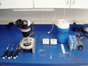
FIGURE 1
Organized drops of media should be placed in a Petri dish at room temperature (Figure 2). This organization of drops allows for rapid media changes in increasing concentrations of cryoprotectants (ethylene glycol and glycerol) and in loading straws [1].
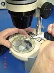
FIGURE 2
Procedure [1,4]:

FIGURE 2
Procedure [1,4]:
- Embryo is removed from EmCare® holding media (Figure 3) and transferred into the first vitrification solution (S1)
a) Embryo should be transferred with minimal residual amount of holding media into S1.
b) Hold embryo in S1 (200 µl) for precisely 5 minutes. - Embryo is removed from S1 and placed into the second vitrification solution (S2) using minimal amount of S1 solution
a) Hold embryo in S2 (200 µl) for precisely 5 minutes. - Embryo is removed from S2 and placed into the third or final vitrification solution using minimal amount of S2 solution.
a) Hold embryo in S3 (30 µl) for no longer than 45 to 60 seconds and this time includes loading the straw and heat sealing it before exposing it to liquid nitrogen vapor. - Loading the 0.25 ml non-irradiated, PVA straw includes (Figure 4):
a) Drawing up 90 µl of dilution solution.
b) Followed by an air bubble (5 to 10 µl).
c) Followed by the embryo in 30 µl of S3.
d) Followed by another air bubble (5 to 10 µl).
e) Finally followed by a second 90 µl of dilution solution - Heat seal the open end of the straw with one to two seals. This takes about 5 seconds (Figure 5).
- Placing the heat sealed straw into a pre-cooled plastic goblet held vertically in liquid nitrogen within an insulated container is the second to last step (Figure 6).
a) The straw is held vertically inside the goblet where only vapor surrounds the straw.
b) The straw is held for 1 minute in vapor before being plunged into the liquid nitrogen (Figure 7).
c) The vitrified embryo can then be transferred into a holding tank.
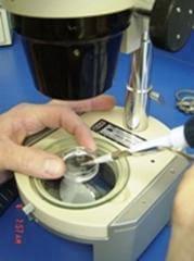
FIGURE 3
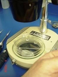
FIGURE 4
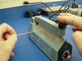
FIGURE 5
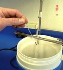
FIGURE 6
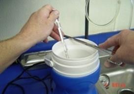
FIGURE 7
Warming Procedure
Warming the embryo is just as critical as vitrifying it. Precise warming time and identification of the embryo within the straw is essential.
Procedure [1,4]:
- Straws are removed from the liquid nitrogen tank.
a) Held in room temperature air for 10 seconds.
b) Place straw into a 20 to 22ºC water bath for 10 seconds.
c) Mix contents of the straw by flicking the straw like a thermometer 5 times. - Straw is held horizontally under the dissecting scope. Note: be sure not to expose it too long over the light source.
a) The embryo is identified and subjectively graded and measured.
b) Lay embryo on a horizontal surface for 6 minutes at room temperature. - After 6 minutes, the heat-sealed end of the straw is cut with scissors.
a) The straw is loaded into a Cassou gun and transferred non-surgically transcervically into an available recipient mare.
All steps should be completed within 8 minutes
References
1. Eldridge-Panuska WD, Caracciolo de Brienza V, Seidel GE Jr, Squires EL, Carnevale EM. Establishment of pregnancies after serial dilutions or direct transfer by vitrified equine embryos. Theriogenology 2005;63:1308-1319.
2. McKinnon AO, Squires EL. Morphological assessment of the equine embryo. J Am Vet Med Assoc 1988;192:401-406.
3. Squires EL, Carnevale EM, McCue PM and Bruemmer JE. Embryo technologies in the horse. Theriogenology 2003;59:151-170.
4. Hudson JJ, Welch SA, Squires EL, Carnevale EM, McCue PM. Vitrification of cooled equine embryos. In: Proceedings of the Annual Conference of the Society for Theriogenology, 2005, p. 783.
5. McCue PM, Farquhar VJ, Carnevale EM, Squires EL. Removal of deslorelin (Ovuplant) implant 48 h after administration results in normal interovulatory intervals in mares. Theriogenology 2002 Sep;58(5):865- 70.
Authors: Jason Hudson1, DVM, MS; Brandi Hudson1, DVM; James Bailey1, DVM; Shelly Williams1; Chris Seagle2, PhD; Travis Meredith2, DVM, MS, DACT
1 Royal Vista Southwest, Purcell, OK, USA
2 Partnar Animal Health, Port Huron, MI, USA
Related topics:
Recommend
Comment
Share

Would you like to discuss another topic? Create a new post to engage with experts in the community.


