Impact of Bio-Mos® on microbial ecology of the pig digestive tract
The performance improvement is greatest during the early days after weaning, and is greater in situations where pigs grow more slowly, but we did not identify other factors consistently related to the Bio-Mos® response. The greater response in slower-growing pigs raises the possibility that the action of Bio-Mos® may be related to improved health.
Bio-Mos® originally resulted from a search for a dietary source of mannose (K. Newman, personal communication). The logic is as follows: A key step in enteric pathogenesis is attachment of the pathogen to the intestinal wall to allow proliferation of the pathogen.
Many pathogens, including several E. coli and salmonella, attach specifically through Type I fimbriae to mannose moieties on the intestinal epithelial cell. If another source of mannose is introduced into the digestive tract, the pathogens may be tricked into binding to that mannose in the lumen of the tract rather than to the mannose binding sites on the epithelial cells, and thus fail to proliferate (Newman, 1994).
Part of this proposed chain of events is known to occur, as there is conclusive evidence that Bio-Mos® agglutinates several strains of E. coli and salmonella (Spring et al., 2000). It is not so clear that this activity is responsible for the benefits of Bio-Mos® in vivo.
Several experiments (Stanley et al., 1996; Finuance et al., 1999; Strickling et al., 2000; Sims et al., 2004; Swanson et al., 2002; Grieshop et al., 2004; Castillo et al., 2005) have measured the effect of Bio-Mos® in the diet on populations of selected bacteria in feces or digesta of pigs and other animal species, but the results taken collectively neither verify nor refute the proposed mode of action. Full understanding of the mode of action of Bio-Mos® awaits further research.
Methods of assessing microbial ecology
Traditionally, measurement of bacterial populations has relied on cultivating them on selective growth media. This method has provided an enormous amount of useful information, but it is now clear that it completely fails to find many bacterial species that inhabit the digestive tract (Tannock, 2001). That realization has led to different types of methods, ones that measure bacterial DNA rather than growth.
A popular one of the molecular (DNA-based) methods is called 16S rDNA PCRDGGE, which focuses on ribosomal DNA (rDNA) found in the 16S ribosome common to bacteria. This particular piece of DNA is chosen because it contains both highly conserved regions (those that are similar in all species) and highly variable regions.
Attention is focused more particularly on one of the variable regions called the V3 region. The importance of this region of this particular strand of ribosomal DNA derives from knowledge of the DNA sequences of this region for many species of bacteria; if the sequence of the V3 region is known, the species can presumptively be identified.
Polymerase chain reaction (PCR) is used to amplify (make many copies of) the V3 region. Focus on this region is provided by selection of both forward and reverse primers specific for the flanking conserved regions. The PCR step produces enough material to separate and visualize using denaturing gradient gel electrophoresis (DGGE).
Electrophoresis is used to pull the DNA (produced by PCR) through a polyacrylamide gel containing an increasing gradient of a denaturant. The susceptibility of each strand of DNA to denaturation is a function of its base composition (not sequence); as each DNA reaches the concentration of denaturant that causes its denaturation, it precipitates in the gel and is visible as a dark band after appropriate treatment of the gel. Each band in the gel may reflect a single species of bacteria, or it may come from multiple species that all happen to denature at the same place.
The laboratory work of 16S rDNA PCR-DGGE can be summarized in three steps, with the products verified visually with the use of gels after each step. First, the DNA is isolated from the sample. Second, PCR is used to amplify the V3 region. Third, the DNA is separated by DGGE.
Special computer programs read the gels, identifying the bands and standardizing their positions from gel to gel. That allows counting of the number of bands in each sample. Note that this is not the total number of bacterial species, but the number of species present in high enough populations to be detected by this method.
However, the number of bands is accepted to be a measure of the diversity of the microbial ecosystem sampled.
Further analyses compute a Sorenson’s Similarity Coefficient (Cs) for each pair of samples. The range of Cs values is from 0 to 100, where two samples with no shared bands would have a Cs value of 0 and two samples with identical bands would have a value of 100. The Cs values for pairs of samples within an experimental treatment show the degree of similarity of animals within treatments. The Cs values for pairs of samples across treatments are usually lower than the intra-treatment values and reflect the degree of difference between the two treatments.
One important result of an experiment using DGGE is identification of bands that are present in animals from some experimental treatments and absent from other treatments. It is possible to retrieve the DNA from those bands, clone and sequence it, and thereby get a presumptive identification of the bacterial species.
An experiment assessing Bio-Mos® response
An experiment was conducted to assess the impact of Bio-Mos® on the microbial ecology of the digestive tract of young pigs using DGGE, and to compare the effect of Bio-Mos® to that of the antibiotic carbadox.
A total of 24 pigs at weaning at 21 days of age were separated into four experimental treatment groups in a 2 x 2 factorial arrangement of Bio-Mos® and carbadox (negative control, Bio-Mos®, carbadox, Bio-Mos® + carbadox). They were fed their appropriate treatment diets until half were sacrificed after seven days and the remainder after an additional 14 days (21 days after weaning, 42 days of age).
At sacrifice, both luminal digesta samples and mucosal scrapings were collected from six sites in the digestive tract: pars esophagea, fundus, jejunum, ileum, proximal colon, distal colon. These samples were subjected to 16S rDNA PCR-DGGE as described above.
RESULTS
Bio-Mos® reduced (P<0.05) the diversity of the bacterial populations (reduced the number of bands) at 21 days post-weaning in 4 of 12 sites (pars esophageal lumen, ileal mucosa, proximal colon mucosa, distal colon lumen), but this effect did not occur by seven days post-weaning. In contrast, carbadox increased the number of bands in some sites and at both ages.
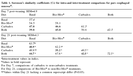
The Cs values suggested that carbadox altered microbial populations at seven days after weaning. Consider the case of the pars esophageal mucosa (Table 1). The four inter-treatment Cs values that test a carbadox-containing treatment against a treatment not containing carbadox are relatively low, ranging from 39.8 to 47.8. Low values indicate differences between the treatments. Contrast these numbers with the higher inter-treatment values that test the two carbadox-containing treatments (50.6) or the two non-carbadox-containing treatments (54.5).
In this site the treatments are not significantly different, but in other cases they are. Of the 12 sites, 10 have on average numerically lower inter-treatment Cs values where the two treatments differ in carbadox than where they differ in Bio-Mos®, suggesting that carbadox exerts a subtle but pervasive influence on microbial ecology at seven days after weaning.

However, the situation was different at 21 days after weaning. In that case, Bio-Mos® seemed to alter microbial populations, whereas carbadox did not, based on inter-treatment Cs values. The pattern can again be seen in the data on the pars esophageal mucosa (Table 1). The four inter-treatment Cs values that test a Bio-Mos®-containing treatment against a treatment not containing Bio-Mos® are relatively low, ranging from 35.2 to 44.7, indicating treatment differences.
In contrast, the inter-treatment Cs values were higher (49.9 and 67.8) in comparisons within Bio-Mos® treatments. Of the 12 sites, nine have on average numerically lower inter-treatment Cs values where the two treatments differ in Bio-Mos® than where they differ in carbadox, suggesting that Bio- Mos® subtly influences microbial ecology at 21 days after weaning.
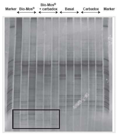
Examination of the gels revealed several bands that were consistently present in the two Bio-Mos® treatments and consistently absent in the two treatments without Bio- Mos®, or the converse. An example of such a gel is shown in Figure 1, where certain bands near the bottom of the gel are present in the two Bio-Mos® treatments and not in the others. At the time of this writing, we have only tentative identification of several of these critical bands, but we will continue our efforts to identify them.

Figure 1.Fundic mucosa 42 days of age. Bands in the black-framed box are present only in pigs fed diets containing Bio-Mos® .
WHAT IT MEANS
These results confirm that Bio-Mos® changes microbial populations in the digestive tract. We make this assertion on the basis of three types of observations:
- The reduction in diversity (number of bands)
- The pattern of inter-treatment Cs values
- The consistent appearance or disappearance of specific bands when Bio-Mos® was added to the diet.
We cannot yet determine whether our data are consistent with the hypothesis that Bio- Mos® acts by binding bacteria bearing Type I fimbriae. That test awaits confirmation of the species that appear or disappear with Bio-Mos® use.
We cannot explain the apparent contradiction in time between the Bio-Mos® effects on growth performance, which occur soon after weaning, and the effects on microbial populations, which occur later.
Certain beneficial effects of Bio-Mos® are well established. However, full exploitation of this product requires further understanding of its mode of action; this experiment has attempted to contribute to that understanding.
Summary
Bio-Mos® improves growth performance of young pigs. There is a widely shared theory of the mechanism through which it exerts its beneficial effects on microbial populations of the digestive tract and thus on performance, but that theory requires further empirical verification.
In this paper we report the results of an experiment that contributes to that verification. We have shown conclusively that Bio-Mos® alters microbial populations in the digestive tract. Further definition of the specific changes is forthcoming.
References
Castillo, M., C. Rodriguez, S.M. Martin-Pelaez, J. Roquet, J.A. Taylor-Pickard, J.F. Perez and S.M. Marin-Orue. 2005. Effect of mannan oligosaccharides and (or) organic zinc on intestinal microbiota and immune response of early-weaned pigs. J. Anim. Sci. 88(Suppl. 1):83 (Abstr.).
Finuance, M.C., K.A. Dawson, P. Spring and K.E. Newman. 1999. The effect of mannan oligosaccharide on the composition of the microflora in turkey poults. Poult. Sci. 78(Suppl. 1):342 (Abstr.).
Grieshop, C.M., E.A. Flickinger, K.J. Bruce, A.R. Patil, G.L. Czarnecki-Mauldin and G.C. Fahey, Jr. 2004. Gastrointestinal and immunological responses of senior dogs to chicory and mannan oligosaccharides. Arch. Anim. Nutr. 58:483-493.
Miguel, J.C., S.L. Rodriguez-Zas and J.E. Pettigrew. 2004. Efficacy of Bio-Mos® in the nursery pig diet: A meta-analysis of the performance response. J. Swine Health Prod. 12:296-307.
Newman, K. 1994. Mannan oligosaccharides: Natural polymers with significant impact on the gastrointestinal microflora and the immune system. In: Biotechnology in the Feed Industry: Proceedings of Alltech’s 10th Annual Symposium (T.P. Lyons and K.A. Jacques, ed). Nottingham University Press, UK, pp. 167-174.
Sims, M.D., K.A. Dawson, K.E. Newman, P. Spring and D. Hooge. 2004. Effects of dietary mannan oligosaccharide, bacitracin methylene disalicylate, or both on the live performance and intestinal microbiology of turkeys. Poult. Sci. 83:1148-1154.
Spring, P., C. Wenk, K.A. Dawson and K.E. Newman. 2000. The effects of dietary mannan oligosaccharides on cecal parameters and the concentrations of enteric bacteria in the ceca of Salmonella-challenged broiler chicks. Poult. Sci. 79:205-211.
Stanley, V.G., H. Chukwu, C. Gray and D. Thomson. 1996. Effects of lactose and Bio- Mos® in dietary application on growth and total coliform bacteria reduction in broiler chicks. Poult. Sci. 75(Suppl. 1):61 (Abstr.).
Strickling, J.A., D.L. Harmon, K.A. Dawson and K.L. Cross. 2000. Evaluation of oligosaccharide addition to dog diets: influences on nutrient digestion and microbial populations. Anim. Feed Sci. Technol. 86:205-219.
Swanson, K.S., C.M. Grieshop, E.A. Flickinger, L.L. Bauer, H.P. Healy, K.A. Dawson, N.R. Merchen and G.C. Fahey, Jr. 2002. Supplemental fructooligosaccharides and mannan oligosaccharides influence immune function, ileal and total tract nutrient digestibilities, microbial populations and concentrations of protein catabolites in the large bowel of dogs. J. Nutr. 132:980-989.
Tannock, G.W. 2001. Normal Microflora. An Introduction to Microbes Inhabiting the Human Body. Chapman and Hall, London.


