Redox Homeostasis in Poultry: Regulatory Roles of NF-kB
Author details:
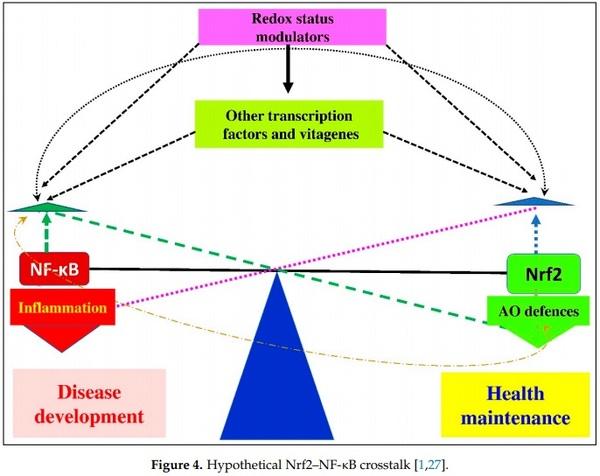
Redox biology is a very quickly developing area of modern biological sciences, and roles of redox homeostasis in health and disease have recently received tremendous attention. There are a range of redox pairs in the cells/tissues responsible for redox homeostasis maintenance/regulation. In general, all redox elements are interconnected and regulated by various means, including antioxidant and vitagene networks. The redox status is responsible for maintenance of cell signaling and cell stress adaptation. Physiological roles of redox homeostasis maintenance in avian species, including poultry, have received limited attention and are poorly characterized. However, for the last 5 years, this topic attracted much attention, and a range of publications covered some related aspects. In fact, transcription factor Nrf2 was shown to be a master regulator of antioxidant defenses via activation of various vitagenes and other protective molecules to maintain redox homeostasis in cells/tissues. It was shown that Nrf2 is closely related to another transcription factor, namely, NF-κB, responsible for control of inflammation; however, its roles in poultry have not yet been characterized. Therefore, the aim of this review is to describe a current view on NF-κB functioning in poultry with a specific emphasis to its nutritional modulation under various stress conditions. In particular, on the one hand, it has been shown that, in many stress conditions in poultry, NF-κB activation can lead to increased synthesis of proinflammatory cytokines leading to systemic inflammation. On the other hand, there are a range of nutrients/supplements that can downregulate NF-κB and decrease the negative consequences of stress-related disturbances in redox homeostasis. In general, vitagene–NF-κB interactions in relation to redox balance homeostasis, immunity, and gut health in poultry production await further research.
Keywords: antioxidants; NF-κB; oxidative stress; poultry; redox balance.
Clostridium perfringens, and Chlamydia psittaci, were shown to induce proinflammatory responses in birds associated with increased NF-κB expression and activity.
1. Surai, P.F. Vitagenes in Avian Biology and Poultry Health; Wageningen Academic Publishers: Wageningen, The Netherlands, 2020.
2. Musaogullari, A.; Chai, Y.C. Redox Regulation by Protein S-Glutathionylation: From Molecular Mechanisms to Implications in
Health and Disease. Int. J. Mol. Sci. 2020, 21, 8113. [CrossRef] [PubMed]
3. Sun, L.; Wang, X.; Saredy, J.; Yuan, Z.; Yang, X.; Wang, H. Innate-adaptive immunity interplay and redox regulation in immune response. Redox. Biol. 2020, 37, 101759. [CrossRef] [PubMed]
4. Francioso, A.; Baseggio Conrado, A.; Mosca, L.; Fontana, M. Chemistry and Biochemistry of Sulfur Natural Compounds: Key
Intermediates of Metabolism and Redox Biology. Oxid. Med. Cell Longev. 2020, 2020, 8294158. [CrossRef] [PubMed]
5. Corkey, B.E.; Deeney, J.T. The Redox Communication Network as a Regulator of Metabolism. Front. Physiol. 2020, 11, 567796.
[CrossRef]
6. Saha, S.; Buttari, B.; Panieri, E.; Profumo, E.; Saso, L. An Overview of Nrf2 Signaling Pathway and Its Role in Inflammation.
Molecules 2020, 25, 5474. [CrossRef]
7. Sies, H.; Jones, D.P. Reactive oxygen species (ROS) as pleiotropic physiological signalling agents. Nat. Rev. Mol. Cell. Biol. 2020,
21, 363–383. [CrossRef]
8. Carvalho, R.H.; Ida, E.I.; Madruga, M.S.; Martínez, S.L.; Shimokomaki, M.; Estévez, M. Underlying connections between the redox system imbalance, protein oxidation and impaired quality traits in pale, soft and exudative (PSE) poultry meat. Food Chem.
2017, 215, 129–137. [CrossRef]
9. Pan, X.; Zhang, L.; Xing, T.; Li, J.; Gao, F. The impaired redox status and activated Nrf2/ARE pathway in wooden breast myopathy in broiler chickens. Asian-Australas. J. Anim. Sci. 2020, in press. [CrossRef]
10. Estevez, M.; Petracci, M. Benefits of Magnesium Supplementation to Broiler Subjected to Dietary and Heat Stress: Improved
Redox Status, Breast Quality and Decreased Myopathy Incidence. Antioxidants 2019, 8, 456. [CrossRef]
11. Dalgaard, T.S.; Briens, M.; Engberg, R.M.; Lauridsen, C. The influence of selenium and selenoproteins on immune responses of poultry and pigs. Anim. Feed Sci. Technol. 2018, 238, 73–83. [CrossRef]
12. Lauridsen, C. From oxidative stress to inflammation: Redox balance and immune system. Poult. Sci. 2019, 98, 4240–4246.
[CrossRef] [PubMed]
13. Surai, P.F. Selenium in Poultry Nutrition and Health; Wageningen Academic Publishers: Wageningen, The Netherlands, 2018.
14. Surai, P.F.; Fisinin, V.I. Antioxidant-Prooxidant Balance in the Intestine: Applications in Chick Placement and Pig Weaning. J. Vet.
Sci. Med. 2015, 3, 16.
15. Mishra, B.; Jha, R. Oxidative Stress in the Poultry Gut: Potential Challenges and Interventions. Front. Vet. Sci. 2019, 6, 60.
[CrossRef]
16. Dong, Y.; Lei, J.; Zhang, B. Effects of dietary quercetin on the antioxidative status and cecal microbiota in broiler chickens fed with oxidized oil. Poult. Sci. 2020, 99, 4892–4903. [CrossRef] [PubMed]
17. Kövesi, B.; Cserháti, M.; Erdélyi, M.; Zándoki, E.; Mézes, M.; Balogh, K. Long-Term Effects of Ochratoxin A on the Glutathione
Redox System and Its Regulation in Chicken. Antioxidants 2019, 8, 178. [CrossRef] [PubMed]
18. Egresi, A.; Süle, K.; Szentmihályi, K.; Blázovics, A.; Fehér, E.; Hagymási, K.; Fébel, H. Impact of milk thistle (Silybum marianum) on the mycotoxin caused redox-homeostasis imbalance of ducks liver. Toxicon 2020, 187, 181–187. [CrossRef]
19. Liu, Y.; Zhao, H.; Wang, Y.; Guo, M.; Mu, M.; Xing, M. Arsenic (III) and/or copper (II) induces oxidative stress in chicken brain and subsequent effects on mitochondrial homeostasis and autophagy. J. Inorg. Biochem. 2020, 211, 111201. [CrossRef]
20. Surai, P.F.; Kochish, I.I. Nutritional modulation of the antioxidant capacities in poultry: The case of selenium. Poult. Sci. 2019, 98,
4231–4239. [CrossRef]
21. Surai, P.F.; Kochish, I.I.; Romanov, M.N.; Griffin, D.K. Nutritional modulation of the antioxidant capacities in poultry: The case of vitamin E. Poult. Sci. 2019, 98, 4030–4041. [CrossRef]
22. Surai, P.F.; Kochish, I.I. Carotenoids in Aviculture. In Pigments from Microalgae Handbook; Springer Nature Switzerland: Cham,
Switzerland, 2020; pp. 515–540.
23. Tolba, S.A.; Magnuson, A.D.; Sun, T.; Lei, X.G. Dietary supplemental microalgal astaxanthin modulates molecular profiles of stress, inflammation, and lipid metabolism in broiler chickens and laying hens under high ambient temperatures. Poult. Sci. 2020,
99, 4853–4860. [CrossRef]
24. Surai, P.F.; Fisinin, V.I. Vitagenes in poultry production. Part 3. Vitagene concept development. Worlds Poult. Sci. J. 2016, 72,
793–804. [CrossRef]
25. Surai, P.F.; Fisinin, V.I. Antioxidant system regulation: From vitamins to vitagenes. In Handbook of Cholesterol; Watson, R.R., de Meester, F., Eds.; Wageningen Academic Publishers: Wageningen, The Netherlands, 2016; pp. 451–481.
26. Surai, P.F.; Kochish, I.I.; Fisinin, V.I. Antioxidant systems in poultry biology: Nutritional modulation of vitagenes. Eur. J. Poult.
Sci. 2017, 81, 1612–9199.
27. Surai, P.F.; Kochish, I.I.; Fisinin, V.I.; Kidd, M.T. Antioxidant Defence Systems and Oxidative Stress in Poultry Biology: An Update.
Antioxidants 2019, 8, 235. [CrossRef] [PubMed]
28. Surai, P.F.; Fisinin, V.I. Vitagenes in poultry production. Part 1. Technological and environmental stresses. Worlds Poult. Sci. J.
2016, 72, 721–733. [CrossRef]
29. Surai, P.F.; Fisinin, V.I. Vitagenes in poultry production. Part 2. Nutritional and internal stresses. Worlds Poult. Sci. J. 2016, 72,
761–772. [CrossRef]
30. Surai, P.F.; Kochish, I.I.; Fisinin, V.I.; Grozina, A.A.; Shatskikh, E.V. Molecular Mechanisms of Chicken Gut Health Maintenance: Role of
Microbiota; Agricultural Technologies: Moscow, Russia, 2018.
31. Schijns, V.E.; van de Zande, S.; Lupiani, B.; Reddy, S.M. Practical aspects of poultry vaccination. In Avian immunology; Academic
Press: Cambridge, MA, USA, 2014; pp. 345–362.
32. Cervantes, H.M. Antibiotic-free poultry production: Is it sustainable? J. Appl. Poult. Res. 2015, 24, 91–97. [CrossRef]
33. Kim, W.H.; Lillehoj, H.S. Immunity, immunomodulation, and antibiotic alternatives to maximize the genetic potential of poultry for growth and disease response. Anim. Feed Sci. Technol. 2019, 250, 41–50. [CrossRef]
34. Desin, T.S.; Köster, W.; Potter, A.A. Salmonella vaccines in poultry: Past, present and future. Expert Rev. Vaccines 2013, 12, 87–96.
[CrossRef]
35. Guillén, S.; Marcén, M.; Álvarez, I.; Mañas, P.; Cebrián, G. Stress resistance of emerging poultry-associated Salmonella serovars.
Int. J. Food Microb. 2020, 335, 108884. [CrossRef]
36. Gast, R.K.; Porter Jr, R.E. Salmonella infections. In Diseases of Poultry; David, E.S., Boulianne, M., Logue, C.M., McDougald, L.R.,
Nair, V., Suarez, D.L., de Wit, S., Grimes, T., Johnson, D., Kromm, M., et al., Eds.; John Wiley & Sons, Inc.: Hoboken, NJ, USA,
2020; pp. 717–753.
37. Iannetti, L.; Neri, D.; Santarelli, G.A.; Cotturone, G.; Vulpiani, M.P.; Salini, R.; Antoci, S.; Di Serafino, G.; Di Giannatale, E.;
Pomilio, F.; et al. Animal welfare and microbiological safety of poultry meat: Impact of different at-farm animal welfare levels on at-slaughterhouse Campylobacter and Salmonella contamination. Food Control 2020, 109, 106921. [CrossRef]
38. Everest, H.; Hill, S.C.; Daines, R.; Sealy, J.E.; James, J.; Hansen, R.; Iqbal, M. The evolution, spread and global threat of H6Nx avian influenza viruses. Viruses 2020, 12, 673. [CrossRef] [PubMed]
39. Wigley, P. Immunity to bacterial infection in the chicken. Dev. Comp. Immunol. 2013, 41, 413–417. [CrossRef] [PubMed]
40. Korver, D.R. Implications of changing immune function through nutrition in poultry. Anim. Feed Sci. Technol. 2012, 173, 54–64.
[CrossRef]
41. Kaspers, B.; Göbel, T.W. The avian immune system. In Encyclopaedia of Immunobiology; Ratcliffe, M.J.H., Ed.; Elsevier: Amsterdam,
The Netherlands, 2016; Volume 1, pp. 498–503.
42. Swaggerty, C.L.; Callaway, T.R.; Kogut, M.H.; Piva, A.; Grilli, E. Modulation of the immune response to improve health and reduce foodborne pathogens in poultry. Microorganisms 2019, 7, 65. [CrossRef] [PubMed]
43. Chen, C.; Li, J.; Zhang, W.; Shah, S.W.A.; Ishfaq, M. Mycoplasma gallisepticum triggers immune damage in the chicken thymus by activating the TLR-2/MyD88/NF-κB signaling pathway and NLRP3 inflammasome. Vet. Res. 2020, 51, 1–13. [CrossRef]
[PubMed]
44. Cardoso Dal Pont, G.; Farnell, M.; Farnell, Y.; Kogut, M.H. Dietary Factors as Triggers of Low-Grade Chronic Intestinal
Inflammation in Poultry. Microorganisms 2020, 8, 139. [CrossRef]
45. Sen, R.; Baltimore, D. Multiple nuclear factors interact with the immunoglobulin enhancer sequences. Cell 1986, 46, 705–716.
[CrossRef]
46. Yu, H.; Lin, L.; Zhang, Z.; Zhang, H.; Hu, H. Targeting NF-κB pathway for the therapy of diseases: Mechanism and clinical study.
Signal Transduct. Target Ther. 2020, 5, 209. [CrossRef]
47. Lambrou, G.I.; Hatziagapiou, K.; Vlahopoulos, S. Inflammation and tissue homeostasis: The NF-κB system in physiology and malignant progression. Mol. Biol. Rep. 2020, 47, 4047–4063. [CrossRef]
48. Chawla, M.; Roy, P.; Basak, S. Role of the NF-κB system in context-specific tuning of the inflammatory gene response. Cur. Opin.
Immunol. 2020, 68, 21–27. [CrossRef]
49. Kopitar-Jerala, N. Innate Immune Response in Brain, NF-Kappa B Signaling and Cystatins. Front. Mol. Neurosci. 2015, 8, 73.
[CrossRef]
50. Sehnert, B.; Burkhardt, H.; Dübel, S.; Voll, R.E. Cell-Type Targeted NF-kappaB Inhibition for the Treatment of Inflammatory
Diseases. Cells 2020, 9, 1627. [CrossRef] [PubMed]
51. Grilli, M.; Memo, M. Transcriptional pharmacology of neurodegenerative disorders: Novel venue towards neuroprotection against excitotoxicity? Mol. Psychiatry. 1997, 2, 192–194. [CrossRef] [PubMed]
52. Li, X.; Zhao, Y.; Tian, B.; Jamaluddin, M.; Mitra, A.; Yang, J.; Rowicka, M.; Brasier, A.R.; Kudlicki, A. Modulation of gene expression regulated by the transcription factor NF-κB/RelA. J. Biol. Chem. 2014, 289, 11927–11944. [CrossRef] [PubMed]
53. Niederberger, E.; Geisslinger, G. Proteomics and NF-κB: An update. Expert Rev. Proteom. 2013, 10, 189–204. [CrossRef] [PubMed]
54. Wu, J.; Ding, J.; Yang, J.; Guo, X.; Zheng, Y. MicroRNA Roles in the Nuclear Factor Kappa B Signaling Pathway in Cancer. Front.
Immunol. 2018, 9, 546. [CrossRef]
55. Thoma, A.; Lightfoot, A.P. NF-kB and Inflammatory Cytokine Signalling: Role in Skeletal Muscle Atrophy. Adv. Exp. Med. Biol.
2018, 1088, 267–279.
56. McGuire, C.; Prinz, M.; Beyaert, R.; van Loo, G. Nuclear factor kappa B (NF-κB) in multiple sclerosis pathology. Trends Mol. Med.
2013, 19, 604–613. [CrossRef]
57. Awasthee, N.; Rai, V.; Chava, S.; Nallasamy, P.; Kunnumakkara, A.B.; Bishayee, A.; Chauhan, S.C.; Challagundla, K.B.; Gupta, S.C.
Targeting IκappaB kinases for cancer therapy. Semin. Cancer Biol. 2019, 56, 12–24. [CrossRef]
58. Baker, R.G.; Hayden, M.S.; Ghosh, S. NF-κB, inflammation, and metabolic disease. Cell. Metab. 2011, 13, 11–22. [CrossRef]
59. Lingappan, K. NF-κB in oxidative stress. Curr. Opin. Toxicol. 2018, 7, 81–86. [CrossRef] [PubMed]
60. Zhang, L.; Yousefzadeh, M.J.; Suh, Y.; Niedernhofer, L.J.; Robbins, P.D. Signal Transduction, Ageing and Disease. Subcell. Biochem.
2019, 91, 227–247. [PubMed]
61. Patel, M.; Horgan, P.G.; McMillan, D.C.; Edwards, J. NF-κB pathways in the development and progression of colorectal cancer.
Transl. Res. 2018, 197, 43–56. [CrossRef] [PubMed]
62. Sivandzade, F.; Prasad, S.; Bhalerao, A.; Cucullo, L. NRF2 and NF-κB interplay in cerebrovascular and neurodegenerative disorders: Molecular mechanisms and possible therapeutic approaches. Redox. Biol. 2019, 21, 101059. [CrossRef]
63. Jones, S.V.; Kounatidis, I. Nuclear Factor-Kappa B and Alzheimer Disease, Unifying Genetic and Environmental Risk Factors from Cell to Humans. Front. Immunol. 2017, 8, 1805. [CrossRef]
64. Hayden, M.S.; Ghosh, S. Regulation of NF-κB by TNF family cytokines. Semin. Immunol. 2014, 26, 253–266. [CrossRef]
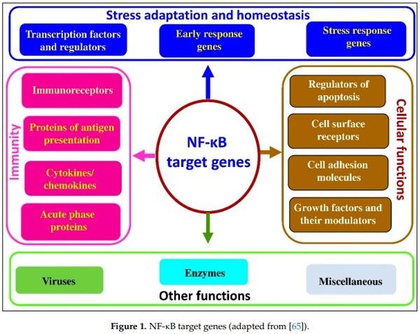
65. NF-kB Target Genes. Available online: www/bu.edu/nf-kb/gene-resources/target-genes/ (accessed on 1 December 2020).
66. Scott, O.; Roifman, C.M. NF-κB pathway and the Goldilocks principle: Lessons from human disorders of immunity and inflammation. J. Allergy Clin. Immunol. 2019, 143, 1688–1701. [CrossRef]
67. Zhang, L.; Xiao, X.; Arnold, P.R.; Li, X.C. Transcriptional and epigenetic regulation of immune tolerance: Roles of the NF-κB family members. Cell Mol. Immunol. 2019, 16, 315–323. [CrossRef]
68. Park, Y.H. The nuclear factor-kappa B pathway and response to treatment in breast cancer. Pharmacogenomics 2017, 18, 1697–1709.
[CrossRef]
69. Sun, S.C. The non-canonical NF-κB pathway in immunity and inflammation. Nat. Rev. Immunol. 2017, 17, 545–558. [CrossRef]
70. Giridharan, S.; Srinivasan, M. Mechanisms of NF-κB p65 and strategies for therapeutic manipulation. J. Inflamm. Res. 2018, 11,
407–419. [CrossRef] [PubMed]
71. Rabie, N.S.; Amin Girh, Z. Bacterial vaccines in poultry. Bull. Natl. Res. Centre 2020, 44, 15. [CrossRef] [PubMed]
72. Gimeno, I.M.; Schat, K.A. Virus-Induced Immunosuppression in Chickens. Avian Dis. 2018, 62, 272–285. [CrossRef] [PubMed]
73. Kim, W.H.; Chaudhari, A.A.; Lillehoj, H.S. Involvement of T Cell Immunity in Avian Coccidiosis. Front. Immunol. 2019, 10, 2732.
[CrossRef] [PubMed]
74. Buelna-Chontal, M.; Zazueta, C. Redox activation of Nrf2 & NF-κB: A double end sword? Cell Signal. 2013, 25, 2548–2557.
75. Pedruzzi, L.M.; Stockler-Pinto, M.B.; Leite, M., Jr.; Mafra, D. Nrf2-keap1 system versus NF-κB: The good and the evil in chronic kidney disease? Biochimie 2012, 94, 2461–2466. [CrossRef]
76. Tkach, K.E.; Oyler, J.E.; Altan-Bonnet, G. Cracking the NF-κB code. Sci. Signal. 2014, 7, pe5. [CrossRef]
77. Pal, S.; Bhattacharjee, A.; Ali, A.; Mandal, N.C.; Mandal, S.C. Chronic inflammation and cancer: Potential chemoprevention through nuclear factor kappa B and p53 mutual antagonism. J. Inflamm. 2014, 11, 23. [CrossRef]
78. Salles, A.; Romano, A.; Freudenthal, R. Synaptic NF-kappa B pathway in neuronal plasticity and memory. J. Physiol. Paris 2014,
108, 256–262. [CrossRef]
79. Tilborghs, S.; Corthouts, J.; Verhoeven, Y.; Arias, D.; Rolfo, C.; Trinh, X.B.; van Dam, P.A. The role of Nuclear Factor-kappa B signaling in human cervical cancer. Crit. Rev. Oncol. Hematol. 2017, 120, 141–150. [CrossRef]
80. Hoffmann, A.; Baltimore, D. Circuitry of nuclear factor κB signaling. Immunol. Rev. 2006, 210, 171–186. [CrossRef] [PubMed]
81. Morgan, M.J.; Liu, Z.G. Crosstalk of reactive oxygen species and NF-κB signaling. Cell Res. 2011, 21, 103–115. [CrossRef]
[PubMed]
82. de Jesús, T.J.; Ramakrishnan, P. NF-κB c-Rel Dictates the Inflammatory Threshold by Acting as a Transcriptional Repressor. iScience 2020, 23, 100876. [CrossRef] [PubMed]
83. Nelson, R.H.; Nelson, D.E. Signal Distortion: How Intracellular Pathogens Alter Host Cell Fate by Modulating NF-κB Dynamics.
Front. Immunol. 2018, 9, 2962. [CrossRef] [PubMed]
84. Fusella, F.; Seclì, L.; Cannata, C.; Brancaccio, M. The one thousand and one chaperones of the NF-κB pathway. Cell. Mol. Life Sci.
2020, 77, 2275–2288. [CrossRef]
85. Zhang, Q.; Lenardo, M.J.; Baltimore, D. 30 years of NF-κB: A blossoming of relevance to human pathobiology. Cell 2017, 168,
37–57. [CrossRef]
86. Lepetsos, P.; Papavassiliou, K.A.; Papavassiliou, A.G. Redox and NF-κB signaling in osteoarthritis. Free Rad. Biol. Med. 2019, 132,
90–100. [CrossRef]
87. Gloire, G.; Legrand-Poels, S.; Piette, J. NF-κB activation by reactive oxygen species: Fifteen years later. Biochem. Pharmacol. 2006,
72, 1493–1505. [CrossRef]
88. Gloire, G.; Piette, J. Redox regulation of nuclear post-translational modifications during NF-kappaB activation. Antioxid. Redox
Signal. 2009, 11, 2209–2222. [CrossRef]
89. Herscovitch, M.; Comb, W.; Ennis, T.; Coleman, K.; Yong, S.; Armstead, B.; Kalaitzidis, D.; Chandani, S.; Gilmore, T.D.
Intermolecular disulfide bond formation in the NEMO dimer requires Cys54 and Cys347. Biochem. Biophys. Res. Commun. 2008,
367, 103–108. [CrossRef]
90. Wu, M.; Bian, Q.; Liu, Y.; Fernandes, A.F.; Taylor, A.; Pereira, P.; Shang, F. Sustained oxidative stress inhibits NF-κB activation partially via inactivating the proteasome. Free Rad. Biol. Med. 2009, 46, 62–69. [CrossRef] [PubMed]
91. Lushchak, V.I. Adaptive response to oxidative stress: Bacteria, fungi, plants and animals. Comp. Biochem. Physiol. C Toxicol.
Pharmacol. 2011, 153, 175–190. [CrossRef] [PubMed]
92. Wang, X.; Hai, C. Novel insights into redox system and the mechanism of redox regulation. Mol. Biol. Rep. 2016, 43, 607–628.
[CrossRef] [PubMed]
93. Dayalan Naidu, S.; Kostov, R.V.; Dinkova-Kostova, A.T. Transcription factors Hsf1 and Nrf2 engage in crosstalk for cytoprotection.
Trends Pharmacol. Sci. 2015, 36, 6–14. [CrossRef]
94. Dengler, V.L.; Galbraith, M.; Espinosa, J.M. Transcriptional regulation by hypoxia inducible factors. Crit. Rev. Biochem. Mol. Biol.
2014, 49, 1–15. [CrossRef]
95. Cai, D.; Khor, S. “Hypothalamic Microinflammation” Paradigm in Aging and Metabolic Diseases. Cell Metab. 2019, 30, 19–35.
[CrossRef]
96. Yan, J.; Zhang, H.; Yin, Y.; Li, J.; Tang, Y.; Purkayastha, S.; Li, L.; Cai, D. Obesity- and aging-induced excess of central transforming growth factor-beta potentiates diabetic development via an RNA stress response. Nat. Med. 2014, 20, 1001–1008. [CrossRef]
97. Zhang, X.; Zhang, G.; Zhang, H.; Karin, M.; Bai, H.; Cai, D. Hypothalamic IKKbeta/NF-kappaB and ER stress link overnutrition to energy imbalance and obesity. Cell 2008, 135, 61–73. [CrossRef]
98. Meng, Q.; Cai, D. Defective hypothalamic autophagy directs the central pathogenesis of obesity via the IkappaB kinase beta (IKKbeta)/NF-kappaB pathway. J. Biol. Chem. 2011, 286, 32324–32332. [CrossRef]
99. Kurata, S.; Matsumoto, M.; Tsuji, Y.; Nakajima, H. Lipopolysaccharide activates transcription of the heme oxygenase gene in mouse M1 cells through oxidative activation of nuclear factor kappa B. Eur. J. Biochem. 1996, 239, 566–571. [CrossRef]
100. Tornatore, L.; Thotakura, A.K.; Bennett, J.; Moretti, M.; Franzoso, G. The nuclear factor kappa B signaling pathway: Integrating metabolism with inflammation. Trends Cell. Biol. 2012, 22, 557–566. [CrossRef] [PubMed]
101. Mauro, C.; Leow, S.C.; Anso, E.; Rocha, S.; Thotakura, A.K.; Tornatore, L.; Moretti, M.; De Smaele, E.; Beg, A.A.; Tergaonkar, V.; et al. NF-κB controls energy homeostasis and metabolic adaptation by upregulating mitochondrial respiration. Nat. Cell Biol.
2011, 13, 1272–1279. [CrossRef] [PubMed]
102. Wakabayashi, N.; Slocum, S.L.; Skoko, J.J.; Shin, S.; Kensler, T.W. When NRF2 talks, who’s listening? Antioxid. Redox Signal. 2020,
13, 1649–1663. [CrossRef] [PubMed]
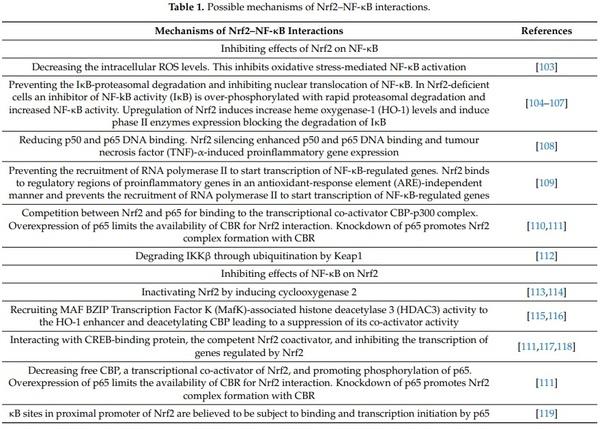
103. Soares, M.P.; Seldon, M.P.; Gregoire, I.P.; Vassilevskaia, T.; Berberat, P.O.; Yu, J.; Tsui, T.Y.; Bach, F.H. Hemeoxygenase-1 modulates the expression of adhesion molecules associated with endothelial cell activation. J. Immunol. 2004, 172, 3553–3563. [CrossRef]
[PubMed]
104. Yerra, V.G.; Negi, G.; Sharma, S.S.; Kumar, A. Potential therapeutic effects of the simultaneous targeting of the Nrf2 and NF-κB pathways in diabetic neuropathy. Redox Biol. 2013, 1, 394–397. [CrossRef] [PubMed]
105. Thimmulappa, R.K.; Lee, H.; Rangasamy, T.; Reddy, S.P.; Yamamoto, M.; Kensler, T.W.; Biswal, S. Nrf2 is a critical regulator of the innate immune response and survival during experimental sepsis. J. Clin. Investig. 2006, 116, 984–995. [CrossRef]
106. Chen, L.G.; Zhang, Y.Q.; Wu, Z.Z.; Hsieh, C.W.; Chu, C.S.; Wung, B.S. Peanut arachidin-1 enhances Nrf2-mediated protective mechanisms against TNF-α-induced ICAM-1 expression and NF-_B activation in endothelial cells. Int. J. Mol. Med. 2018,
41, 541–547.
107. Bellezza, I.; Tucci, A.; Galli, F.; Grottelli, S.; Mierla, A.L.; Pilolli, F.; Minelli, A. Inhibition of NF-κB nuclear translocation via HO-1 activation underlies α-tocopheryl succinate toxicity. J. Nutr. Biochem. 2012, 23, 1583–1591. [CrossRef]
108. Rushworth, S.A.; Shah, S.; MacEwan, D.J. TNF mediates the sustained activation of Nrf2 in human monocytes. J. Immunol. 2011,
187, 702–707. [CrossRef]
109. Kobayashi, E.H.; Suzuki, T.; Funayama, R.; Nagashima, T.; Hayashi, M.; Sekine, H.; Tanaka, N.; Moriguchi, T.; Motohashi, H.;
Nakayama, K.; et al. Nrf2 suppresses macrophage inflammatory response by blocking proinflammatory cytokine transcription.
Nat. Commun. 2016, 7, 11624. [CrossRef]
110. Kim, S.W.; Lee, H.K.; Shin, J.H.; Lee, J.K. Up-down regulation of HO-1 and iNOS gene expressions by ethyl pyruvate via recruiting p300 to Nrf2 and depriving It from p65. Free Radic Biol Med. 2013, 65, 468–476. [CrossRef] [PubMed]
111. Liu, G.H.; Qu, J.; Shen, X. NF-kappaB/p65 antagonizes Nrf2-ARE pathway by depriving CBP from Nrf2 and facilitating recruitment of HDAC3 to MafK. Biochim. Biophys. Acta 2008, 1783, 713–727. [CrossRef] [PubMed]
112. Lee, D.F.; Kuo, H.P.; Liu, M.; Chou, C.K.; Xia, W.; Du, Y.; Shen, J.; Chen, C.T.; Huo, L.; Hsu, M.C.; et al. KEAP1 E3 ligase-mediated downregulation of NF-kappaB signaling by targeting IKKbeta. Mol. Cell 2009, 36, 131–140. [CrossRef] [PubMed]
113. Banning, A.; Brigelius-Flohé, R. NF-kappaB, Nrf2, and HO-1 interplay in redox-regulated VCAM-1 expression. Antioxid. Redox
Signal. 2005, 7, 889–899. [CrossRef]
114. Brigelius-Flohé, R.; Flohé, L. Basic principles and emerging concepts in the redox control of transcription factors. Antioxid. Redox
Signal. 2011, 15, 2335–2381. [CrossRef]
115. Chuang, H.C.; Chang, C.W.; Chang, G.D.; Yao, T.P.; Chen, H. Histone deacetylase 3 binds to and regulates the GCMa transcription factor. Nucl. Acids Res. 2006, 34, 1459. [CrossRef]
116. Hung, H.L.; Kim, A.Y.; Hong, W.; Rakowski, C.; Blobel, G.A. Stimulation of NF-E2 DNA binding by CREB-binding protein (CBP)-mediated a117. Ahmed, S.M.U.; Luo, L.; Namani, A.; Wang, X.J.; Tang, X. Nrf2 signaling pathway: Pivotal roles in inflammation. Biochim. Biophys.
Acta 2017, 1863, 585–597. [CrossRef]
118. Yang, H.; Magilnick, N.; Ou, X.; Lu, S.C. Tumour necrosis factor α induces co-ordinated activation of rat GSH synthetic enzymes via nuclear factor κB and activator protein-1. Biochem. J. 2005, 391, 399–408. [CrossRef]
119. Rushworth, S.A.; Zaitseva, L.; Murray, M.Y.; Shah, N.M.; Bowles, K.M.; MacEwan, D.J. The high Nrf2 expression in human acute myeloid leukaemia is driven by NF-kappaB and underlies its chemo-resistance. Blood 2012, 120, 5188–5198. [CrossRef]
120. Wu, Y.; Lin, Z.; Yan, Z.; Wang, Z.; Fu, X.; Yu, K. Sinomenine contributes to the inhibition of the inflammatory response and the improvement of osteoarthritis in mouse-cartilage cells by acting on the Nrf2/HO-1 and NF-κB signaling pathways. Int.
Immunopharmacol. 2019, 75, 105715. [CrossRef] [PubMed]
121. Lee, W.; Yang, S.; Lee, C.; Park, E.K.; Kim, K.M.; Ku, S.K.; Bae, J.S. Aloin reduces inflammatory gene iNOS via inhibition activity and p-STAT-1 and NF-κB. Food Chem. Toxicol. 2019, 126, 67–71. [CrossRef]
122. Wang, J.; Chen, G.; Shi, T.; Wang, Y.; Guan, C. Possible treatment for cutaneous lichen planus: An in vitro anti-inflammatory role of Angelica polysaccharide in human keratinocytes HaCaT. Int. J. Immunopathol. Pharmacol. 2019, 33, 2058738418821837.
[CrossRef] [PubMed]
123. Lee, G.; Park, J.S.; Lee, E.J.; Ahn, J.H.; Kim, H.S. Anti-inflammatory and antioxidant mechanisms of urolithin B in activated microglia. Phytomedicine 2019, 55, 50–57. [CrossRef] [PubMed]
124. Zhao, D.R.; Jiang, Y.S.; Sun, J.Y.; Li, H.H.; Luo, X.L.; Zhao, M.M. Anti-inflammatory Mechanism Involved in 4-EthylguaiacolMediated Inhibition of LPS-Induced Inflammation in THP-1 Cells. J Agric. Food Chem. 2019, 67, 1230–1243. [CrossRef] [PubMed]
125. Ren, J.; Li, L.; Wang, Y.; Zhai, J.; Chen, G.; Hu, K. Gambogic acid induces heme oxygenase-1 through Nrf2 signaling pathway and inhibits NF-κB and MAPK activation to reduce inflammation in LPS-activated RAW264.7 cells. Biomed. Pharmacother. 2019, 109,
555–562. [CrossRef] [PubMed]
126. G ˛egotek, A.; Ambrozewicz, E.; Jastrz ˛ab, A.; Jarocka-Karpowicz, I.; Skrzydlewska, E. Rutin and ascorbic acid cooperation in ˙ antioxidant and antiapoptotic effect on human skin keratinocytes and fibroblasts exposed to UVA and UVB radiation. Arch.
Dermatol. Res. 2019, 311, 203–219. [CrossRef]
127. Luo, Z.; Zheng, B.; Jiang, B.; Xue, X.; Xue, E.; Zhou, Y. Peiminine inhibits the IL-1β induced inflammatory response in mouse articular chondrocytes and ameliorates murine osteoarthritis. Food Funct. 2019, 10, 2198–2208. [CrossRef]
128. Muhammad, T.; Ikram, M.; Ullah, R.; Rehman, S.U.; Kim, M.O. Hesperetin, a Citrus Flavonoid, Attenuates LPS-Induced
Neuroinflammation, Apoptosis and Memory Impairments by Modulating TLR4/NF-κB Signaling. Nutrients 2019, 11, 648.
[CrossRef]
129. Jia, Y.N.; Peng, Y.L.; Zhao, Y.P.; Cheng, X.F.; Zhou, Y.; Chai, C.L.; Zeng, L.S.; Pan, M.H.; Xu, L. Comparison of the Hepatoprotective
Effects of the Three Main Stilbenes from Mulberry Twigs. J Agric. Food Chem. 2019, 67, 5521–5529. [CrossRef]
130. Zhang, H.F.; Wang, J.H.; Wang, Y.L.; Gao, C.; Gu, Y.T.; Huang, J.; Wang, J.H.; Zhang, Z. Salvianolic Acid A Protects the Kidney against Oxidative Stress by Activating the Akt/GSK-3β/Nrf2 Signaling Pathway and Inhibiting the NF-κB Signaling Pathway in
5/6 Nephrectomized Rats. Oxid. Med. Cell Longev. 2019, 2019, 2853534. [CrossRef] [PubMed]
131. Abdel-Magied, N.; Shedid, S.M. The effect of naringenin on the role of nuclear factor (erythroid-derived 2)-like2 (Nrf2) and haem oxygenase 1 (HO-1) in reducing the risk of oxidative stress-related radiotoxicity in the spleen of rats. Environ. Toxicol. 2019, 34,
788–795. [CrossRef] [PubMed]
132. Wang, G.W.; Zhang, X.L.; Wu, Q.H.; Jin, Y.B.; Ning, C.T.; Wang, R.; Mao, J.X.; Chen, M. The hepatoprotective effects of Sedum sarmentosum extract and its isolated major constituent through Nrf2 activation and NF-κB inhibition. Phytomedicine 2019, 53,
263–273. [CrossRef] [PubMed]
133. He, Y.; Xia, Z.; Yu, D.; Wang, J.; Jin, L.; Huang, D.; Ye, X.; Li, X.; Zhang, B. Hepatoprotective effects and structure-activity relationship of five flavonoids against lipopolysaccharide/d-galactosamine induced acute liver failure in mice. Int. Immunopharmacol.
2019, 68, 171–178. [CrossRef]
134. Li, Z.; Feng, H.; Wang, Y.; Shen, B.; Tian, Y.; Wu, L.; Zhang, Q.; Jin, M.; Liu, G. Rosmarinic acid protects mice from lipopolysaccharide/d-galactosamine-induced acute liver injury by inhibiting MAPKs/NF-κB and activating Nrf2/HO-1 signaling pathways. Int. Immunopharmacol. 2019, 67, 465–472. [CrossRef]
135. Liu, T.G.; Sha, K.H.; Zhang, L.G.; Liu, X.X.; Yang, F.; Cheng, J.Y. Protective effects of alpinetin on lipopolysaccharide/dGalactosamine-induced liver injury through inhibiting inflammatory and oxidative responses. Microb. Pathog. 2019, 126, 239–244.
[CrossRef]
136. Tang, F.; Fan, K.; Wang, K.; Bian, C. Amygdalin attenuates acute liver injury induced by D-galactosamine and lipopolysaccharide by regulating the NLRP3, NF-κB and Nrf2/NQO1 signalling pathways. Biomed. Pharmacother. 2019, 111, 527–536. [CrossRef]
137. Li, Q.; Tian, Z.; Wang, M.; Kou, J.; Wang, C.; Rong, X.; Li, J.; Xie, X.; Pang, X. Luteoloside attenuates neuroinflammation in focal cerebral ischemia in rats via regulation of the PPARγ/Nrf2/NF-κB signaling pathway. Int. Immunopharmacol. 2019, 66, 309–316.
[CrossRef] [PubMed]
138. Bian, X.; Liu, X.; Liu, J.; Zhao, Y.; Li, H.; Zhang, L.; Li, P.; Gao, Y. Hepatoprotective effect of chiisanoside from Acanthopanax sessiliflorus against LPS/D-GalN-induced acute liver injury by inhibiting NF-κB and activating Nrf2/HO-1 signaling pathways.
J. Sci. Food Agric. 2019, 99, 3283–3290. [CrossRef]
139. Ye, J.; Guan, M.; Lu, Y.; Zhang, D.; Li, C.; Zhou, C. Arbutin attenuates LPS-induced lung injury via Sirt1/ Nrf2/ NF-κBp65 pathway. Pulm. Pharmacol. Ther. 2019, 54, 53–59. [CrossRef]cetylation. J. Biol. Chem. 2001, 276, 10715. [CrossRef]
140. Ding, H.; Ci, X.; Cheng, H.; Yu, Q.; Li, D. Chicoric acid alleviates lipopolysaccharide-induced acute lung injury in mice through anti-inflammatory and anti-oxidant activities. Int. Immunopharmacol. 2019, 66, 169–176. [CrossRef] [PubMed]
141. Alam, J.; Stewart, D.; Touchard, C.; Boinapally, S.; Choi, A.M.; Cook, J.L. Nrf2, a Cap’n’Collar transcription factor, regulates induction of the heme oxygenase-1 gene. J. Biol. Chem. 1999, 274, 26071–26078. [CrossRef] [PubMed]
142. Lavrovsky, Y.; Schwartzman, M.L.; Levere, R.D.; Kappas, A.; Abraham, N.G. Identification of binding sites for transcription factors NF-kappa B and AP-2 in the promoter region of the human heme oxygenase 1 gene. Proc. Natl. Acad. Sci. USA 1994, 91,
5987–5991. [CrossRef] [PubMed]
143. Mulcahy, R.T.; Wartman, M.A.; Bailey, H.H.; Gipp, J.J. Constitutive and beta-naphthoflavone-induced expression of the human gamma-glutamylcysteine synthetase heavy subunit gene is regulated by a distal antioxidant response element = TRE sequence.
J. Biol. Chem. 1997, 272, 7445–7454. [CrossRef]
144. Kimura, T.; Kawasaki, Y.; Okumura, F.; Sone, T.; Natsuki, R.; Isobe, M. Ethanol-induced expression of glutamate-cysteine ligase catalytic subunit gene is mediated by NF-kappaB. Toxicol. Lett. 2009, 185, 110–115. [CrossRef]
145. Muri, J.; Kopf, M. Redox regulation of immunometabolism. Nat. Rev. Immunol. 2020, in press. [CrossRef] [PubMed]
146. Kairisalo, M.; Korhonen, L.; Blomgren, K.; Lindholm, D. X-linked inhibitor of apoptosis protein increases mitochondrial antioxidants through NF-kappaB activation. Biochem. Biophys. Res. Commun. 2007, 364, 138–144. [CrossRef]
147. Djavaheri-Mergny, M.; Javelaud, D.; Wietzerbin, J.; Besançon, F. NF-kappaB activation prevents apoptotic oxidative stress via an increase of both thioredoxin and MnSOD levels in TNFalpha-treated Ewing sarcoma cells. FEBS Lett. 2004, 578, 111–115.
[CrossRef]
148. Anrather, J.; Racchumi, G.; Iadecola, C. NF-kappaB regulates phagocytic NADPH oxidase by inducing the expression of gp91phox.
J. Biol. Chem. 2006, 281, 5657–5667. [CrossRef]
149. Reuter, S.; Gupta, S.C.; Chaturvedi, M.M.; Aggarwal, B.B. Oxidative stress, inflammation, and cancer: How are they linked? Free
Radic. Biol. Med. 2010, 49, 1603–1616. [CrossRef] [PubMed]
150. Moldogazieva, N.T.; Mokhosoev, I.M.; Feldman, N.B.; Lutsenko, S.V. ROS and RNS signalling: Adaptive redox switches through oxidative/nitrosative protein modifications. Free Radic. Res. 2018, 52, 507–543. [CrossRef] [PubMed]
151. Ikeda, T.; Honjo, K.; Hirota, Y.; Onodera, T. Isolation of the chicken NF-kappa B p65 subunit-encoding cDNA and characterization of its products. Gene 1993, 133, 237–242. [PubMed]
152. Ikeda, T.; Hirota, Y.; Onodera, T. Isolation of a cDNA encoding the chicken p50B/p97 (Lyt-10) transcription factor. Gene 1994, 138,
193–196. [PubMed]
153. Krishnan, V.A.; Schatzle, J.D.; Hinojos, C.M.; Bose, H.R., Jr. Structure and regulation of the gene encoding avian inhibitor of nuclear factor kappa B-alpha. Gene 1995, 166, 261–266. [CrossRef]
154. Van Phi, L. Transcriptional activation of the chicken lysozyme gene by NF-kappa Bp65 (RelA) and c-Rel, but not by NF-kappa
Bp50. Biochem. J. 1996, 313, 39–44. [CrossRef] [PubMed]
155. Piffat, K.A.; Hrdlicková, R.; Nehyba, J.; Ikeda, T.; Liss, A.; Huang, S.; Sif, S.; Gilmore, T.D.; Bose, H.R., Jr. The chicken RelB transcription factor has transactivation sequences and a tissue-specific expression pattern that are distinct from mammalian RelB.
Mol. Cell. Biol. Res. Commun. 2001, 4, 266–275. [CrossRef]
156. Qiu, Y.; Shen, Y.; Li, X.; Ding, C.; Ma, Z. Molecular cloning and functional characterization of a novel isoform of chicken myeloid differentiation factor 88 (MyD88). Dev. Comp. Immunol. 2008, 32, 1522–1530. [CrossRef]
157. Zhou, P.; Zeng, Y.; Rao, Z.; Li, Y.; Zheng, H.; Luo, R. Molecular characterization and functional analysis of duck IKKα. Dev. Comp.
Immunol. 2021, 115, 103880. [CrossRef]
158. Kim, Y.; Tian, M. NF-kappaB family of transcription factor facilitates gene conversion in chicken B cells. Mol. Immunol. 2009, 46,
3283–3291. [CrossRef]
159. Chen, K.; Luo, Z.; Zheng, S.J. Gallus Heat shock cognate protein 70, a novel binding partner of Apoptin. Virol. J. 2011, 8, 324.
[CrossRef]
160. Kogut, M.H.; He, H.; Genovese, K.J. Bacterial toll-like receptor agonists induce sequential NF-κB-mediated leukotriene B4 and prostaglandin E2 production in chicken heterophils. Vet. Immunol. Immunopathol. 2012, 145, 159–170. [CrossRef]
161. Shinohara, H.; Behar, M.; Inoue, K.; Hiroshima, M.; Yasuda, T.; Nagashima, T.; Kimura, S.; Sanjo, H.; Maeda, S.; Yumoto, N.; et al.
Positive feedback within a kinase signaling complex functions as a switch mechanism for NF-κB activation. Science 2014, 344,
760–764. [CrossRef] [PubMed]
162. Tang, X.; Zhang, L.; Wei, W. Roles of TRAFs in NF-κB signaling pathways mediated by BAFF. Immunol. Lett. 2018, 196, 113–118.
[CrossRef] [PubMed]
163. Yang, H.L.; Feng, Z.Q.; Zeng, S.Q.; Li, S.M.; Zhu, Q.; Liu, Y.P. Molecular cloning and expression analysis of TRAF3 in chicken.
Genet. Mol. Res. 2015, 14, 4408–4419. [CrossRef] [PubMed]
164. Zhai, Y.; Luo, F.; Chen, Y.; Zhou, S.; Li, Z.; Liu, M.; Bi, D.; Jin, H. Molecular characterization and functional analysis of duck
TRAF6. Dev. Comp. Immunol. 2015, 49, 1–6. [CrossRef] [PubMed]
165. Guo, Y.; Xu, Y.; Kang, X.; Meng, C.; Gu, D.; Zhou, Y.; Xiong, D.; Geng, S.; Jiao, X.; Pan, Z. Molecular cloning and functional analysis of TRAF6 from Yangzhou great white goose Anser anser. Dev. Comp. Immunol. 2019, 101, 103435. [CrossRef] [PubMed]
166. Guo, Y.; Xu, Y.; Xiong, D.; Zhou, Y.; Kang, X.; Meng, C.; Gu, D.; Jiao, X.; Pan, Z. Molecular characterisation, expression and functional feature of TRAF6 in the King pigeon (Columba livia). Innate Immun. 2020, 26, 490–504. [CrossRef]
167. Zhou, Y.; Kang, X.; Xiong, D.; Zhu, S.; Zheng, H.; Xu, Y.; Guo, Y.; Pan, Z.; Jiao, X. Molecular and functional characterization of pigeon (Columba livia) tumor necrosis factor receptor-associated factor 3. Dev. Comp. Immunol. 2017, 69, 51–59. [CrossRef]
168. Kang, Y.; Nii, T.; Isobe, N.; Yoshimura, Y. Effects of TLR Ligands on the Expression of Cytokines and Possible Role of NFκB in its
Process in the Theca of Chicken Follicles. J. Poult. Sci. 2018, 55, 288–300. [CrossRef]
169. Chen, S.; Cheng, A.; Wang, M. Innate sensing of viruses by pattern recognition receptors in birds. Vet. Res. 2013, 44, 82. [CrossRef]
170. Downing, T.; Lloyd, A.T.; O’Farrelly, C.; Bradley, D.G. The differential evolutionary dynamics of avian cytokine and TLR gene classes. J. Immunol. 2010, 184, 6993–7000. [CrossRef] [PubMed]
171. Gillespie, M.; Shamovsky, V.; D’Eustachio, P. Human and chicken TLR pathways: Manual curation and computer-based orthology analysis. Mamm. Genome 2011, 22, 130–138. [CrossRef]
172. Farnell, M.B.; Crippen, T.L.; He, H.; Swaggerty, C.L.; Kogut, M.H. Oxidative burst mediated by toll like receptors (TLR) and CD14 on avian heterophils stimulated with bacterial toll agonists. Dev. Comp. Immunol. 2003, 27, 423–429. [CrossRef]
173. Kannaki, T.R.; Reddy, M.R.; Verma, P.C.; Shanmugam, M. Differential Toll-like receptor (TLR) mRNA expression patterns during chicken embryological development. Anim. Biotechnol. 2015, 26, 130–135. [CrossRef] [PubMed]
174. Yang, Y.; Jiang, Y.; Yin, Q.; Liang, H.; She, R. Chicken intestine defensins activated murine peripheral blood mononuclear cells through the TLR4-NF-kappaB pathway. Vet. Immunol. Immunopathol. 2010, 133, 59–65. [CrossRef]
175. Yu, H.; Lu, Y.; Qiao, X.; Wei, L.; Fu, T.; Cai, S.; Wang, C.; Liu, X.; Zhong, S.; Wang, Y. Novel Cathelicidins from Pigeon Highlights
Evolutionary Convergence in Avain Cathelicidins and Functions in Modulation of Innate Immunity. Sci. Rep. 2015, 5, 11082.
[CrossRef]
176. Ishige, T.; Hara, H.; Hirano, T.; Kono, T.; Hanzawa, K. Basic characterization of avian NK-lysin (NKL) from the Japanese quail,
Coturnix japonica. Anim. Sci. J. 2014, 85, 90–95. [CrossRef] [PubMed]
177. Kamimura, T.; Isobe, N.; Yoshimura, Y. Effects of inhibitors of transcription factors, nuclear factor-κB and activator protein 1, on the expression of proinflammatory cytokines and chemokines induced by stimulation with Toll-like receptor ligands in hen vaginal cells. Poult. Sci. 2017, 96, 723–730. [CrossRef]
178. Yong, Y.H.; Liu, S.F.; Hua, G.H.; Jia, R.M.; Gooneratne, R.; Zhao, Y.T.; Liao, M.; Ju, X.H. Goose toll-like receptor 3 (TLR3) mediated
IFN-γ and IL-6 in anti-H5N1 avian influenza virus response. Vet. Immunol. Immunopathol. 2018, 197, 31–38. [CrossRef] [PubMed]
179. Gao, M.; Guo, Y.; Du, J.; Song, Z.; Luo, X.; Wang, J.; Han, W. Evolutional conservation of molecular structure and antiviral function of a type I interferon, IFN-kappa, in poultry. Dev. Comp. Immunol. 2018, 89, 44–53. [CrossRef]
180. Jia, H.; Li, G.; Li, J.; Tian, Y.; Wang, D.; Shen, J.; Tao, Z.; Xu, J.; Lu, L. Cloning, expression and bioinformatics analysis of the duck
TLR 4 gene. Br. Poult. Sci. 2012, 53, 190–197. [CrossRef]
181. Fang, Q.; Pan, Z.; Geng, S.; Kang, X.; Huang, J.; Sun, X.; Li, Q.; Cai, Y.; Jiao, X. Molecular cloning, characterization and expression of goose Toll-like receptor 5. Mol. Immunol. 2012, 52, 117–124. [CrossRef] [PubMed]
182. Yong, Y.; Liu, S.; Hua, G.; Jia, R.; Zhao, Y.; Sun, X.; Liao, M.; Ju, X. Identification and functional characterization of Toll-like receptor 2-1 in geese. BMC Vet. Res. 2015, 11, 108. [CrossRef] [PubMed]
183. Xiong, D.; Song, L.; Pan, Z.; Jiao, X. Molecular cloning, characterization, and functional analysis of pigeon (Columba livia)
Toll-like receptor 5. Poult. Sci. 2018, 97, 4031–4039. [CrossRef] [PubMed]
184. Li, S.; Wang, Y.; Zhao, H.; Shao, Y.; Liu, J.; Xing, M. Characterization, functional and signaling elucidation of pigeon (Columba livia) interferon-α: Knockdown p53 negatively modulates antiviral response. Dev. Comp. Immunol. 2019, 90, 29–40. [CrossRef]
[PubMed]
185. Xiong, D.; Song, L.; Pan, Z.; Chen, X.; Geng, S.; Jiao, X. Identification and immune functional characterization of pigeon TLR7. Int.
J. Mol. Sci. 2015, 16, 8364–8381. [CrossRef] [PubMed]
186. Tao, Z.Y.; Zhu, C.H.; Shi, Z.H.; Song, C.; Xu, W.J.; Song, W.T.; Zou, J.M.; Qin, A.J. Molecular characterization, expression, and functional analysis of NOD1 in Qingyuan partridge chicken. Genet. Mol. Res. 2015, 14, 2691–2701. [CrossRef]
187. Li, H.; Jin, H.; Li, Y.; Liu, D.; Foda, M.F.; Jiang, Y.; Luo, R. Molecular cloning and functional characterization of duck nucleotidebinding oligomerization domain 1 (NOD1). Dev. Comp. Immunol. 2017, 74, 82–89. [CrossRef]
188. Truong, A.D.; Hong, Y.; Hoang, C.T.; Lee, J.; Hong, Y.H. Chicken IL-26 regulates immune responses through the JAK/STAT and
NF-κB signaling pathways. Dev. Comp. Immunol. 2017, 73, 10–20. [CrossRef]
189. Truong, A.D.; Hong, Y.; Rengaraj, D.; Lee, J.; Lee, K.; Hong, Y.H. Identification and functional characterization, including cytokine production modulation, of the novel chicken Interleukin-11. Dev. Comp. Immunol. 2018, 87, 51–63. [CrossRef]
190. Hoang, C.T.; Hong, Y.; Truong, A.D.; Lee, J.; Lee, K.; Hong, Y.H. Molecular cloning of chicken interleukin-17B, which induces proinflammatory cytokines through activation of the NF-κB signaling pathway. Dev. Comp. Immunol. 2017, 74, 40–48. [CrossRef]
191. Rohde, F.; Schusser, B.; Hron, T.; Farkašová, H.; Plachý, J.; Härtle, S.; Hejnar, J.; Elleder, D.; Kaspers, B. Characterization of Chicken
Tumor Necrosis Factor-α, a Long Missed Cytokine in Birds. Front. Immunol. 2018, 9, 605. [CrossRef] [PubMed]
192. Ji, G.G.; Shu, J.T.; Zhang, M.; Ju, X.J.; Shan, Y.J.; Liu, Y.F.; Tu, Y.J. Transcriptional regulatory region and DNA methylation analysis of TNNI1 gene promoters in Gaoyou duck skeletal muscle (Anas platyrhynchos domestica). Br. Poult. Sci. 2019, 60, 202–208.
[CrossRef] [PubMed]
193. Barjesteh, N.; Taha-Abdelaziz, K.; Kulkarni, R.R.; Sharif, S. Innate antiviral responses are induced by TLR3 and TLR4 ligands in chicken tracheal epithelial cells: Communication between epithelial cells and macrophages. Virology 2019, 534, 132–142.
194. Sutton, K.M.; Hu, T.; Wu, Z.; Siklodi, B.; Vervelde, L.; Kaiser, P. The functions of the avian receptor activator of NF-κB ligand (RANKL) and its receptors, RANK and osteoprotegerin, are evolutionarily conserved. Dev. Comp. Immunol. 2015, 51, 170–184.
[CrossRef]
195. Truong, A.D.; Hong, Y.; Nguyen, H.T.; Nguyen, C.T.; Chu, N.T.; Tran, H.T.T.; Dang, H.V.; Lillehoj, H.S.; Hong, Y.H. Molecular identification and characterisation of a novel chicken leukocyte immunoglobulin-like receptor A5. Br. Poult. Sci. 2020, in press.
196. Paital, B.; Panda, S.K.; Hati, A.K.; Mohanty, B.; Mohapatra, M.K.; Kanungo, S.; Chainy, G.B. Longevity of animals under reactive oxygen species stress and disease susceptibility due to global warming. World J. Biol. Chem. 2016, 7, 110–127. [CrossRef]
197. Vandana, G.D.; Sejian, V.; Lees, A.M.; Pragna, P.; Silpa, M.V.; Maloney, S.K. Heat stress and poultry production: Impact and amelioration. Int. J. Biometeorol. 2020, in press. [CrossRef]
198. Wasti, S.; Sah, N.; Mishra, B. Impact of Heat Stress on Poultry Health and Performances, and Potential Mitigation Strategies.
Animals 2020, 10, 1266. [CrossRef]
199. Pu, S.; Usuda, K.; Nagaoka, K.; Gore, A.; Crews, D.; Watanabe, G. The relation between liver damage and reproduction in female
Japanese quail (Coturnix japonica) exposed to high ambient temperature. Poult. Sci. 2020, 99, 4586–4597. [CrossRef]
200. Nawab, A.; Li, G.; An, L.; Wu, J.; Chao, L.; Xiao, M.; Zhao, Y.; Birmani, M.W.; Ghani, M.W. Effect of curcumin supplementation on
TLR4 mediated non-specific immune responses in liver of laying hens under high-temperature conditions. J. Therm. Biol. 2019, 84,
384–397. [CrossRef] [PubMed]
201. Liu, L.; Fu, C.; Yan, M.; Xie, H.; Li, S.; Yu, Q.; He, S.; He, J. Resveratrol modulates intestinal morphology and HSP70/90, NF-κB and EGF expression in the jejunal mucosa of black-boned chickens on exposure to circular heat stress. Food Funct. 2016, 7,
1329–1338. [CrossRef] [PubMed]
202. Hangalapura, B.N.; Kaiser, M.G.; Poel, J.J.; Parmentier, H.K.; Lamont, S.J. Cold stress equally enhances in vivo pro-inflammatory cytokine gene expression in chicken lines divergently selected for antibody responses. Dev. Comp. Immunol. 2006, 30, 503–511.
[CrossRef] [PubMed]
203. Zhao, F.Q.; Zhang, Z.W.; Wang, C.; Zhang, B.; Yao, H.D.; Li, S.; Xu, S.W. The role of heat shock proteins in inflammatory injury induced by cold stress in chicken hearts. Cell Stress Chaperones 2013, 18, 773–783. [CrossRef] [PubMed]
204. Fu, J.; Liu, C.P.; Zhang, Z.W.; Xing, M.W.; Xu, S.W. Influence of inflammatory pathway markers on oxidative stress induced by cold stress in intestine of quails. Res. Vet. Sci. 2013, 95, 495–501. [CrossRef] [PubMed]
205. Ren, J.; Liu, C.; Zhao, D.; Fu, J. The role of heat shock protein 70 in oxidant stress and inflammatory injury in quail spleen induced by cold stress. Environ. Sci. Pollut. Res. Int. 2018, 25, 21011–21023. [CrossRef]
206. Wei, H.; Zhang, R.; Su, Y.; Bi, Y.; Li, X.; Zhang, X.; Li, J.; Bao, J. Effects of Acute Cold Stress After Long-Term Cold Stimulation on
Antioxidant Status, Heat Shock Proteins, Inflammation and Immune Cytokines in Broiler Heart. Front. Physiol. 2018, 9, 1589.
[CrossRef]
207. Surai, P.F.; Dvorska, J.E. Effects of Mycotoxins on Antioxidant Status and Immunity. In The Mycotoxin Blue Book; Diaz, D.E., Ed.;
Nottingham University Press: Nottingham, UK, 2005; pp. 93–137.
208. Gao, Y.; Meng, L.; Liu, H.; Wang, J.; Zheng, N. The Compromised Intestinal Barrier Induced by Mycotoxins. Toxins 2020, 12, 619.
[CrossRef]
209. Li, Y.; Ma, Q.G.; Zhao, L.H.; Wei, H.; Duan, G.X.; Zhang, J.Y.; Ji, C. Effects of lipoic acid on immune function, the antioxidant defense system, and inflammation-related genes expression of broiler chickens fed aflatoxin contaminated diets. Int. J. Mol. Sci.
2014, 15, 5649–5662. [CrossRef]
210. Ma, Q.; Li, Y.; Fan, Y.; Zhao, L.; Wei, H.; Ji, C.; Zhang, J. Molecular Mechanisms of Lipoic Acid Protection against Aflatoxin
B1 -Induced Liver Oxidative Damage and Inflammatory Responses in Broilers. Toxins 2015, 7, 5435–5447. [CrossRef]
211. Rajput, S.A.; Sun, L.; Zhang, N.Y.; Khalil, M.M.; Ling, Z.; Chong, L.; Wang, S.; Rajput, I.R.; Bloch, D.M.; Khan, F.A.; et al. Grape
Seed Proanthocyanidin Extract Alleviates AflatoxinB1 -Induced Immunotoxicity and Oxidative Stress via Modulation of NF-κB and Nrf2 Signaling Pathways in Broilers. Toxins 2019, 11, 23. [CrossRef] [PubMed]
212. Du, Y.; Zhu, Y.; Teng, X.; Zhang, K.; Teng, X.; Li, S. Toxicological Effect of Manganese on NF-κB/iNOS-COX-2 Signaling Pathway in Chicken Testes. Biol. Trace Elem. Res. 2015, 168, 227–234. [CrossRef] [PubMed]
213. Li, S.; Zhao, H.; Wang, Y.; Shao, Y.; Li, J.; Liu, J.; Xing, M. The inflammatory responses in Cu-mediated elemental imbalance is associated with mitochondrial fission and intrinsic apoptosis in Gallus gallus heart. Chemosphere 2017, 189, 489–497. [CrossRef]
[PubMed]
214. Yang, F.; Liao, J.; Yu, W.; Pei, R.; Qiao, N.; Han, Q.; Hu, L.; Li, Y.; Guo, J.; Pan, J.; et al. Copper induces oxidative stress with triggered NF-κB pathway leading to inflammatory responses in immune organs of chicken. Ecotoxicol. Environ. Saf. 2020, 200,
110715. [CrossRef] [PubMed]
215. Zhang, K.; Zhao, P.; Guo, G.; Guo, Y.; Tian, L.; Sun, X.; Li, S.; He, Y.; Sun, Y.; Chai, H.; et al. Arsenic Trioxide Attenuates NF-κB and Cytokine mRNA Levels in the Livers of Cocks. Biol. Trace Elem. Res. 2016, 170, 432–437. [CrossRef] [PubMed]
216. Li, S.W.; Guo, Y.; He, Y.; Sun, X.; Zhao, H.J.; Wang, Y.; Wang, Y.J.; Xing, M.W. Assessment of arsenic trioxide toxicity on cock muscular tissue: Alterations of oxidative damage parameters, inflammatory cytokines and heat shock proteins. Ecotoxicology
2017, 26, 1078–1088. [CrossRef]
217. Li, S.; Wang, Y.; Zhao, H.; He, Y.; Li, J.; Jiang, G.; Xing, M. NF-κB-mediated inflammation correlates with calcium overload under arsenic trioxide-induced myocardial damage in Gallus gallus. Chemosphere 2017, 185, 618–627. [CrossRef]
218. Sun, X.; He, Y.; Guo, Y.; Li, S.; Zhao, H.; Wang, Y.; Zhang, J.; Xing, M. Arsenic affects inflammatory cytokine expression in Gallus gallus brain tissues. BMC Vet. Res. 2017, 13, 157. [CrossRef]
219. Li, S.W.; Sun, X.; He, Y.; Guo, Y.; Zhao, H.J.; Hou, Z.J.; Xing, M.W. Assessment of arsenic trioxide in the heart of Gallus gallus:
Alterations of oxidative damage parameters, inflammatory cytokines, and cardiac enzymes. Environ. Sci. Pollut. Res. Int. 2017, 24,
5781–5790. [CrossRef]
220. Wang, Y.; Zhao, H.; Guo, M.; Shao, Y.; Liu, J.; Jiang, G.; Xing, M. Arsenite renal apoptotic effects in chickens co-aggravated by oxidative stress and inflammatory response. Metallomics 2018, 10, 1805–1813. [CrossRef]
221. Zhao, H.; Wang, Y.; Shao, Y.; Liu, J.; Wang, S.; Xing, M. Oxidative stress-induced skeletal muscle injury involves in NF-κB/p53- activated immunosuppression and apoptosis response in copper (II) or/and arsenite-exposed chicken. Chemosphere 2018, 210,
76–84. [CrossRef]
222. Sun, X.; Li, J.; Zhao, H.; Wang, Y.; Liu, J.; Shao, Y.; Xue, Y.; Xing, M. Synergistic effect of copper and arsenic upon oxidative stress, inflammation and autophagy alterations in brain tissues of Gallus gallus. J. Inorg. Biochem. 2018, 178, 54–62. [CrossRef] [PubMed]
223. Liu, J.; Zhao, H.; Wang, Y.; Shao, Y.; Zhang, L.; Xing, M. Impacts of simultaneous exposure to arsenic (III) and copper (II) on inflammatory response, immune homeostasis, and heat shock response in chicken thymus. Int. Immunopharmacol. 2018, 64, 60–68.
[CrossRef] [PubMed]
224. Liu, J.; Zhao, H.; Wang, Y.; Shao, Y.; Li, J.; Xing, M. Alterations of antioxidant indexes and inflammatory cytokine expression aggravated hepatocellular apoptosis through mitochondrial and death receptor-dependent pathways in Gallus gallus exposed to arsenic and copper. Environ. Sci. Pollut. Res. Int. 2018, 25, 15462–15473. [CrossRef] [PubMed]
225. Guo, M.; Zhao, H.; Wang, Y.; Liu, J.; Fei, D.; Yang, X.; Mu, M.; Xing, M. Elemental imbalance elicited by arsenic and copper exposures leads to oxidative stress and immunotoxicity in chicken gizzard, activating the protective effects of heat shock proteins.
Environ. Sci. Pollut. Res. Int. 2019, 26, 36343–36353. [CrossRef]
226. Liu, J.; Wang, Y.; Zhao, H.; Mu, M.; Guo, M.; Nie, X.; Sun, Y.; Xing, M. Arsenic (III) or/and copper (II) exposure induce immunotoxicity through trigger oxidative stress, inflammation and immune imbalance in the bursa of chicken. Ecotoxicol. Environ.
Saf. 2020, 190, 110127. [CrossRef]
227. Wang, D.; Zong, C.; Cheng, K. Chicken thalamic injury induced by copper (II) or / and arsenite exposure involves oxidative stress and inflammation-induced apoptosis. Ecotoxicol. Environ. Saf. 2020, 197, 110554. [CrossRef]
228. Nie, X.; Wang, Y.; Zhao, H.; Guo, M.; Liu, Y.; Xing, M. As3+ or/and Cu2+ exposure triggers oxidative stress imbalance, induces inflammatory response and apoptosis in chicken brain. Ecotoxicol. Environ. Saf. 2020, 203, 110993. [CrossRef]
229. Wang, H.; Li, S.; Teng, X. The antagonistic effect of selenium on lead-induced inflammatory factors and heat shock proteins mRNA expression in chicken livers. Biol. Trace Elem. Res. 2016, 171, 437–444. [CrossRef]
230. Li, X.; Xing, M.; Chen, M.; Zhao, J.; Fan, R.; Zhao, X.; Cao, C.; Yang, J.; Zhang, Z.; Xu, S. Effects of selenium-lead interaction on the gene expression of inflammatory factors and selenoproteins in chicken neutrophils. Ecotoxicol. Environ. Saf. 2017, 139, 447–453.
[CrossRef]
231. Xie, W.; Ge, M.; Li, G.; Zhang, L.; Tang, Z.; Li, R.; Zhang, R. Astragalus Polysaccharide Protect against Cadmium-Induced
Cytotoxicity through the MDA5/NF-κB Pathway in Chicken Peripheral Blood Lymphocytes. Molecules 2017, 22, 1610. [CrossRef]
[PubMed]
232. Chen, M.; Li, X.; Fan, R.; Yang, J.; Jin, X.; Hamid, S.; Xu, S. Cadmium induces BNIP3-dependent autophagy in chicken spleen by modulating miR-33-AMPK axis. Chemosphere 2018, 194, 396–402. [CrossRef] [PubMed]
233. Wang, Y.; Liu, J.; Chen, R.; Qi, M.; Tao, D.; Xu, S. The Antagonistic Effects of Selenium Yeast (SeY) on Cadmium-Induced
Inflammatory Factors and the Heat Shock Protein Expression Levels in Chicken Livers. Biol. Trace Elem. Res. 2020, 198, 260–268.
[CrossRef] [PubMed]
234. Cao, H.; Gao, F.; Xia, B.; Zhang, M.; Liao, Y.; Yang, Z.; Hu, G.; Zhang, C. Alterations in trace element levels and mRNA expression of Hsps and inflammatory cytokines in livers of duck exposed to molybdenum or/and cadmium. Ecotoxicol. Environ. Saf. 2016,
125, 93–101. [CrossRef]
235. Guo, H.; Deng, H.; Cui, H.; Peng, X.; Fang, J.; Zuo, Z.; Deng, J.; Wang, X.; Wu, B.; Chen, K. Nickel chloride (NiCl2)-caused inflammatory responses via activation of NF-κB pathway and reduction of anti-inflammatory mediator expression in the kidney.
Oncotarget 2015, 6, 28607–28620. [CrossRef] [PubMed]
236. Xie, W.; Lv, A.; Li, R.; Tang, Z.; Ma, D.; Huang, X.; Zhang, R.; Ge, M. Agaricus blazei Murill Polysaccharides Protect Against
Cadmium-Induced Oxidative Stress and Inflammatory Damage in Chicken Spleens. Biol. Trace Elem. Res. 2018, 184, 247–258.
[CrossRef]
237. Yin, J.; Duan, J.; Cui, Z.; Ren, W.; Li, T.; Yin, Y. Hydrogen peroxide-induced oxidative stress activates NF-κB and Nrf2/Keap1 signals and triggers autophagy in piglets. RSC Advances 2015, 5, 15479–15486. [CrossRef]
238. Chen, Z.; Xing, T.; Li, J.; Zhang, L.; Jiang, Y.; Gao, F. Hydrogen peroxide-induced oxidative stress impairs redox status and damages aerobic metabolism of breast muscle in broilers. Poult. Sci. 2020, in press. [CrossRef]
239. Hofmann, T.; Schmucker, S.S.; Bessei, W.; Grashorn, M.; Stefanski, V. Impact of Housing Environment on the Immune System in
Chickens: A Review. Animals 2020, 10, 1138. [CrossRef]
240. Chen, X.; Zhang, L.; Li, J.; Gao, F.; Zhou, G. Hydrogen Peroxide-Induced Change in Meat Quality of the Breast Muscle of Broilers
Is Mediated by ROS Generation, Apoptosis, and Autophagy in the NF-κB Signal Pathway. J. Agric. Food Chem. 2017, 65, 3986–3994.
[CrossRef]241. Chen, X.; Gu, R.; Zhang, L.; Li, J.; Jiang, Y.; Zhou, G.; Gao, F. Induction of nuclear factor-κB signal-mediated apoptosis and autophagy by reactive oxygen species is associated with hydrogen peroxide-impaired growth performance of broilers. Animal
2018, 12, 2561–2570. [CrossRef] [PubMed]
242. Sies, H. Hydrogen peroxide as a central redox signaling molecule in physiological oxidative stress: Oxidative eustress. Redox Biol.
2017, 11, 613–619. [CrossRef]
243. Shi, Q.; Wang, W.; Chen, M.; Zhang, H.; Xu, S. Ammonia induces Treg/Th1 imbalance with triggered NF-κB pathway leading to chicken respiratory inflammation response. Sci. Total Environ. 2019, 659, 354–362. [CrossRef] [PubMed]
244. An, Y.; Xing, H.; Zhang, Y.; Jia, P.; Gu, X.; Teng, X. The evaluation of potential immunotoxicity induced by environmental pollutant ammonia in broilers. Poult. Sci. 2019, 98, 3165–3175. [CrossRef] [PubMed]
245. Han, Q.; Tong, J.; Sun, Q.; Teng, X.; Zhang, H.; Teng, X. The involvement of miR-6615-5p/Smad7 axis and immune imbalance in ammonia-caused inflammatory injury via NF-κB pathway in broiler kidneys. Poult. Sci. 2020, 99, 5378–5388. [CrossRef]
246. Shah, S.; Ishfaq, M.; Nasrullah, M.; Qayum, A.; Akhtar, M.U.; Jo, H.; Hussain, M.; Teng, X. Ammonia inhalation-induced inflammation and structural impairment in the bursa of fabricius and thymus of broilers through NF-κB signaling pathway.
Environ. Sci. Pollut. Res. Int. 2020, 27, 11596–11607. [CrossRef]
247. Chen, D.; Ning, F.; Zhang, J.; Tang, Y.; Teng, X. NF-κB pathway took part in the development of apoptosis mediated by miR-15a and oxidative stress via mitochondrial pathway in ammonia-treated chicken splenic lymphocytes. Sci. Total Environ. 2020, 729,
139017. [CrossRef]
248. Zhao, F.; Qu, J.; Wang, W.; Li, S.; Xu, S. The imbalance of Th1/Th2 triggers an inflammatory response in chicken spleens after ammonia exposure. Poult. Sci. 2020, 99, 3817–3822. [CrossRef]
249. Wang, W.; Chen, M.; Jin, X.; Li, X.; Yang, Z.; Lin, H.; Xu, S. H(2)S induces Th1/Th2 imbalance with triggered NF-κB pathway to exacerbate LPS-induce chicken pneumonia response. Chemosphere 2018, 208, 241–246. [CrossRef]
250. Chi, Q.; Chi, X.; Hu, X.; Wang, S.; Zhang, H.; Li, S. The effects of atmospheric hydrogen sulfide on peripheral blood lymphocytes of chickens: Perspectives on inflammation, oxidative stress and energy metabolism. Environ. Res. 2018, 167, 1–6. [CrossRef]
251. Chi, Q.; Wang, D.; Hu, X.; Li, S.; Li, S. Hydrogen Sulfide Gas Exposure Induces Necroptosis and Promotes Inflammation through the MAPK/NF-κB Pathway in Broiler Spleen. Oxid. Med. Cell Longev. 2019, 2019, 8061823. [CrossRef] [PubMed]
252. Hu, X.; Chi, Q.; Liu, Q.; Wang, D.; Zhang, Y.; Li, S. Atmospheric H2S triggers immune damage by activating the TLR7/MyD88/NF-κB pathway and NLRP3 inflammasome in broiler thymus. Chemosphere 2019, 237, 124427. [CrossRef] [PubMed]
253. Scott, T.; Owens, M.D. Thrombocytes respond to lipopolysaccharide through Toll-like receptor-4, and MAP kinase and NF-kappaB pathways leading to expression of interleukin-6 and cyclooxygenase-2 with production of prostaglandin E2. Mol. Immunol. 2008,
45, 1001–1008. [CrossRef] [PubMed]
254. Winkler, C.; Ferdous, F.; Dimmick, M.; Scott, T. Lipopolysaccharide induced Interleukin-6 production is mediated through activation of ERK 1/2, p38 MAPK, MEK, and NFκB in chicken thrombocytes. Dev. Comp. Immunol. 2017, 73, 124–130. [CrossRef]
[PubMed]
255. Kogut, M.H.; Genovese, K.J.; He, H.; Kaiser, P. Flagellin and lipopolysaccharide up-regulation of IL-6 and CXCLi2 gene expression in chicken heterophils is mediated by ERK1/2-dependent activation of AP-1 and NF-kappaB signaling pathways. Innate Immun.
2008, 14, 213–222. [CrossRef]
256. Ariyadi, B.; Isobe, N.; Yoshimura, Y. Toll-like receptor signaling for the induction of mucin expression by lipopolysaccharide in the hen vagina. Poult. Sci. 2014, 93, 673–679. [CrossRef]
257. Liu, L.; Shen, J.; Zhao, C.; Wang, X.; Yao, J.; Gong, Y.; Yang, X. Dietary Astragalus polysaccharide alleviated immunological stress in broilers exposed to lipopolysaccharide. Int. J. Biol. Macromol. 2015, 72, 624–632. [CrossRef]
258. Huang, X.Y.; Ansari, A.R.; Huang, H.B.; Zhao, X.; Li, N.Y.; Sun, Z.J.; Peng, K.M.; Zhong, J.; Liu, H.Z. Lipopolysaccharide mediates immuno-pathological alterations in young chicken liver through TLR4 signaling. BMC Immunol. 2017, 18, 12. [CrossRef]
259. Li, N.; Ansari, A.R.; Sun, Z.; Huang, H.; Cui, L.; Hu, Y.; Zhao, X.; Zhong, J.; Abdel-Kafy, E.M.; Liu, H. Toll like receptor 4 signaling pathway participated in Salmonella lipopolysaccharide-induced spleen injury in young chicks. Microb. Pathog. 2017, 112, 288–294.
[CrossRef]
260. Chang, Y.; Yuan, L.; Liu, J.; Muhammad, I.; Cao, C.; Shi, C.; Zhang, Y.; Li, R.; Li, C.; Liu, F. Dihydromyricetin attenuates
Escherichia coli lipopolysaccharide-induced ileum injury in chickens by inhibiting NLRP3 inflammasome and TLR4/NF-κB signalling pathway. Vet. Res. 2020, 51, 72. [CrossRef]
261. Yang, L.; Liu, G.; Lian, K.; Qiao, Y.; Zhang, B.; Zhu, X.; Luo, Y.; Shang, Y.; Gu, X.L. Dietary leonurine hydrochloride supplementation attenuates lipopolysaccharide challenge-induced intestinal inflammation and barrier dysfunction by inhibiting the
NF-κB/MAPK signaling pathway in broilers. J. Anim. Sci. 2019, 97, 1679–1692. [CrossRef] [PubMed]
262. Velleman, S.G. Pectoralis Major (Breast) Muscle Extracellular Matrix Fibrillar Collagen Modifications Associated With the Wooden
Breast Fibrotic Myopathy in Broilers. Front. Physiol. 2020, 11, 461. [CrossRef] [PubMed]
263. Xing, T.; Luo, D.; Zhao, X.; Xu, X.; Li, J.; Zhang, L.; Gao, F. Enhanced cytokine expression and upregulation of inflammatory signaling pathways in broiler chickens affected by wooden breast myopathy. J. Sci. Food Agric. 2020, in press. [CrossRef]
[PubMed]
264. Fancher, C.A.; Zhang, L.; Kiess, A.S.; Adhikari, P.A.; Dinh, T.; Sukumaran, A.T. Avian Pathogenic Escherichia coli and Clostridium perfringens: Challenges in No Antibiotics Ever Broiler Production and Potential Solutions. Microorganisms 2020, 8, 1533. [CrossRef]
[PubMed]
265. Nhung, N.T.; Chansiripornchai, N.; Carrique-Mas, J.J. Antimicrobial Resistance in Bacterial Poultry Pathogens: A Review. Front.
Vet. Sci. 2017, 4, 126. [CrossRef]
266. Peng, L.Y.; Cui, Z.Q.; Wu, Z.M.; Fu, B.D.; Yi, P.F.; Shen, H.Q. RNA-seq profiles of chicken type II pneumocyte in response to
Escherichia coli infection. PLoS ONE 2019, 14, e0217438. [CrossRef] [PubMed]
267. Peng, L.Y.; Yuan, M.; Wu, Z.M.; Song, K.; Zhang, C.L.; An, Q.; Xia, F.; Yu, J.L.; Yi, P.F.; Fu, B.D.; et al. Anti-bacterial activity of baicalin against APEC through inhibition of quorum sensing and inflammatory responses. Sci. Rep. 2019, 9, 4063. [CrossRef]
268. Peng, L.Y.; Yuan, M.; Song, K.; Yu, J.L.; Li, J.H.; Huang, J.N.; Yi, P.F.; Fu, B.D.; Shen, H.Q. Baicalin alleviated APEC-induced acute lung injury in chicken by inhibiting NF-κB pathway activation. Int. Immunopharmacol. 2019, 72, 467–472. [CrossRef]
269. Jiao, J.; Yang, Y.; Liu, M.; Li, J.; Cui, Y.; Yin, S.; Tao, J. Artemisinin and Artemisia annua leaves alleviate Eimeria tenella infection by facilitating apoptosis of host cells and suppressing inflammatory response. Vet. Parasitol. 2018, 254, 172–177. [CrossRef]
270. Yuan, M.; Peng, L.Y.; Wu, S.C.; Li, J.H.; Song, K.; Chen, S.; Huang, J.N.; Yu, J.L.; An, Q.; Yi, P.F.; et al. Schizandrin attenuates inflammation induced by avian pathogenic Escherichia coli in chicken type II pneumocytes. Int. Immunopharmacol. 2020, 81,
106313. [CrossRef]
271. Wang, W.; Li, Z.; Han, Q.; Guo, Y.; Zhang, B.; D’inca, R. Dietary live yeast and mannan-oligosaccharide supplementation attenuate intestinal inflammation and barrier dysfunction induced by Escherichia coli in broilers. Br. J. Nutr. 2016, 116, 1878–1888.
[CrossRef] [PubMed]
272. Thames, H.T.; Sukumaran, A.T. A Review of Salmonella and Campylobacter in Broiler Meat: Emerging Challenges and Food Safety
Measures. Foods 2020, 9, 776. [CrossRef]
273. Keestra, A.M.; de Zoete, M.R.; van Aubel, R.A.; van Putten, J.P. Functional characterization of chicken TLR5 reveals species-specific recognition of flagellin. Mol. Immunol. 2008, 45, 1298–1307. [CrossRef] [PubMed]
274. Chiang, H.I.; Berghman, L.R.; Zhou, H. Inhibition of NF-kB 1 (NF-kBp50) by RNA interference in chicken macrophage HD11 cell line challenged with Salmonellaenteritidis. Genet. Mol. Biol. 2009, 32, 507–515. [CrossRef] [PubMed]
275. Zhao, H.; Chen, Z.; Xie, L.J.; Liu, G.F. Suppression of TLR4/NFκB signaling pathway improves cerebral ischemia-reperfusion injury in rats. Mol. Neurobiol. 2018, 55, 4311–4319. [CrossRef] [PubMed]
276. Liu, X.; Ma, T.; Wang, H.; Sheng, Z.; Dou, X.; Wang, K.; Li, Z.; Pan, Z.; Chang, G.; Chen, G. Up-regulation of NLRC5 and NF-κB signaling pathway in carrier chickens challenged with Salmonella enterica Serovar Pullorum at different persistence periods.
Indian J. Biochem. Biophys. 2015, 52, 132–139. [PubMed]
277. Geng, S.; Wang, Y.; Xue, Y.; Wang, H.; Cai, Y.; Zhang, J.; Barrow, P.; Pan, Z.; Jiao, X. The SseL protein inhibits the intracellular
NF-κB pathway to enhance the virulence of Salmonella Pullorum in a chicken model. Microb. Pathog. 2019, 129, 1–6. [CrossRef]
278. He, Y.; Yang, Y.; Dong, Y.; Yan, C.; Zhang, B. The Effects of Flavomycin and Colistin Sulfate Pre-Treatment on Ileal Bacterial
Community Composition, the Response to Salmonella typhimurium and Host Gene Expression in Broiler Chickens. Microorganisms
2019, 7, 574. [CrossRef]
279. He, Y.; Yang, Y.; Dong, Y.; Ito, K.; Zhang, B. Highly nutritious diet resists Salmonella Typhimurium infections by improving intestinal microbiota and morphology in broiler chickens. Poult. Sci. 2020, 99, 7055–7065. [CrossRef]
280. Li, Q.; Xu, L.; Yin, C.; Liu, Z.; Li, Y.; Yuan, Y.; Hu, Y.; Jiao, X. The Invasion Plasmid Antigen J (IpaJ) from Salmonella Inhibits NF-κB
Activation by Suppressing IκBα Ubiquitination. Infect. Immun. 2020, 88, e00875-19. [CrossRef]
281. Ishfaq, M.; Hu, W.; Khan, M.Z.; Ahmad, I.; Guo, W.; Li, J. Current status of vaccine research, development, and challenges of vaccines for Mycoplasma gallisepticum. Poult. Sci. 2020, 99, 4195–4202. [CrossRef] [PubMed]
282. Majumder, S.; Zappulla, F.; Silbart, L.K. Mycoplasma gallisepticum lipid associated membrane proteins up-regulate inflammatory genes in chicken tracheal epithelial cells via TLR-2 ligation through an NF-κB dependent pathway. PLoS ONE 2014, 9, e112796.
[CrossRef] [PubMed]
283. Tian, W.; Zhao, C.; Hu, Q.; Sun, J.; Peng, X. Roles of Toll-like receptors 2 and 6 in the inflammatory response to Mycoplasma gallisepticum infection in DF-1 cells and in chicken embryos. Dev. Comp. Immunol. 2016, 59, 39–47. [CrossRef] [PubMed]
284. Sheedy, F.J. Turning 21: Induction of miR-21 as a Key Switch in the Inflammatory Response. Front. Immunol. 2015, 6, 19. [CrossRef]
[PubMed]
285. Krichevsky, A.M.; Gabriely, G. miR-21: A small multi-faceted RNA. J. Cell. Mol. Med. 2009, 13, 39–53. [CrossRef] [PubMed]
286. Zhao, Y.; Zou, M.; Sun, Y.; Zhang, K.; Peng, X. gga-miR-21 modulates Mycoplasma gallisepticum (HS strain)-Induced inflammation via targeting MAP3K1 and activating MAPKs and NF-κB pathways. Vet. Microbiol. 2019, 237, 108407. [CrossRef]
287. Zhang, K.; Han, Y.; Wang, Z.; Zhao, Y.; Fu, Y.; Peng, X. gga-miR-146c Activates TLR6/MyD88/NF-κB Pathway through Targeting
MMP16 to Prevent Mycoplasma Gallisepticum (HS Strain) Infection in Chickens. Cells 2019, 8, 501. [CrossRef]
288. Zhang, K.; Han, Y.; Zhao, Y.; Sun, Y.; Zou, M.; Fu, Y.; Peng, X. Upregulated gga-miR-16-5p Inhibits the Proliferation Cycle and
Promotes the Apoptosis of MG-Infected DF-1 Cells by Repressing PIK3R1-Mediated the PI3K/Akt/NF-κB Pathway to Exert
Anti-Inflammatory Effect. Int. J. Mol. Sci. 2019, 20, 1036. [CrossRef]
289. Yuan, B.; Zou, M.; Zhao, Y.; Zhang, K.; Sun, Y.; Peng, X. Up-Regulation of miR-130b-3p Activates the PTEN/PI3K/AKT/NF-κB
Pathway to Defense against Mycoplasma gallisepticum (HS Strain) Infection of Chicken. Int. J. Mol. Sci. 2018, 19, 2172. [CrossRef]
290. Yu, Y.; Chen, Y.; Wang, Y.; Li, Y.; Zhang, L.; Xin, J. TLR2/MyD88/NF-κB signaling pathway regulates IL-1β production in
DF-1cells exposed to Mycoplasma gallisepticum LAMPs. Microb. Pathog. 2018, 117, 225–231. [CrossRef]
291. Beaudet, J.; Tulman, E.R.; Pflaum, K.; Liao, X.; Kutish, G.F.; Szczepanek, S.M.; Silbart, L.K.; Geary, S.J. Transcriptional Profiling of the Chicken Tracheal Response to Virulent Mycoplasma gallisepticum Strain Rlow. Infect. Immun. 2017, 85, e00343-17. [CrossRef]
[PubMed]
292. Zou, M.; Yang, W.; Niu, L.; Sun, Y.; Luo, R.; Wang, Y.; Peng, X. Polydatin attenuates Mycoplasma gallisepticum (HS strain)-induced inflammation injury via inhibiting the TLR6/ MyD88/NF-κB pathway. Microb. Pathog. 2020, 149, 104552. [CrossRef]
293. Wu, Z.; Chen, C.; Miao, Y.; Liu, Y.; Zhang, Q.; Li, R.; Ding, L.; Ishfaq, M.; Li, J. Baicalin Attenuates Mycoplasma gallisepticumInduced Inflammation via Inhibition of the TLR2-NF-κB Pathway in Chicken and DF-1 Cells. Infect. Drug Resist. 2019, 12,
3911–3923. [CrossRef]
294. Ishfaq, M.; Chen, C.; Bao, J.; Zhang, W.; Wu, Z.; Wang, J.; Liu, Y.; Tian, E.; Hamid, S.; Li, R.; et al. Baicalin ameliorates oxidative stress and apoptosis by restoring mitochondrial dynamics in the spleen of chickens via the opposite modulation of NF-κB and
Nrf2/HO-1 signaling pathway during Mycoplasma gallisepticum infection. Poult. Sci. 2019, 98, 6296–6310. [CrossRef] [PubMed]
295. Zou, M.; Yang, L.; Niu, L.; Zhao, Y.; Sun, Y.; Fu, Y.; Peng, X. Baicalin ameliorates Mycoplasma gallisepticum-induced lung inflammation in chicken by inhibiting TLR6-mediated NF-κB signalling. Br. Poult. Sci. 2020, in press. [CrossRef] [PubMed]
296. Niu, L.; Luo, R.; Zou, M.; Sun, Y.; Fu, Y.; Wang, Y.; Peng, X. Puerarin inhibits Mycoplasma gallisepticum (MG-HS)-induced inflammation and apoptosis via suppressing the TLR6/MyD88/NF-κB signal pathway in chicken. Int. Immunopharmacol. 2020,
88, 106993. [CrossRef] [PubMed]
297. Fatoba, A.J.; Adeleke, M.A. Diagnosis and control of chicken coccidiosis: A recent update. J. Parasit. Dis. 2018, 42, 483–493.
[CrossRef] [PubMed]
298. Jin, H.; Haicheng, Y.; Caiyun, Z.; Yong, Z.; Jinrong, W. The Expression of NF-kB Signaling Pathway Was Inhibited by Silencing
TGF-b4 in Chicken IECs Infected with E. tenella. Braz. J. Poult. Sci. 2020, in press. [CrossRef]
299. Timbermont, L.; Haesebrouck, F.; Ducatelle, R.; Van Immerseel, F. (Necrotic enteritis in broilers: An updated review on the pathogenesis. Avian Pathol. 2011, 40, 341–347. [CrossRef]
300. Mora, Z.V.; Macías-Rodríguez, M.E.; Arratia-Quijada, J.; Gonzalez-Torres, Y.S.; Nuño, K.; Villarruel-López, A. Clostridium perfringens as Foodborne Pathogen in Broiler Production: Pathophysiology and Potential Strategies for Controlling Necrotic
Enteritis. Animals 2020, 10, 1718. [CrossRef]
301. Van Immerseel, F.; De Buck, J.; Pasmans, F.; Huyghebaert, G.; Haesebrouck, F.; Ducatelle, R. Clostridium perfringens in poultry:
An emerging threat for animal and public health. Avian Pathol. 2004, 33, 537–549. [CrossRef] [PubMed]
302. Guo, S.; Li, C.; Liu, D.; Guo, Y. Inflammatory responses to a Clostridium perfringens type A strain and α-toxin in primary intestinal epithelial cells of chicken embryos. Avian Pathol. 2015, 44, 81–91. [CrossRef] [PubMed]
303. Athanasiadou, S.; Russell, K.M.; Kaiser, P.; Kanellos, T.; Burgess, S.T.; Mitchell, M.; Clutton, E.; Naylor, S.W.; Low, C.J.; Hutchings,
M.R.; et al. Genome wide transcriptomic analysis identifies pathways affected by the infusion of Clostridium perfringens culture supernatant in the duodenum of broilers in situ. J. Anim. Sci. 2015, 93, 3152–3163. [CrossRef] [PubMed]
304. Guo, S.; Liu, D.; Zhang, B.; Li, Z.; Li, Y.; Ding, B.; Guo, Y. Two Lactobacillus Species Inhibit the Growth and α-Toxin Production of
Clostridium perfringens and Induced Proinflammatory Factors in Chicken Intestinal Epithelial Cells in Vitro. Front. Microbiol. 2017,
8, 2081. [CrossRef] [PubMed]
305. Li, Z.; Wang, W.; Liu, D.; Guo, Y. Effects of Lactobacillus acidophilus on the growth performance and intestinal health of broilers challenged with Clostridium perfringens. J. Anim. Sci. Biotechnol. 2018, 9, 25. [CrossRef] [PubMed]
306. Knittler, M.R.; Sachse, K. Chlamydia psittaci: Update on an underestimated zoonotic agent. Pathog. Dis. 2015, 73, 1–15. [CrossRef]
307. Chu, J.; Li, X.; Qu, G.; Wang, Y.; Li, Q.; Guo, Y.; Hou, L.; Liu, J.; Eko, F.O.; He, C. Chlamydia psittaci PmpD-N Exacerbated Chicken
Macrophage Function by Triggering Th2 Polarization and the TLR2/MyD88/NF-κB Signaling Pathway. Int. J. Mol. Sci. 2020, 21,
2003. [CrossRef]
308. Chen, Q.; Li, Y.; Yan, X.; Sun, Z.; Wang, C.; Liu, S.; Xiao, J.; Lu, C.; Wu, Y. Chlamydia psittaci Plasmid-Encoded CPSIT_P7 Elicits
Inflammatory Response in Human Monocytes via TLR4/Mal/MyD88/NF-κB Signaling Pathway. Front Microbiol. 2020, 11,
578009. [CrossRef]
309. López-Osorio, S.; Chaparro-Gutiérrez, J.J.; Gómez-Osorio, L.M. Overview of Poultry Eimeria Life Cycle and Host-Parasite
Interactions. Front. Vet. Sci. 2020, in press.
310. Rehman, Z.U.; Meng, C.; Sun, Y.; Safdar, A.; Pasha, R.H.; Munir, M.; Ding, C. Oxidative Stress in Poultry: Lessons from the Viral
Infections. Oxid. Med. Cell Longev. 2018, 2018, 5123147. [CrossRef]
311. Khatri, M.; Sharma, J.M. Infectious bursal disease virus infection induces macrophage activation via p38 MAPK and NF-kappaB pathways. Virus Res. 2006, 118, 70–77. [CrossRef] [PubMed]
312. Wong, R.T.; Hon, C.C.; Zeng, F.; Leung, F.C. Screening of differentially expressed transcripts in infectious bursal disease virusinduced apoptotic chicken embryonic fibroblasts by using cDNA microarrays. J. Gen. Virol. 2007, 88, 1785–1796. [CrossRef]
[PubMed]
313. Cubas-Gaona, L.L.; Diaz-Beneitez, E.; Ciscar, M.; Rodríguez, J.F.; Rodríguez, D. Exacerbated Apoptosis of Cells Infected with
Infectious Bursal Disease Virus upon Exposure to Interferon Alpha. J. Virol. 2018, 92, e00364-18. [CrossRef] [PubMed]
314. Ou, C.; Wang, Q.; Wei, X.; Liu, M.; Liu, X.; He, C. Pro-apoptosis effects of protocatechuic acid in the early stage of infectious bursal disease virus infection. Microb. Pathog. 2018, 124, 216–222. [CrossRef] [PubMed]
315. Bello, M.B.; Yusoff, K.; Ideris, A.; Hair-Bejo, M.; Peeters, B.P.H.; Omar, A.R. Diagnostic and Vaccination Approaches for Newcastle
Disease Virus in Poultry: The Current and Emerging Perspectives. Biomed. Res. Int. 2018, 2018, 7278459. [CrossRef]
316. Mayers, J.; Mansfield, K.L.; Brown, I.H. The role of vaccination in risk mitigation and control of Newcastle disease in poultry.
Vaccine 2017, 35, 5974–5980. [CrossRef]
317. Qu, Y.; Zhan, Y.; Yang, S.; Ren, S.; Qiu, X.; Rehamn, Z.U.; Tan, L.; Sun, Y.; Meng, C.; Song, C.; et al. Newcastle disease virus infection triggers HMGB1 release to promote the inflammatory response. Virology 2018, 525, 19–31. [CrossRef]
318. Park, J.; Kim, M.; Na, G.; Jeon, I.; Kwon, Y.K.; Kim, J.H.; Youn, H.; Koo, Y. Glucocorticoids modulate NF-kappaB-dependent gene expression by up-regulating FKBP51 expression in Newcastle disease virus-infected chickens. Mol. Cell. Endocrinol. 2007, 278,
7–17. [CrossRef]
319. Qian, J.; Xu, X.; Ding, J.; Yin, R.; Sun, Y.; Xue, C.; Wang, J.; Ding, C.; Yu, S.; Liu, X.; et al. Newcastle disease virus-like particles induce DC maturation through TLR4/NF-κB pathway and facilitate DC migration by CCR7-CCL19/CCL21 axis. Vet. Microbiol.
2017, 203, 158–166. [CrossRef]
320. Chen, Y.; Liu, W.; Xu, H.; Liu, J.; Deng, Y.; Cheng, H.; Zhan, T.; Lu, X.; Liao, T.; Guo, L.; et al. Gga-miR-19b-3p Inhibits Newcastle
Disease Virus Replication by Suppressing Inflammatory Response via Targeting RNF11 and ZMYND11. Front Microbiol. 2019, 10,
2006. [CrossRef]
321. Li, J.J.; Yin, Y.; Yang, H.L.; Yang, C.W.; Yu, C.L.; Wang, Y.; Yin, H.D.; Lian, T.; Peng, H.; Zhu, Q.; et al. mRNA expression and functional analysis of chicken IFIT5 after infected with Newcastle disease virus. Infect. Genet. Evol. 2020, 86, 104585. [CrossRef]
[PubMed]
322. Bertzbach, L.D.; Conradie, A.M.; You, Y.; Kaufer, B.B. Latest Insights into Marek’s Disease Virus Pathogenesis and Tumorigenesis.
Cancers 2020, 12, 647. [CrossRef] [PubMed]
323. Kumar, S.; Kunec, D.; Buza, J.J.; Chiang, H.I.; Zhou, H.; Subramaniam, S.; Pendarvis, K.; Cheng, H.H.; Burgess, S.C. Nuclear
Factor kappa B is central to Marek’s disease herpesvirus induced neoplastic transformation of CD30 expressing lymphocytes in-vivo. BMC Syst. Biol. 2012, 6, 123. [CrossRef] [PubMed]
324. Liu, Y.; Gao, L.; Xu, Z.; Luo, D.; Zhang, Y.; Gao, Y.; Liu, C.; Zhang, Y.; Qi, X.; Cui, H.; et al. Marek’s Disease Virus RLORF4 Inhibits
Type I Interferon Production by Antagonizing NF-κB Activation. J. Virol. 2019, 93, e01037-19. [CrossRef] [PubMed]
325. Benavente, J.; Martínez-Costas, J. Avian reovirus: Structure and biology. Virus Res. 2007, 123, 105–119. [CrossRef] [PubMed]
326. De Carli, S.; Wolf, J.M.; Gräf, T.; Lehmann, F.K.; Fonseca, A.S.; Canal, C.W.; Lunge, V.R.; Ikuta, N. Genotypic characterization and molecular evolution of avian reovirus in poultry flocks from Brazil. Avian Pathol. 2020, 49, 611–620. [CrossRef] [PubMed]
327. Lin, P.Y.; Liu, H.J.; Liao, M.H.; Chang, C.D.; Chang, C.I.; Cheng, H.L.; Lee, J.W.; Shih, W.L. Activation of PI 3-kinase/Akt/NFkappaB and Stat3 signaling by avian reovirus S1133 in the early stages of infection results in an inflammatory response and delayed apoptosis. Virology 2010, 400, 104–114. [CrossRef]
328. Xie, L.; Xie, Z.; Wang, S.; Huang, J.; Deng, X.; Xie, Z.; Luo, S.; Zeng, T.; Zhang, Y.; Zhang, M. Altered gene expression profiles of the MDA5 signaling pathway in peripheral blood lymphocytes of chickens infected with avian reovirus. Arch. Virol. 2019, 164,
2451–2458. [CrossRef]
329. Yun, T.; Hua, J.; Ye, W.; Ni, Z.; Chen, L.; Zhang, C. The phosphoproteomic responses of duck (Cairna moschata) to classical/novel duck reovirus infections in the spleen tissue. Sci. Rep. 2020, 10, 15315. [CrossRef]
330. Campbell, L.K.; Magor, K.E. Pattern Recognition Receptor Signaling and Innate Responses to Influenza A Viruses in the Mallard
Duck, Compared to Humans and Chickens. Front. Cell. Infect. Microbiol. 2020, 10, 209. [CrossRef]
331. Li, N.; Zhao, J.; Yang, Y.; Zeng, Y.; Liu, S. Innate immune responses to duck Tembusu virus infection. Vet. Res. 2020, 51, 87.
[CrossRef] [PubMed]
332. Nettleford, S.K.; Prabhu, K.S. Selenium and Selenoproteins in Gut Inflammation-A Review. Antioxidants 2018, 7, 36. [CrossRef]
[PubMed]
333. Zhang, J.L.; Xu, B.; Huang, X.D.; Gao, Y.H.; Chen, Y.; Shan, A.S. Selenium Deficiency Affects the mRNA Expression of
Inflammatory Factors and Selenoprotein Genes in the Kidneys of Broiler Chicks. Biol. Trace Elem. Res. 2016, 171, 201–207.
[CrossRef] [PubMed]
334. Liu, Z.; Qu, Y.; Wang, J.; Wu, R. Selenium Deficiency Attenuates Chicken Duodenal Mucosal Immunity via Activation of the
NF-κb Signaling Pathway. Biol. Trace Elem. Res. 2016, 172, 465–473. [CrossRef] [PubMed]
335. Wang, J.; Liu, Z.; He, X.; Lian, S.; Liang, J.; Yu, D.; Sun, D.; Wu, R. Selenium deficiency induces duodenal villi cell apoptosis via an oxidative stress-induced mitochondrial apoptosis pathway and an inflammatory signaling-induced death receptor pathway.
Metallomics 2018, 10, 1390–1400. [CrossRef] [PubMed]
336. Zhang, R.; Guo, R.; Liu, Q.; Li, G.; Sun, B.; Huang, X. Selenium Deficiency via the TLR4/TRIF/NF-κB Signaling Pathway Leading to Inflammatory Injury in Chicken Spleen. Biol. Trace Elem. Res. 2020, in press. [CrossRef]
337. Wang, Y.; Jiang, L.; He, J.; Hu, M.; Zeng, F.; Li, Y.; Tian, H.; Luo, X. The Adverse Effects of Se Toxicity on Inflammatory and
Immune Responses in Chicken Spleens. Biol. Trace Elem. Res. 2018, 185, 170–176. [CrossRef] [PubMed]
338. Huang, H.; Chen, J.; Sun, Q.; Liu, Y.; Tang, Y.; Teng, X. NLRP3 inflammasome is involved in the mechanism of mitigative effect of selenium on lead-induced inflammatory damage in chicken kidneys. Environ. Sci. Pollut. Res. Int. 2020, in press. [CrossRef]
339. Jiayong, Z.; Shengchen, W.; Xiaofang, H.; Gang, S.; Shiwen, X. The antagonistic effect of selenium on lead-induced necroptosis via
MAPK/NF-κB pathway and HSPs activation in the chicken spleen. Ecotoxicol. Environ. Saf. 2020, 204, 111049. [CrossRef]
340. Zhang, J.; Hao, X.; Xu, S. Selenium Prevents Lead-Induced Necroptosis by Restoring Antioxidant Functions and Blocking
MAPK/NF-κB Pathway in Chicken Lymphocytes. Biol. Trace Elem. Res. 2020, 198, 644–653. [CrossRef]341. Wang, X.; Bao, R.; Fu, J. The Antagonistic Effect of Selenium on Cadmium-Induced Damage and mRNA Levels of Selenoprotein
Genes and Inflammatory Factors in Chicken Kidney Tissue. Biol. Trace Elem. Res. 2018, 181, 331–339. [CrossRef] [PubMed]
342. Zhang, R.; Liu, Y.; Xing, L.; Zhao, N.; Zheng, Q.; Li, J.; Bao, J. The protective role of selenium against cadmium-induced hepatotoxicity in laying hens: Expression of Hsps and inflammation-related genes and modulation of elements homeostasis.
Ecotoxicol. Environ. Saf. 2018, 159, 205–212. [CrossRef]
343. Tang, K.K.; Li, H.Q.; Qu, K.C.; Fan, R.F. Selenium alleviates cadmium-induced inflammation and meat quality degradation via antioxidant and anti-inflammation in chicken breast muscles. Environ. Sci. Pollut. Res. Int. 2019, 26, 23453–23459. [CrossRef]
[PubMed]
344. Zhang, W.; Zhang, R.; Wang, T.; Jiang, H.; Guo, M.; Zhou, E.; Sun, Y.; Yang, Z.; Xu, S.; Cao, Y.; et al. Selenium inhibits LPS-induced pro-inflammatory gene expression by modulating MAPK and NF-κB signaling pathways in mouse mammary epithelial cells in primary culture. Inflammation 2014, 37, 478–485. [CrossRef] [PubMed]
345. Liu, J.; Wang, S.; Zhang, Q.; Li, X.; Xu, S. Selenomethionine alleviates LPS-induced chicken myocardial inflammation by regulating the miR-128-3p-p38 MAPK axis and oxidative stress. Metallomics 2020, 12, 54–64. [CrossRef]
346. Qu, J.; Wang, W.; Zhang, Q.; Li, S. Inhibition of Lipopolysaccharide-Induced Inflammation of Chicken Liver Tissue by Selenomethionine via TLR4-NF-κB-NLRP3 Signaling Pathway. Biol. Trace Elem. Res. 2020, 195, 205–214. [CrossRef] [PubMed]
347. Shi, X.; Wang, W.; Zheng, S.; Zhang, Q.; Xu, S. Selenomethionine relieves inflammation in the chicken trachea caused by LPS though inhibiting the NF-κB pathway. Biol. Trace Elem. Res. 2020, 194, 525–535. [CrossRef] [PubMed]
348. Tan, J.; Liu, S.; Guo, Y.; Applegate, T.J.; Eicher, S.D. Dietary L-arginine supplementation attenuates lipopolysaccharide-induced inflammatory response in broiler chickens. Br. J. Nutr. 2014, 111, 1394–1404. [CrossRef] [PubMed]
349. Bi, Y.; Nan, X.M.; Zheng, S.S.; Jiang, L.S.; Xiong, B.H. Effects of dietary threonine and immune stress on growth performance, carcass trait, serum immune parameters, and intestinal muc2 and NF-κb gene expression in Pekin ducks from hatch to 21 days.
Poult. Sci. 2018, 97, 177–187. [CrossRef]
350. Liu, S.Q.; Wang, L.Y.; Liu, G.H.; Tang, D.Z.; Fan, X.X.; Zhao, J.P.; Jiao, H.C.; Wang, X.J.; Sun, S.H.; Lin, H. Leucine alters immunoglobulin a secretion and inflammatory cytokine expression induced by lipopolysaccharide via the nuclear factor-κB pathway in intestine of chicken embryos. Animal 2018, 12, 1903–1911. [CrossRef]
351. Surai, P.F. Polyphenol compounds in the chicken/animal diet: From the past to the future. J. Anim. Physiol. Anim. Nutr. 2014, 98,
19–31. [CrossRef] [PubMed]
352. Surai, P.F. Silymarin as a Natural Antioxidant: An Overview of the Current Evidence and Perspectives. Antioxidants 2015, 4,
204–247. [CrossRef] [PubMed]
353. Zhang, C.; Tian, Y.; Yan, F.; Kang, X.; Han, R.; Sun, G.; Zhang, H. Modulation of growth and immunity by dietary supplementation with resveratrol in young chickens receiving conventional vaccinations. Am. J. Vet. Res. 2014, 75, 752–759. [CrossRef] [PubMed]
354. Sahin, K.; Orhan, C.; Akdemir, F.A.; Tuzcu, M.; Iben, C.; Sahin, N. Resveratrol protects quail hepatocytes against heat stress:
Modulation of the Nrf2 transcription factor and heat shock proteins. J. Anim. Physiol. Anim. Nutr. 2012, 96, 66–74. [CrossRef]
[PubMed]
355. Fan, H.; Lv, Z.; Gan, L.; Guo, Y. Transcriptomics-Related Mechanisms of Supplementing Laying Broiler Breeder Hens with Dietary
Daidzein to Improve the Immune Function and Growth Performance of Offspring. J. Agric. Food Chem. 2018, 66, 2049–2060.
[CrossRef] [PubMed]
356. Yang, J.X.; Maria, T.C.; Zhou, B.; Xiao, F.L.; Wang, M.; Mao, Y.J.; Li, Y. Quercetin improves immune function in Arbor Acre broilers through activation of NF-κB signaling pathway. Poult. Sci. 2020, 99, 906–913. [CrossRef]
357. Ge, C.; Zhang, C.; Ye, J.; Tang, X.; Wu, Y. Ginbaltosides promote proliferation of chicken primordial germ cells via PKC-involved activation of NF-kappaB. Cell. Biol. Int. 2007, 31, 1251–1256. [CrossRef]
358. Lee, M.T.; Lin, W.C.; Wang, S.Y.; Lin, L.J.; Yu, B.; Lee, T.T. Evaluation of potential antioxidant and anti-inflammatory effects of
Antrodia cinnamomea powder and the underlying molecular mechanisms via Nrf2- and NF-κB-dominated pathways in broiler chickens. Poult. Sci. 2018, 97, 2419–2434. [CrossRef]
359. Chen, H.; Muhammad, I.; Zhang, Y.; Ren, Y.; Zhang, R.; Huang, X.; Diao, L.; Liu, H.; Li, X.; Sun, X.; et al. Antiviral Activity Against
Infectious Bronchitis Virus and Bioactive Components of Hypericum perforatum L.. Front. Pharmacol. 2019, 10, 1272. [CrossRef]
360. Yu, M.; Ge, C.; Zeng, W.; Mi, Y.; Zhang, C. Retinoic acid promotes proliferation of chicken primordial germ cells via activation of
PI3K/Akt-mediated NF-κB signalling cascade. Cell Biol. Int. 2012, 36, 705–712. [CrossRef]
361. Zhang, X.; Zhong, X.; Zhou, Y.; Wang, G.; Du, H.; Wang, T. Dietary RRR-α-tocopherol succinate attenuates lipopolysaccharideinduced inflammatory cytokines secretion in broiler chicks. Br. J. Nutr. 2010, 104, 1796–1805. [CrossRef] [PubMed]
362. Zhan, T.; Han, Y.; Tang, C.; Zhao, Q.; Sun, D.; Li, Y.; Jia, X.; Zhou, L.; Zhang, J. Metabolism and biological activity of α-tocopherol derived from vitamin E-enriched transgenic maize in broilers. J. Sci. Food. Agric. 2020, 100, 4319–4328. [CrossRef] [PubMed]
363. Ma, Z.; Zhang, J.; Ma, H.; Dai, B.; Zheng, L.; Miao, J.; Zhang, Y. The influence of dietary taurine and reduced housing density on hepatic functions in laying hens. Poult. Sci. 2014, 93, 1724–1736. [CrossRef] [PubMed]
364. Dai, B.; Zhang, Y.S.; Ma, Z.L.; Zheng, L.H.; Li, S.J.; Dou, X.H.; Gong, J.S.; Miao, J.F. Influence of dietary taurine and housing density on oviduct function in laying hens. J. Zhejiang Univ.-SCIENCE B 2015, 16, 456–464. [CrossRef]
365. Ma, Z.L.; Gao, Y.; Ma, H.T.; Zheng, L.H.; Dai, B.; Miao, J.F.; Zhang, Y.S. Effects of taurine and housing density on renal function in laying hens. J. Zhejiang Univ.-SCIENCE B 2016, 17, 952–964. [CrossRef]
366. Wu, Y.; Zhen, W.; Geng, Y.; Wang, Z.; Guo, Y. Pretreatment with probiotic Enterococcus faecium NCIMB 11181 ameliorates necrotic enteritis-induced intestinal barrier injury in broiler chickens. Sci. Rep. 2019, 9, 10256. [CrossRef]
367. Sun, Y.; Qian, J.; Xu, X.; Tang, Y.; Xu, W.; Yang, W.; Jiang, Y.; Yang, G.; Ding, Z.; Cong, Y.; et al. Dendritic cell-targeted recombinant
Lactobacilli induce DC activation and elicit specific immune responses against G57 genotype of avian H9N2 influenza virus infection. Vet. Microbiol. 2018, 223, 9–20. [CrossRef]
368. Lan, D.; Xun, X.; Hu, Y.; Li, N.; Yang, C.; Jiang, X.; Liu, Y. Research on the Effect of Pediococcus pentosaceus on Salmonella enteritidis-Infected Chicken. BioMed Res. Int. 2020, 2020, 6416451. [CrossRef]
369. Weavers, H.; Martin, P. The cell biology of inflammation: From common traits to remarkable immunological adaptations. J. Cell
Biol. 2020, 219, e202004003. [CrossRef]
370. Pflug, K.M.; Sitcheran, R. Targeting NF-κB-Inducing Kinase (NIK) in Immunity, Inflammation, and Cancer. Int. J. Mol. Sci. 2020,
21, 8470. [CrossRef]
371. de Gregorio, E.; Colell, A.; Morales, A.; Marí, M. Relevance of SIRT1-NF-κB Axis as Therapeutic Target to Ameliorate Inflammation in Liver Disease. Int. J. Mol. Sci. 2020, 21, 3858. [CrossRef] [PubMed]
372. Zhou, Y.; Cui, C.; Ma, X.; Luo, W.; Zheng, S.G.; Qiu, W. Nuclear Factor κB (NF-κB)-Mediated Inflammation in Multiple Sclerosis.
Front. Immunol. 2020, 11, 391. [CrossRef] [PubMed]
373. Afonina, I.S.; Zhong, Z.; Karin, M.; Beyaert, R. Limiting inflammation-the negative regulation of NF-κB and the NLRP3 inflammasome. Nat. Immunol. 2017, 18, 861–869. [CrossRef]
374. Kunnumakkara, A.B.; Shabnam, B.; Girisa, S.; Harsha, C.; Banik, K.; Devi, T.B.; Choudhury, R.; Sahu, H.; Parama, D.; Sailo,
B.L.; et al. Inflammation, NF-κB, and Chronic Diseases: How are They Linked? Crit. Rev. Immunol. 2020, 40, 1–39. [CrossRef]
[PubMed]
375. Serhan, C.N. Treating inflammation and infection in the 21st century: New hints from decoding resolution mediators and mechanisms. FASEB J. 2017, 31, 1273–1288. [CrossRef]
376. Rothschild, D.E.; McDaniel, D.K.; Ringel-Scaia, V.M.; Allen, I.C. Modulating inflammation through the negative regulation of
NF-κB signaling. J. Leukoc. Biol. 2018, 103, 1131–1150. [CrossRef]
377. Kogut, M.H.; Genovese, K.J.; Swaggerty, C.L.; He, H.; Broom, L. Inflammatory phenotypes in the intestine of poultry: Not all inflammation is created equal. Poult. Sci. 2018, 97, 2339–2346. [CrossRef]
378. Julian, R.J. Production and growth-related disorders and other metabolic diseases of poultry—A review. Vet. J. 2005, 169, 350–369.
[CrossRef]
379. Kiero ´nczyk, B.; Rawski, M.; Józefiak, D.; Swi ˛atkiewicz, S. Infectious and non-infectious factors associated with leg disorders in ´ poultry—A review. Ann. Anim. Sci. 2017, 17, 645–669. [CrossRef]
380. Ducatelle, R.; Goossens, E.; De Meyer, F.; Eeckhaut, V.; Antonissen, G.; Haesebrouck, F.; Van Immerseel, F. Biomarkers for monitoring intestinal health in poultry: Present status and future perspectives. Vet. Res. 2018, 49, 43. [CrossRef]
381. Golonka, R.M.; Xiao, X.; Abokor, A.A.; Joe, B.; Vijay-Kumar, M. Altered nutrient status reprograms host inflammation and metabolic health via gut microbiota. J. Nutr. Biochem. 2020, in press. [CrossRef] [PubMed]
382. Yang, Q.; Wang, Y.; Jia, A.; Wang, Y.; Bi, Y.; Liu, G. The crosstalk between gut bacteria and host immunity in intestinal inflammation.
J. Cell. Physiol. 2020, in press. [CrossRef] [PubMed]
383. Tilg, H.; Zmora, N.; Adolph, T.E.; Elinav, E. The intestinal microbiota fuelling metabolic inflammation. Nat. Rev. Immunol. 2020,
20, 40–54. [CrossRef] [PubMed]
384. Broom, L.J. Host–Microbe Interactions and Gut Health in Poultry—Focus on Innate Responses. Microorganisms 2019, 7, 139.
[CrossRef] [PubMed]
385. Oviedo-Rondón, E.O. Holistic view of intestinal health in poultry. Anim. Feed Sci. Technol. 2019, 250, 1–8. [CrossRef]
386. Rius-Pérez, S.; Pérez, S.; Martí-Andrés, P.; Monsalve, M.; Sastre, J. NF-κB Signaling complexes in acute inflammation. Antioxid.
Redox Signal. 2019, in press.
387. Farooqui, A.A. Inflammation and Oxidative Stress in Neurological Disorders; Springer: Cham, Switzerland; Heidelberg, Germany;
New York, NY, USA; Dordrecht, The Netherlands; London, UK, 2014.
388. Kircheis, R.; Haasbach, E.; Lueftenegger, D.; Heyken, W.T.; Ocker, M.; Planz, O. NF-κB Pathway as a Potential Target for Treatment of Critical Stage COVID-19 Patients. Front. Immunol. 2020, 11, 598444. [CrossRef]
389. Sha, W.C.; Liou, H.C.; Tuomanen, E.I.; Baltimore, D. Targeted disruption of the p50 subunit of NF-κB leads to multifocal defects in immune responses. Cell 1995, 80, 321–330. [CrossRef]
390. Surai, P.F. Antioxidants in Poultry Nutrition and Reproduction: An Update. Antioxidants 2020, 9, 105. [CrossRef]
391. Surai, P.F.; Kochish, I.I.; Kidd, M.T. Taurine in Poultry Nutrition. Anim. Feed Sci. Technol. 2020, 260, 114339. [CrossRef]
392. Surai, P.F. Antioxidant action of carnitine: Molecular mechanisms and practical applications. EC Vet. Sci. 2015, 2, 66–84.
393. Surai, P.F. Carnitine enigma: From antioxidant action to vitagene regulation. Part 1. Absorption, metabolism and antioxidant activities. J. Vet. Sci. Med. 2015, 3, 14. [CrossRef]
394. Surai, P.F. Carnitine enigma: From antioxidant action to vitagene regulation. Part 2. Transcription factors and practical applications. J. Vet. Sci. Med. 2015, 3, 17. [CrossRef]


.jpg&w=3840&q=75)