Performance of broiler chickens and turkeys exposed to Histomonas meleagridis (blackhead)-infected litter while fed diets supplemented with Natustat™ or Histostat®
Histomoniasis (infectious enterohepatitis) is a disease of broiler chickens on litter or exposed to range and is caused by the fragile protozoa Histomonas meleagridis (Skinner, 2000). First symptoms are a decline in feed consumption (loss of appetite), inactivity, drooping wings (drowsiness) and unkempt feathers, followed by a drop in body weight and appearance of diarrhea with sulfur-colored droppings (Renwald, 1970).
Other symptoms include poor feed utilization, increased thirst, darkening of the facial regions (‘blackhead’ in turkeys, actually a misnomer), and sometimes death (Hall et al., 1975). Morbidity and mortality are variable, and mortality including culls may reach 20%. With mild infections, the chickens will show no overt symptoms except that they will expel the cecal cores. Infected farms may suffer clinical outbreaks year after year (McDougald, 1997).
In chickens, the disease primarily affects the ceca, and is characterized by cecal inflammation, roughness and thickening of the cecal wall and large cheese-like (caseous) cecal cores. Chickens are more resistant to histomoniasis than turkeys, and consequently chickens are less likely to experience liver mottling deep into the tissue and head discoloration (Renwald, 1970; McDougald, 1997).
The virulence of Histomonas varies greatly in infections in chickens. Some strains of the parasite produce mild to severe gross lesions in the ceca whereas others produce moderate to severe lesions in both the ceca and liver. Extra mortality occurs in some outbreaks and not in others (McDougald, 1997).
Based on the chronology of pathology in turkeys, after a bird becomes severely infected the histomonads first congregate in the epithelial cells lining the ceca to live and multiply. About the third day after entering the digestive tract, the organisms cause small hemorrhagic patches in the interior lining of the cecal wall. Careful observation is needed to see these lesions.
On the fourth day, more extensive hemorrhaging will be noted along with thickening of the cecal lining. Small, scattered dead areas may be seen. The bird may show signs of being sick on the third day or fourth day. On the sixth day, the ceca will be almost filled with whitish, cheesy material and sometimes contains blood. Liver lesions, as pinpoint spots, will appear about this time.
The liver lesions are caused by organisms reaching the bloodstream from the ceca. In a short time, the organisms destroy large sections of the liver. On the seventh and eighth days, the cecal walls are more thickened. The interior is filled with cheesy material called the cecal core. The liver is sometimes enlarged and size of lesions increases.
By the eighth day, the lesions are well defined having oval, concave depressions (pitted surface lesions) or dark-red, gray, yellowish or yellow-green subsurface mottling that may extend deep into liver tissue. On the ninth day, the ceca will be greatly enlarged, with spongy, fibrous cores. The liver has many advanced lesions as described above.
Sulfur-colored droppings may be seen, and a few birds may die. Usually, if mortality occurs, it will not be heavy until the 14th or 15th day (Hall and Wehe, 1953; Salsbury Laboratories Inc., 1982).
Histomoniasis may occur in association with roundworm and cecal worm (nematode) parasites, enteritis, coccidiosis, or combinations of these, sometimes making the individual diseases difficult to distinguish.
Occasionally, acute attacks may cause cecal bleeding similar to that in cecal coccidiosis (Renwald, 1970), and histomoniasis may be misdiagnosed as cecal coccidiosis (McDougald and Casey, 1982). Piperazine, commonly used for large roundworms, is not effective against cecal worms (McDougald and Casey, 1982) although Hygromycin® B in feed is effective for preventive use (North, 1972; McDougald, 1997).
Histomonads are round, except for small pseudopodia, and approximately 10 microns in diameter, being smaller in size than Eimeria mitis, the smallest coccidium, which measures about 14 x 15 microns. Under a microscope, the cells change shape in an amoeboid fashion and possess a flagellum (McDougald, 1997).
The histomonad is a parasite of worms and is typically spread in any of three ways: 1) in droppings of cecalinfected birds (coprophagy), 2) in infected cecal worm eggs, and/or 3) by infected earthworms (especially with dirt floors). Unprotected histomonads dessicate readily and their existence on the litter is probably no longer than a few hours to one day, but ingestion of histomonads directly from the droppings of infected birds is possible during this time.
Because histomonads are spread directly (feces) or indirectly (cecal worms or earthworms), young flocks should not be reared near infected older flocks and an all-in and all-out (similar age) production system is preferred (Salisbury Laboratories Inc., 1982). Recovered birds may become carriers of the histomonads (Hall and Wehe, 1953). Freeflying birds may also introduce the infection anew (Hall et al., 1975).
Cecal worms (Heterakis gallinarum; small roundworm), found in the lumen of the ceca, are small, white, and measure about 0.90 to 1.25 cm (0.35 to 0.50 inch) in length (Hall et al., 1975). Cecal worms are difficult to detect except by postmortem examination (Renwald, 1970). Both chickens and turkeys can carry cecal worms, and in some cases, turkeys may be the main source of infection for broiler chickens, Leghorn breeders, or broiler breeder chickens. Histomonads protected within cecal worm eggs can remain in the soil for four or more years and still infect birds. After about two weeks outside the bird’s body, histomonadinfected cecal worm eggs can develop into larvae that are eaten by other birds, so spreading the disease.
Earthworms consume embryonated cecal worm eggs, which hatch in their intestine. The released larvae burrow through the intestinal wall and can live for a year or more in the body cavity of the earthworm host. If parasitized earthworms are eaten by chickens, the worm eggs hatch inside the host, and cecal worms containing histomonads develop, and a histomoniasis outbreak may result.
“The protozoan parasites are thus delivered right to the very door of their new home” (McDougald, 1997). Cleanout of litter between flocks does not ensure removal of the parasites if there is exposed soil unless treatment with cresylic acid in diesel oil or with other chemicals is done. Deeper litter is helpful to avoid bare spots exposing dirt (McDougald, 1997).
There is little or no evidence that birds develop a protective immunity against Histomonas meleagridis, even following recovery from an active illness, so immunization against the organism is not an option, according to McDougald (1997). Histomonads are difficult to eliminate from farms with a history of the disease, and preventive medication is the best way to control the disease.
Nitroimidazoles such as ronidazole, ipronidazole, and dimetridazole are effective for treatment or prevention but are not available in the US according to the Merck Veterinary Manual (2003). The only approved drug in the US for the control (prophylaxis) of histomoniasis is Histostat® (4-nitrophenylarsonic acid, sold under the trade name of Histostat® by Alpharma Inc., Fort Lee, NJ) at 0.01875% (170.3 g nitarsone/ton) in the feed until five days before marketing. Histostat® may be used in combination with bacitracin methylene disalicylate or zinc bacitracin (Skinner, 2000). There has apparently never been a known case of drug resistant Histomonas meleagridis.
Natustat™ is a natural, plant-derived, blended proprietary product used as an alternative supplement in poultry feeds. Its proposed modes of action include decreasing the level of protozoa present via antiprotozoal activity, improving immune competence, and aiding tissue repair in the bird following infection.
Recent research
Two floor pen studies, one with broilers and another with turkeys, were conducted to observe bird performance when exposed to litter contaminated with Heterakis gallinarum eggs carrying Histomonas meleagridis. Test animals were fed a basal diet supplemented with Natustat™ (NAT+h), 1.925 kg/ton, or Histostat® (NIT+h) at 170.3 g/ton. Exposed controls (CON+h) and unexposed controls (CON) were fed an unsupplemented diet. Performance (weights, feed efficiency and mortality) and lesions (cecal and liver) from the challenge were monitored.
Prior to the start of each study, a large quantity (>1 ton) of litter was collected from a local broiler breeder flock verified to contain Heterakis gallinarum eggs by microscopic observation. This litter was mixed with feed and fed to 50 7-day old turkey poults for five days. Mortality from histomoniasis was confirmed within 14 days; and on the 21st day after feeding, all remaining poults were euthanized and more than 75% had lesions of liver necrosis and cecal cores confirming the presence of H. meleagridis. This exposure litter was used in both the broiler and turkey experiments.
METHODS
Experiment 1: Performance of chickens exposed to Histomonas meleagridis-infected litter while fed diets supplemented with Natustat™ or Histostat®
A total of 600 1-day old, Hubbard Hi-Y broilers were randomly distributed to four groups resulting in 150 straight run broilers of similar health per group. Each group was weighed and placed into one of four study pens (not replicated). Initial live weights averaged 40 g per broiler chick.
Each pen contained four water fountains and four feed tubes with a capacity of 22 kg (50 lb). The dimensions of each pen were 10 X 12 feet, which provided an initial stocking density of 0.8 ft2/broiler.
The NIT+h, CON+h and NAT+h pens had 2 inches of new wood shavings for bedding at day 0, which topdressed approximately 1 inch of old bedding from a commercial broiler breeder flock (see exposure system).
The CON pens had 3 inches of new wood shavings. Room temperature was checked and recorded at least daily. Heat was provided by propane-fueled brooders and negative pressure air exchange was used for ventilation.
Basal feeds for starter and grower diets were corn and soy-based. Starter and grower feed formulas were used from day 0 to 21 and day 22 to 42, respectively (Table 1).
Broilers were exposed by top-dressing one inch of the exposure litter with new wood shavings at study start and raking it to the top at 7 days. In addition, approximately 2 lbs of exposure litter was poured around the feeder of the three exposure pens on a daily basis beginning between 7 and 12 days.
All broilers and feeds were weighed by pen at 0, 28, 35, and 42 days. On the 28th, 35th and 42nd day, 25 broilers from each of the four pens were randomly caught, weighed and euthanized. The cecas and livers of the chickens and turkeys of the two experiments were scored using the following schedule:
Cecal scores
0 - Normal – no lesions observed;
1 - Slight thickening of cecal wall(s);
2 - Moderately thickened cecal wall(s) or slight/moderate cecal core;
3 - Moderate thickening of cecal wall(s) and cecal cores;
4 - Ceca grossly enlarged due to presence of large cecal core;
Table 1.Ingredient and nutrient composition of the broiler basal diets.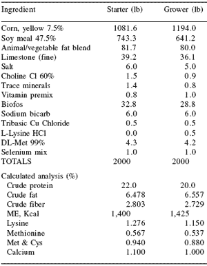
Liver scores
0 - Normal liver;
1 - Slight mottling of liver observed;
2 - Moderate mottling of liver observed;
3 - Concentrated areas of necrotic liver tissue;
All deaths were presented for necropsy. The livers and ceca of all animals that died after exposure to challenge litter were scored as listed above.
Experiment 2: Performance of turkeys exposed to Histomonas meleagridis-infected litter while fed diets supplemented with Natustat™ or Histostat® A total of 1125 1-day old, tom Hybrid turkeys were randomly distributed to 32 groups with 35 per group.
Each group of 35 was weighed and placed into one of 32 study pens. Initial live weights averaged 55 g per poult. Each pen contained one water fountain and one feed tube with a capacity of 22 kg (50 lb). The dimensions of each pen were 4 X 6 feet, which provided an initial stocking density of 0.7 ft2/poult. All pens had 3 inches of new wood shavings for bedding at Day 0. Heat was provided by propane-fueled brooders and negative pressure air exchange was used for ventilation. Basal feeds for starter and grower diets were corn and soy-based. Starter feed was fed from Day 0 to 21 and grower feed from Day 22 to 42 (Table 2).
Table 2.Ingredient and nutrient composition of the turkey basal diets.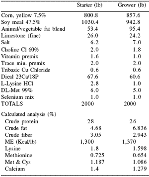
Turkeys in the CON+h, NIT+h and NAT+h designated pens were exposed to H. meleagridis beginning on the 7th day by adding up to 1 lb of the exposure litter to each pen for 21 days. On the 28th and 35th days 5 turkeys per pen were randomly caught, euthanized and their ceca and livers were scored in same manner as broilers.
All deaths were presented for necropsy. The livers and ceca of all animals that died after exposure to challenge litter were lesion scored and that data included in the analysis. All poults and feeds were weighed by pen at 28 and 42 days.
RESULTS
Experiment 1: Performance of chickens exposed to Histomonas meleagridis-infected litter while fed diets supplemented with Natustat™ or Histostat®
Because the broiler study had only one pen per treatment, live performance data were obtained but no statistical analysis was possible. The live performance response variables were 42-day body weight, feed conversion ratio, mortality-adjusted feed conversion ratio, and mortality.
Cecal lesion scores and liver lesion scores from 25 birds per pen (treatment) at 28, 35, and 42 days of age were statistically evaluated by one-way ANOVA. Arc sine transformed data were used for cecal lesion scores and liver lesion scores. When ANOVA was significant (P<0.05), means were separated by Tukey’s test.
Body weights were variable by pen with CON+h lowest (1,612 g), NIT+h highest (1,860 g), and the other two treatments intermediate at 42 days (Table 3).
Table 3. Effects of Histostat® and Natustat™ on the body weights (g) of histomoniasis-challenged broilers.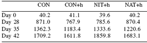
There was a large numerical difference between the NAT+h feed conversion ratio (2.125 g/g) and the other treatments (2.547 to 3.195) at 42 days, indicating a possible improvement by Natustat™, which deserves further evaluation (Table 4).
Table 4.Effects of Histostat® and Natustat™ on feed efficiency of histomoniasis-challenged broilers.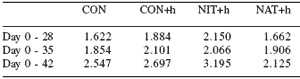
Mortality-adjusted feed conversion ratio (Table 5) followed a similar trend, with NAT+h substantially lower (2.086 g/g) than the other treatments (2.347 to 2.672 g/ g). Total mortality was 3.33% for NAT+h compared to 8.00, 16.67, and 26.67% in the CON, CON+h, and NIT+h groups (Table 6). These results indicate that the challenge model with infected broiler breeder litter was working to lower body weight and worsen feed conversion ratio and mortality in the CON+h group compared to the other treatments.
A loss of appetite and a reduction in body weight are among the first symptoms observed in histomoniasis (Renwald, 1970). Poor feed utilization has been reported as a symptom of histomoniasis (Hall et al., 1975).
Skinner (2000) observed that feed conversion ratios of female turkeys were better in uninfected control and infected Histostat® treated birds than in nonsupplemented, infected birds. Morbidity and extra mortality of turkeys are commonly observed in a histomoniasis outbreak (McDougald, 1997). Skinner (2000) reported that female turkeys challenged with histomonads carried by cecal worms had 15.12% mortality at 70 days compared to 3.30% in uninfected control and 2.35% in infected Histostat® treated birds.
Table 5. Effects of Histostat® and Natustat™ on feed efficiency adjusted for mortality weights of histomoniasis-challenged broilers.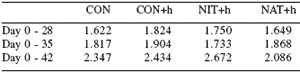
Table 6.Effects of Histostat® and Natustat™ on the mortality (%) of histomoniasis-challenged broilers.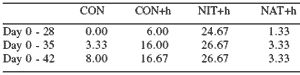
Histomoniasis is typically characterized by cecal inflammation, roughness and thickening of the cecal wall, and large cheese-like (caseous) cecal cores.
Occasionally, acute attacks may cause cecal bleeding similar to that in cecal coccidiosis (Renwald, 1970). As shown in Table 7, cecal lesion scores were higher (P<0.001) for the CON+h broiler chickens at 28, 35, and 42 days of age compared to the CON chickens, indicating that the Heterakis egg infected litter did cause histomoniasis. At 28 and 35 days, the NIT+h and NAT+h chickens had cecal lesion scores that were statistically equivalent and lower than CON+h birds. At 42 days, the NAT+h birds had significantly lower cecal lesion scores (0.44) than the NIT+h (0.96) birds.
Table 7.Effects of Histostat® and Natustat™ on the cecal lesion scores of histomoniasis-challenged broilers.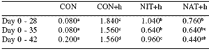
abcMeans within a row differ (P<0.05).
Liver mottling deep into tissue is associated with moderate to severe histomoniasis (Salsbury Laboratories Inc., 1982). As presented in Table 8, although liver lesion scores in broiler chickens were relatively low (0 to 1.16 range), significant treatment differences were detected.
Table 8.Effects of Histostat® and Natustat™ on the liver lesion scores of histomoniasis-challenged broilers.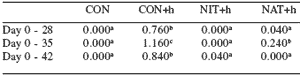
abMeans within a row differ (P<0.05)
At 28, 35, and 42 days, broiler chickens in the CON+h group had higher (P<0.05) liver lesion scores (0.76, 1.16, and 0.84, respectively) than the CON group (0 at each sampling). The NIT+h treatment (0, 0, and 0.04) and the NAT+h treatment (0.04, 0.24, and 0) results were statistically equivalent, indicating that for broiler chicken liver protection during histomoniasis, the Natustat™ and Histostat® performed similarly (P>0.05).
It is important to note that the investigators observed ‘enteritis-like’ lesions among several of the deaths, which were a by-product of the natural challenge model. Of this mortality and among the three challenged groups, the fewest deaths occurred in the NAT+h group (0.67%), followed by 4.00 and 16.67% in the NIT+h and CON groups, respectively. This suggests that Natustat™ may play a role in controlling this type of mortality and should be further investigated.
Experiment 2: Performance of turkeys exposed to Histomonas meleagridis-infected litter while fed diets supplemented with Natustat™ or Histostat®
Each of the four treatments of this turkey experiment had eight pen replicates nested within blocks throughout the turkey research building. The live performance response variables were 42 day body weight, feed conversion ratio, mortality-adjusted feed conversion ratio and mortality. Cecal lesion scores and liver lesion scores from five birds per pen (40 per treatment) at 28, and 42 days of age were statistically evaluated by oneway ANOVA. Arc sine transformed data were used for cecal lesion scores and liver lesion scores. When ANOVA was significant (P<0.05), means were separated by Tukey’s test.
Body weights (Table 9) were lowest for the CON+h group, which was significantly lowest at 28 and 42 days but not different for the CON, NIT+h and NAT+h groups. Feed efficiency (Table 10) and feed efficiency adjusted for mortality (Table 11) at 42 days of the NAT+h group were numerically lower than that of the NIT+h group following a trend observed in the broiler experiment. The exposure to H. meleagridis did not appear to affect the feed efficiency values of the NIT+h and NAT+h groups while adverse to the CON+h group at both 28 and 42 days.
Table 9. Effects of Histostat® and Natustat™ on the body weights (g) of histomoniasis-challenged tom turkeys.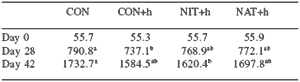
abMeans within a row differ (P<0.05)
Table 10.Effects of Histostat® and Natustat™ on the feed efficiency of histomoniasis-challenged tom turkeys.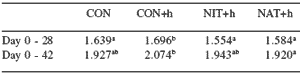
abMeans within a row differ (P<0.05)
Table 11.Effects of Histostat® and Natustat™ on the feed efficiency adjusted for mortality weights of histomoniasischallenged tom turkeys.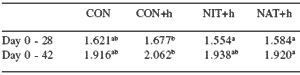
abMeans within a row differ (P<0.05)
Mortality (Table 12) was not a factor for determination in this trial as both of the treated groups had fewer deaths than the non-infected controls. Postmortem observations revealed lesions having characteristics of necrotic enteritis in 7 of the 26; lack of food consumption with no other lesions (starve-outs) for 5 of the 26; 2 of the 26 were culled for severe leg problems, 4 of the 26 succumbed to air sacculitis; another 5 died from cardiac disorder; 1 death was from peritonitis; and 1 death from pericarditis.
Table 12. Effects of Histostat® and Natustat™ on the mortality (%) of histomoniasis-challenged tom turkeys.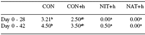
abMeans within a row differ (P<0.05)
Cecal lesion scores (Table 13) of the NIT+h group at 28 days was lower (P=0.05) than that of the NAT+h group, with the 35 day cecal lesion scores indicating no differences (P>0.05) between the two groups. Cecal lesions were lowest (P<0.05) in the CON groups and highest (P<0.05) in the CON+h group at 28 and 35 days. These cecal lesion score results indicate that the natural exposure Histomoniasis disease model employed provided a mild to moderate challenge in this experiment.
Table 13. Effects of Histostat® and Natustat™ on the cecal lesion scores of histomoniasis-challenged tom turkeys.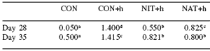
abcdMeans within a row differ (P<0.05)
Liver lesions (Table 14) were not observed at 28 days suggesting that this was too early in the life cycle of the histomonads for these clinical signs. This is further supported by comparing the 28 and 35 day cecal lesion scores that showed a progression in severity giving clear evidence that disease was emerging at time of scoring.
At 35 days, liver lesion scores of the NIT+h and NAT+h groups were not different (P>0.05). Liver lesion scores were lowest (P<0.05) in the CON groups and highest (P<0.05) in the CON+h group at 35 days.
Table 14.Effects of Histostat® and Natustat™ on the liver lesion scores of histomoniasis-challenged tom turkeys.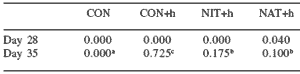
abMeans within a row differ (P<0.05).
Summary
The performance of both turkeys and chickens in the two experiments suggests that Natustat™ may play an important role as a dietary supplement in poultry susceptible to histomoniasis. This is supported by the cecal and liver lesion scores in both species, which were generally not different between Natustat™ and Histostat®- supplemented birds (Tables 7 and 13).
In addition, feed efficiency values of broilers and turkeys (Tables 5, 6, 11 and 12) receiving Natustat™ were less affected by the histomoniasis challenge than those receiving Histostat®. Data from both experiments showed that H. meleagridis-challenged birds fed Natustat™ performed similar to (or intermediate to, depending on parameter) non-challenged, non-supplemented birds and challenged birds supplemented with Histostat®.
References
Alpharma Inc. Technical manual: Histostat® prevents blackhead in yurkeys - challenge trial with histomonas carried by nematodes. http://www.alpharma.com/ahd/ pdf/pd0051.pdf.
Hall, C.F., R.R. Bell, T.B. Blount, W.O. Cawley, S.E. Glass, J.E. Grimes, L.C. Grumbles and S.A. Naqi. 1975. A Manual of Poultry Diseases (B-1031). Texas A&M University, College Station, TX, USA, p. 46.
Hall, W.J. and E.E. Wehe. 1953. Diseases and parasites of poultry. Farmers Bulletin 1652, U.S. Department of Agriculture, Washington, DC, pp. 46-48.
McDougald, L.R. 1997. Blackhead disease (histomoniasis) in chickens. Poult. Digest 56(9):8, 10-11.
McDougald, L.R. and J.M. Casey. 1982. Blackhead disease in chickens. In: Poultry Tips (Feb.). Cooperative Extension Service, University of Georgia, Athens, GA, USA.
North, M.O. 1972. Commercial Chicken Production Manual. The AVI Publishing Co., Inc., Westport, CT, USA, p. 602.
Renwald, R.T. 1970. Gland-O-Lac Poultry Health Book. Omaha, NE, USA, pp. 8, 27.
Salsbury Laboratories Inc. 1982. Salsbury Manual of Poultry Diseases. 7th Ed. Charles City, IA, USA, pp. 7-9.
Skinner, J.T. 2000. Effects of nitarsone as an aid in prevention of blackhead in turkeys challenged with Histomonas meleagridis carried by nematodes. In: Proc. Arkansas Poult. Symp., Springdale, pp. 97-99.
The Merck Veterinary Manual. Histomoniasis, © 2003, Merck & Co., Inc. in cooperation with Merial Ltd. http://www.merckvetmanual.com/mvm/ index.jsp?cfile=htm/bc/203000.htm.
Authors: M.D. SIMS1, C. DUFFY2, R.F. POWER2 and D. HOOGE3
1 Virginia Diversified Research Corporation, Harrisonburg, Virginia, USA
2 European Biosciences Centre, Alltech Inc., Dunboyne, County Meath, Ireland
3 Hooge Consulting Service Inc., Eagle Mountain, Utah, USA


.jpg&w=3840&q=75)