Explore
Communities in English
Advertise on Engormix

Identification and Comparative Expression Analysis of Interleukin 2/15 Receptor b Chain in Chickens Infected with E. tenella

Published: May 12, 2020
By: Jipseol Jeong 1, Woo H. Kim 1, Jeongmi Yoo 1, Changhwan Lee 1, Suk Kim 1, Jae-Hyeon Cho 1, Hyung Kwan Jang 2, Dong W. Kim 3, Hyun S. Lillehoj 4, Wongi Min 1.
Summary

Author details:

1 College of Veterinary Medicine and Research Institute of Life Science, Gyeongsang National University, Jinju, Korea, 2 Departments of Infectious Diseases and Avian Diseases, College of Veterinary Medicine and Korea Zoonosis Research Institute, Chonbuk National University, Jeonju, Korea, 3 National Institute of Animal Science, RDA, Cheonan, Chungnam, Korea, 4 Animal Parasitic Diseases Laboratory, Animal and Natural Resources Institute, Agricultural Research Service, United States Department of Agriculture, Beltsville, Maryland, United States of America.


Background: Interleukin (IL) 2 and IL15 receptor b chain (IL2/15Rb, CD122) play critical roles in signal transduction for the biological activities of IL2 and IL15. Increased knowledge of non-mammalian IL2/15Rb will enhance the understanding of IL2 and IL15 functions.

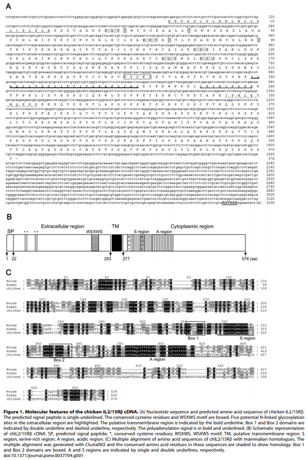
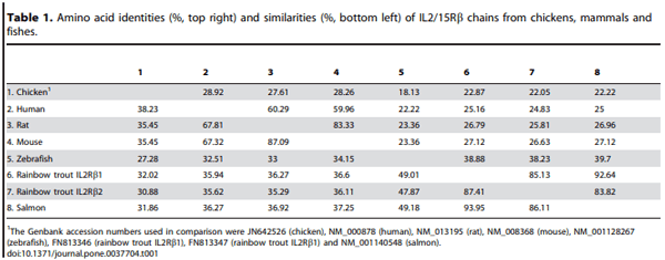
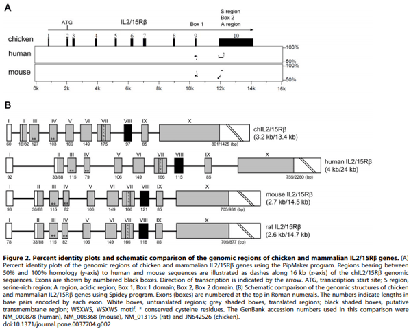
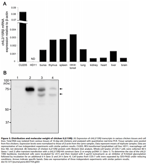
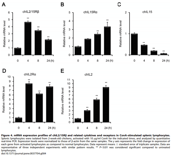
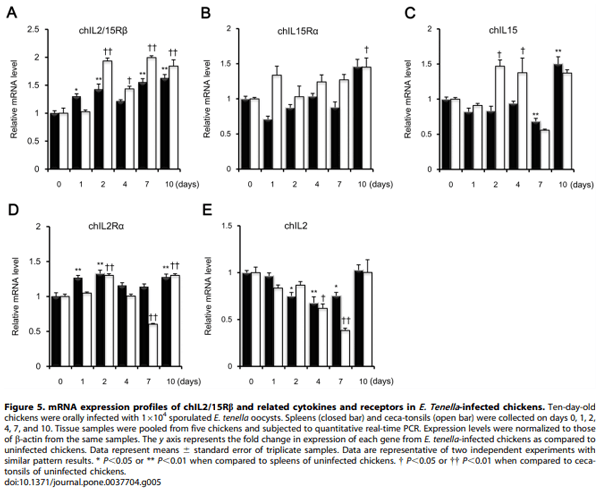
Methology/Principal Findings: Chicken IL2/15Rb (chIL2/15Rb) cDNA was cloned using 59/39-RACE. The predicted protein sequence contained 576 amino acids and typical features of the type-I cytokine receptor family. COS-7 cells transfected with chIL2/15Rb produced proteins of approximately 75 and 62.5 kDa under normal and tunicamycin-treated conditions, respectively. The genomic structure of chIL2/15Rb was similar to its mammalian counterparts. chIL2/15Rb transcripts were detected in the lymphoblast cell line CU205 and in normal lymphoid organs and at moderate levels in bursa samples. Expression profiles of chIL2/15Rb and its related cytokines and receptors were examined in ConA-stimulated splenic lymphocytes and in ceca-tonsils of Eimeria tenella-infected chickens using quantitative real-time PCR. Expression levels of chIL2/15Rb, chIL2Ra, and chIL15Ra were generally elevated in ceca-tonsils and ConA-activated splenic lymphocytes. However, chIL2 and chIL15 expression levels were differentially regulated between the samples. chIL2 expression was upregulated in ConA-activated splenic lymphocytes, but not in ceca-tonsils. In constrast, chIL15 expression was upregulated in ceca-tonsils, but not in ConA-activated splenic lymphocytes.

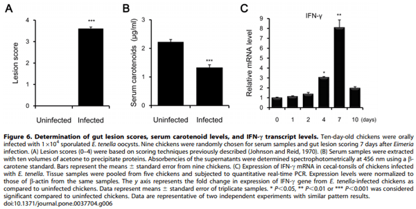
Conclusions/Significance: We identified an avian form of IL2/15Rb and compared its gene expression pattern with those of chIL2, chIL15, chIL2Ra, and chIL15Ra. Our observations suggest that chIL15 and its receptors, including chIL2/15Rb, play important roles in mucosal immunity to intestinal intracellular parasites such as Eimeria.

Introduction
Interleukin 2 (IL2) and IL15 play key roles in the regulation of homeostasis and function of T cells and natural killer (NK) cells. The effects of these cytokines on target cells are mediated by their heterotrimeric receptors that consist of a specific a-subunit and two shared subunits, IL2 and IL15 receptor b (IL2/15Rb, CD122) and a common cytokine receptor γ (γc) [1,2]. Although the asubunits of the IL2 and IL15 receptors define the binding specificity of the cytokines, the subunits do not participate directly in intracellular signaling due to their short cytoplasmic tails. Thus, the cytoplasmic regions of IL2/15Rb and γc, members of the typeI cytokine receptor family that display a characteristic spacing of four conserved cysteine residues and a WSXWS motif, bind to intracellular signaling molecules and transmit intracellular signals [3–5].
The gene encoding IL2/15Rb is composed of ten exons and nine introns, resulting in an 80 kDa transmembrane receptor. IL2/15Rb is expressed on NK cells and CD8+ T cells and is present on activated CD4+ T cells, B cells, and monocytes [6–8]. IL2/15Rb plays critical functions in the regulation of lymphoid development, differentiation, and homeostasis in both innate and adaptive immunity. Expression of IL2/15Rb has been identified in normal, non-immune tissues, such as neurons and glial cells of adult rat brains, suggesting involvement in nerve regeneration [9]. Furthermore, soluble or truncated IL2/15Rb forms have been detected in sera from healthy individuals and patients with inflammatory bowel disease and in lymphoid cell lines [10–12]. IL2/15Rb-related disorders have been reported in which defective IL2/15Rb expression leads to abnormal development of NK cells and intestinal intraepithelial lymphocytes [13,14] and autoimmunity [15–17]. Blockade of IL-2/15Rb results in splenic NK cell deficiency [18] and effectively resolves autoimmune intestinal damage induced by elevated levels of IL15 in IL15-transgenic mice [19].
The avian immune system provides an important model for the study of basic and applied immunology. Despite the general lack of cross-reactivity and the low level of sequence conservation between avian and mammalian cytokines, many chicken genes are homologous to their mammalian counterparts, including IL2 [20], IL15 [21], IL2 receptor a chain (IL2Ra) [22], and IL15Ra [23]. Avian species, unlike mammals, express two different cc transcripts due to alternative splicing [24,25]. Little information, however, is available on chicken IL2/15Rb (chIL2/15Rb). Therefore, we examined full-length cDNA and the genomic structure encoding a chicken homologue of mammalian IL2/ 15Rb. Through quantitative real-time PCR and Western blot analyses, tissue distribution of chIL2/15Rb transcripts and molecular weights were analyzed. Furthermore, quantitative real-time PCR was used to evaluate expression profiles of chIL2/15Rb and related cytokines and receptors in ConAstimulated splenic lymphocytes and ceca-tonsils and spleens from chickens infected with E. tenella.
Results
Cloning and Characterization of chIL2/15Rb cDNA
The full-length cDNA of chIL2/15Rb was cloned using 5’/3’- RACE based on the sequence information of an EST fragment (accession No. ENSGALG00000012472). The chIL2/15Rb cDNA was approximately 3.2 kb and contained a 1731 bp open reading frame (ORF) predicted to encode a putative 576 amino acid protein with a molecular weight of 61.4 kDa (non-glycosylated) and a predicted isoelectric point of 5.15. The predicted chIL2/15Rb amino acid sequence contained a leader sequence (amino acids 1–22), an extracellular domain (amino acids 23–253), a transmembrane domain (amino acids 254–276), and a cytoplasmic domain (amino acids 277–576) (Fig. 1A). Amino acid sequence comparison using ClustalW (www.ebi.ac.uk/Tools/ clustalw2) indicated that chIL2/15Rb shared 27–29% identity with its human, mouse, and rat counterparts [26–28], and 18– 22% identity with those from zebrafish, salmon, and rainbow trout [29] (Table 1).
As shown in Figure 1A and 1B, chIL2/15Rb contained four conserved cysteine residues and a WSXWS motif, both hallmarks of the type-I cytokine receptor superfamily [30]. The translated chIL2/15Rb sequence contained five potential N-linked glycosylation sites (Asn-X-Ser/Thr) in the extracellular region. The large cytoplasmic region harbored signaling molecule binding sites, including PX(I/V)PXP(E/K) (Box 1) and (V/L)E(V/L)L (Box 2) motifs that are common to most hematopoietic cytokine receptors, and serine-rich (S region) and acidic-rich (A region) regions [30– 32]. The S region (amino acids 301–354) contained nine serine residues. The A region (amino acids 347–416) contained 10 negatively-charged amino acids and one positively-charged amino acid. Amino acids 286–294 and 330–340 included Box 1 (LKIHIPDPE) and Box 2 (ISMLEVMQKND), respectively. Interestingly, the highest level of homology is located in the amino acid sequence of the cytoplasmic region with approximately 50% identity and 55–61% similarity (Fig. 1C). By percent identity plots of the genomic regions of chicken and mammalian IL2/ 15Rb genes using the PipMaker program (http://pipmaker.bx. psu.edu/pipmaker/), regions bearing between 50% and 100% homology to human and mouse sequences are located in exons 9 and 10 including Box 1, Box 2, S and A regions (Fig. 2A).
Genomic Structure of the chIL2/15Rb Gene
The genomic clone spanned approximately 13.4 kb and consisted of ten exons and nine introns (Fig. 2B). The exon/ intron organization of the chIL2/15Rb gene was remarkably similar to its mammalian counterparts [33,34], and all exon/ intron boundaries contained consensus splice donor and acceptor sites. The extracellular domain was coded within exons 2–8, the transmembrane domain within exon 8, and the intracellular domain within exons 8–10. The four consensus cysteine residues were located in exons 3 and 4, two in each exon, and the WSXWS motif resided in exon 7. Interestingly, when compared with the genomic structures of the human, rat and mouse IL2/15Rb genes, exons 6 and 9 were identical in length in all four sequences.
Distribution of chIL2/15Rb mRNA in Normal Tissues and Cell Lines
Quantitative real-time PCR analysis was used to examine the expression of chIL2/15Rb transcripts in various normal tissues and two chicken cell lines, the REV-transformed lymphoblast cell line CU205 and the macrophage cell line HD11 (Fig. 3A). The expression levels of chIL2/15Rb transcripts were relatively high in thymus, spleen and ceca-tonsil. Moderate expression was also identified in bursa, lung and liver with low expression levels identified in kidney, heart and brain. Of the two cell lines, only CU205 expressed a high level of chIL2/15Rb transcripts.
Molecular Weight of chIL2/15Rb
Chicken IL2/15Rb gene included eight N-glycosylation sites including five N-glycosylation sites in the extracellular region compared to four N-glycosylation sites in mammalian IL2/15Rb genes such as human, mouse and rat. Thus, molecular weights of chIL2/15Rb were identified in tunicamycin-treated COS-7 cells transfected with a chIL2/15Rb-HA construct. As shown in Figure 3B, molecular weights of chIL2/15Rb were similar to those observed in mammals. The 62.5 kDa protein (lower arrow) is likely the backbone of chIL2/15Rb, whereas the 75 kDa protein (upper arrow) represents an N-linked glycosylated form of the protein.
Quantitative Analysis of chIL2/15Rb mRNA Expression in ConA-activated Splenic Lymphocytes and Tissues from E. tenella-infected Chickens
Using quantitative real-time PCR, expression profiles of chIL2/ 15Rb, IL2, IL15, IL2Ra, and IL15Ra were determined in ConAstimulated splenic lymphocytes (Fig. 4) and in the ceca-tonsils and spleens of chickens infected with E. tenella (Fig. 5). As shown in Figure 4, expression levels of the three receptors, chIL2/15Rb, chIL2Ra, and chIL15Ra, were always elevated in ConA-activated splenic lymphocytes compared to normal splenic lymphocytes. The expression level of chIL2/15Rb peaked after 4 h of ConA stimulation and then gradually decreased (Fig. 4A). Expression levels of chIL2Ra were higher than those of chIL15Ra (Fig. 4B, D). Interestingly, chIL2 and chIL15 mRNA showed inverse expression patterns in splenic lymphocytes following ConA activation; chIL2 mRNA expression increased while chIL15 mRNA levels decreased in time-dependent manners. Compared with control splenic lymphocytes, chIL2 and chIL15 transcript levels 24 h after ConA activation were 9.1 and 0.08 times higher, respectively (Fig. 4C, E), showing an approximately 114-fold difference in expression levels between the two cytokines.
Identification and Comparative Expression Analysis of Interleukin 2/15 Receptor b Chain in Chickens Infected with E. tenella - Image 1
In E. tenella-infected chickens (Fig. 5), expression levels of chIL2/15Rb and chIL2Ra transcripts were, in general, upregulated or unchanged in both ceca-tonsils and spleens compared to those of healthy controls. However, chIL2Ra mRNA expression was down-regulated in ceca-tonsils on day 7 (Fig. 5A, D). Expression of chIL15Ra and chIL15 transcripts was increased in ceca-tonsils, but not in spleens, where chIL15Ra and chIL15 mRNA levels decreased slightly except on day 10 after infection (Fig. 5B, C). chIL2 mRNA expression was remarkably reduced in ceca-tonsils and spleens until day 7, after which chIL2 mRNA levels returned to control levels by day 10 post-infection (Fig. 5E). Collectively, these results suggest that IL15 and its receptors play an imortant function in Eimeria infections.
On day 7, expression levels of chIL15 mRNAs, but not chIL15Ra, were dramatically reduced in ceca-tonsils of E. tenellainfected chickens (Fig. 5B, C). Thus, intestinal lesion scores, serum carotenoid levels, and interferon (IFN)-c transcript levels were additionally monitored post-infection. As shown in Figure 6A, intestinal lesion scores increased significantly (P,0.001) in E. tenellainfected chickens as compared to uninfected chickens. Serum carotenoid levels were reduced significantly in infected chickens (P,0.001) (Fig. 6B). IFN-c transcript expression was approximately 8.1-fold higher in infected chickens on day 7 as compared to control chickens (Fig. 6C). Given that the infections with E. tenella were successful and that the samples were prepared properly, the reduction of chIL15 mRNA may be caused by significant apoptosis of damaged intraepithelial cells (IECs), although additional studies would be needed to determine if the changes seen here are due to changes in transcription or transcript stability.
Discussion
IL2/15Rb functions as the signal-transducing component of the IL2/15R complex. Here, a full-length cDNA encoding chIL2/ 15Rb was cloned. When compared to mammalian sequences, chIL2/15Rb showed higher conservation levels than did chIL2Ra and chcc. When compared to their mammalian counterparts, chIL2Ra and chcc had different molecular characteristics, especially with regard to exon numbers and alternative splicing [22,24]. In contrast, the exon/intron structure was very similar between mammalian and chicken IL2/15Rbs [33,34]. The number of conserved features and domains of the cytoplasmic region of IL2/15Rb between different species suggest that chIL2/ 15Rb plays an important role in IL2- and IL15-mediated signaling events. First, the large cytoplasmic region (300 amino acids) of chIL2/15Rb, as compared to those of chIL2Ra (4 amino acids) and chcc (113 amino acids), provide binding sites for signaling molecules. Second, chIL2/15Rb possesses the hallmark characteristics of mammalian IL2/15Rb, such as the S and A regions and the Box 1 and Box 2 domains, whose locations were highly conserved. Cytoplasmic S and A regions are responsible for IL2- induced mitotic signaling and the physical association of p56lck, a src-family protein tyrosine kinase, respectively [35,36]. The Box 1 and Box 2 domains play a crucial role in the association with Jak1 [32]. Using Multiple alignment and PipMaker analysis, comparison of nucleotide sequences indicated that Box 1, Box 2, S and A regions of chIL2/15Rb gene shared with 60–75% identity to their mammalian counterparts, suggesting that signaling pathways engaged by IL2/15Rb are similar between chickens and mammals. Third, a high degree of synteny was found between chicken and mammalian genomes, in which MPST, KCTD17, TMPRSS6, IL2/15Rb, and C1QTNF6 were present in the same order on chicken chromosome 1, human chromosome 22, and mouse chromosome 15 (data not shown).
Expression of chIL2/15Rb transcripts was high in lymphoid organs, including spleen, ceca-tonsil, and thymus. Interestingly, the CU205 cell line that expresses chicken T-cell receptor 3 (TCR3) and NK cell marker 28–4 antigen constitutively expressed chIL2/15Rb [37,38]. In the bursa, the site of B-cell development in chickens, only moderate levels of chIL2/15Rb transcripts were detected. No chIL2/15Rb transcripts were detected in the HD11 macrophage cell line with quantitative real-time PCR analysis suggesting a minimal interaction of IL2 with chIL2/15Rb as seen in mammals [39,40]. Prior studies have shown that although murine primary macrophages and cell lines express IL2/15Rb in their resting states, IL2 concentrations that are necessary for biological activity are at the nanomolar level (Kd > 10 nM), indicating an absence of high-affinity IL2R complexes in macrophages [39,40]. Because chIL2/15Rb transcripts were mainly expressed in lymphoid organs and cell lines composed of NK and T cells, not B cells and macrophages, tissue-specific expression patterns of IL2/15Rb are similar between chickens and mammals [6,7].
Identification and Comparative Expression Analysis of Interleukin 2/15 Receptor b Chain in Chickens Infected with E. tenella - Image 2
 
Identification and Comparative Expression Analysis of Interleukin 2/15 Receptor b Chain in Chickens Infected with E. tenella - Image 3
Human IL2/15Rb has a calculated molecular size of 58.3 kDa and an actual translated size of 65–77 kDa that increases to 85– 92 kDa upon cross-linking with IL2 [41,8]. N-linked glycosylation and sialylation of human IL2/15Rb are well established and supported in that endoglycosidase F and neuraminidase treatments diminish the molecular weight of human IL2/15Rb from 70–82 kDa to a broader range of 60–78 kDa [42]. Our data suggest post-translational modification of chIL2/15Rb due to the calculated molecular size of 61.4 kDa. As shown in Figure 3B, a shift in chIL2/15Rb size was observed after treatment with tunicamycin, an inhibitor of glycosylation.
chIL2 induces T-cell proliferative activity, and its transcripts are expressed in ConA-activated spleen T cells [20]. ConA activation of T cells also induces cell-surface expression of chIL2R [43]. The interaction between IL2 and its receptors stimulates the growth, differentiation, and survival of antigen-selected cytotoxic T cells by autocrine or paracrine methods [1]. Considering the critical role of IL2/15Rb in IL2-mediated signal transduction and the formation of high- and intermediate-affinity IL2R complexes, increased chIL2/15Rb transcript levels may be necessary to ensure a continued supply of chIL2/15Rb for the formation of higher affinity receptor complexes during mitogenic activation. In contrast, chIL15 is down-regulated in spleen cells activated with ConA [21]. In our data, chIL2 and chIL15 expression levels were inversely related as chIL2 expression increased and chIL15 expression decreased following ConA activation of splenic lymphocytes. These findings suggest that chIL2/15Rb signaling was driven by chIL2 rather than chIL15 in ConA-activated splenic lymphocytes. Interestingly, the expression kinetics of the components of the chIL2/15R complex were quite different. The expression of chIL2/15Rb increased 4.6 times after 4 h of mitogen stimulation with a subsequent decline, whereas transcript levels of chIL2Ra, chIL15Ra, and chIL2 steadily increased indicating independent gene regulation of chIL2/15Rb.
The mRNA expression profiles of the chIL2/15R complex were examined in ceca-tonsils and spleens following oral challenge with live E. tenella oocysts. Because E. tenella mainly infects the cecum, local immunity mediated by cecal tonsil lymphocytes is important in coccidiosis [44,45]. Our data demonstrated the possibility that lymphocytes in ceca-tonsils were more responsive to IL15 than spleen lymphocytes from E. tenella-infected chickens. In cecatonsils, overall mRNA levels of chIL2/15Rb, chIL15Ra, and chIL15 were elevated, suggesting that the interactions of these molecules in local hosts play an important role in immune responses to intracellular parasites.
Identification and Comparative Expression Analysis of Interleukin 2/15 Receptor b Chain in Chickens Infected with E. tenella - Image 4
Interestingly, chIL15 expression levels were significantly downregulated 7 days post-infection with E. tenella, whereas chIL2/ 15Rb and chIL15Ra levels steadily rose. Intraepithelial cells (IECs) constitutively express IL15 mRNA and protein [46]. IL15 synthesis in IECs is up-regulated in L. monocytogenes-infected rats [47], and intestinal cd T cells express high levels of IL15Ra mRNA and proliferate in response to IL15 in mice [48]. In addition, IL2/15Rb expression is induced on CD8ab+ NK1.1+ T cells in the lamina propria (LP) of transgenic mice; IL15 is preferentially expression in small IECs [49]. During E. tenella infection, IECs represent the first line of defense and undergo severe damage, especially 6–8 days after infection [50]. The reduction of chIL15 seven days post-infection may be caused by significant apoptosis of damaged IECs rather than intestinal intraepithelial lymphocytes (IELs) or LP lymphocytes.
Identification and Comparative Expression Analysis of Interleukin 2/15 Receptor b Chain in Chickens Infected with E. tenella - Image 5
The intestinal damage induced by E. tenella infection was accompanied by changes in the expression of chIL2Ra or chIL2. These observations can be explained by the immunophysiological regulation of IL2 production that can prevent autoimmunity [1]. Indeed, when chickens are given an external source of chIL15, but not chIL2, levels of CD3+ T cells are elevated and accompanied by reduced oocyst shedding and body weight loss [51,52]. Taken together, the growth and activation of IELs or LP lymphocytes in response to signaling through chIL2/15Rb is preferentially regulated by chIL15 rather than chIL2 during E. tenella infection.
Here, we cloned chIL2/15Rb and examined its expression patterns and functions. Molecular analysis indicated that chIL2/ 15Rb possesses a number of conserved features and cytoplasmic domains as compared to mammalian IL2/15Rb. Furthermore, chIL2/15Rb transcripts are mainly expressed by NK cells and T-cell subsets, but not on B cells and macrophages. Transcription of chIL2/15Rb was elevated in the cecum during E. tenella infection, indicating an important role of IL15-mediated immunoregulation in local defenses against intracellular parasitism. The availability of recombinant chIL2/15Rb and chIL2/15Rbspecific antibodies for detection will enhance future study of the role of the IL2/15R complex in chickens and its evolutionary relationship among its mammalian counterparts.
Materials and Methods
Animals and Infections
Male cobb 500 chickens (Harim, Korea) were given unlimited access to feed and water. Constant light was provided for the duration of the experiments. Ten-day-old chickens were orally infected with 1x10⁴ sporulated E. tenella oocysts (Korean isolate 291–7) and transferred to disposable cages [53]. E. tenella were cleaned by flotation on 5.25% sodium hypochlorite and washed three times with PBS. All animal experiments protocols were approved by the Institutional Animal Care and Use Committee (IACUC) at Gyeongsang National University, Jinju, Republic of Korea (Approval Number: GNU-LA-34).
Identification and Comparative Expression Analysis of Interleukin 2/15 Receptor b Chain in Chickens Infected with E. tenella - Image 6
 
Identification and Comparative Expression Analysis of Interleukin 2/15 Receptor b Chain in Chickens Infected with E. tenella - Image 7
Cloning of chIL2/15Rb cDNA
Chicken spleens were dilacerated with a syringe plunger through a cell strainer (SPL Life Sciences, Korea) to obtain single-cell suspensions in Hank’s balanced salt solution (HBSS) (Sigma-Aldrich, USA). Total RNA was extracted from splenic lymphocytes using RiboEx reagent (Geneall, Korea) and treated with RNase-free DNase I (Fermentas, Canada). Single-stranded cDNA was synthesized from total RNA with oligo dT primers using a Transcriptor First Strand cDNA Synthesis Kit (Roche Applied Science, Germany).
Based on a chIL2/15Rb EST sequence (http://www.ensembl. org/; accession number: ENSGALG00000012472), 59/39-Rapid Amplification of cDNA Ends (RACE) was performed with chIL2/ 15Rb-specific primers (for 59 RACE, 59-TCTTCTGCATCACCTCCAGC-39; for 39 RACE, 59-CCTCCCCATTCTCCACATCT-39) with splenic lymphocyte cDNA using a 59/39 RACE kit (59/39 RACE 2nd Generation; Roche Applied Science) according to the manufacturer’s protocol. PCR products were cloned into TA vectors (RBC, Taiwan) and sequenced (Macrogen, Korea). PCR was performed on a DNA Engine thermocycler (BioRad, USA) as follows: 5 min at 95uC, 30 cycles of 1 min at 95uC, 1 min at 55uC, and 2 min at 72uC, and a final 5 min extension at 72uC. The cDNA sequence was submitted to GenBank with accession number JN642526.
Cell Culture
Chicken lymphoblast cell line CU205 [37,54], macrophage cell line HD11 [37,55], and splenic lymphocytes were cultured in Dulbecco’s modified eagle’s medium (DMEM) (Hyclone, USA) supplemented with 10% FBS and penicillin/streptomycin (10,000 unit/ml) (Hyclone) at 41°C in 5% CO2. Splenic lymphocytes were resuspended to 5 x 10⁶ cells/ml and stimulated with 10 mg/ml ConA (Amersham Bioscience, Sweden).
Expression of chIL2/15Rb in COS-7 Cells
Full-length chIL2/15Rb cDNA was amplified by PCR from single-stranded cDNA from splenic lymphocytes using the following primers: 59-GATCAAGCTTCCAGAACAGATGAAGCCCTCCT-39 and 59-GATCTCTAGACTAAGCGTAATCTGGAACATCGTATGGGTAGACAGAGCCATGGCTGTATTG-39 containing Hind III and Xba I restriction enzyme sites (single underline) and the influenza virus hemagglutinin (HA) sequence (double underline). PCR products were digested with Hind III and Xba I and cloned into the corresponding restriction sites of pcDNA3.1 (Invitrogen, USA). COS-7 cells [24] were transiently transfected with 10 mg constructs using Lipofectamine Reagent (Invitrogen) and incubated for 5 h in serum-free DMEM at 37uC in 5% CO2. FBS was then added to the growth medium to a final concentration of 10%. To determine the size of the chIL2/15Rb backbone, transfected cells were incubated for 24 h and then treated with 5 mg/ml tunicamycin (Sigma–Aldrich) as an inhibitor of N-linked glycosylation followed by incubation for an additional 6 h and 24 h.
Western Blot Analysis
Protein samples were mixed with equal volumes of sample buffer (0.125 M Tris-HCl, pH 6.8, 4% SDS, 20% glycerol, 10% 2-mercaptoethanol, and 0.004% bromophenol blue), heated for 4 min at 94uC, resolved on 10% SDS-polyacrylamide gels, and electroblotted onto polyvinyl difluoride (PVDF) membranes (BioRad). Membranes were blocked with PBS containing 1% nonfat dry milk for 16 h at 4uC, incubated with monoclonal anti-HA antibody (Sigma–Aldrich) for 1 h, washed three times with PBS containing 0.05% Tween 20 (PBS-T), and incubated with horseradish peroxidase-conjugated goat anti-mouse IgG antibody (Promega, USA) in PBS containing 1% nonfat dry milk for 40 min at room temperature. Membranes were washed five times with PBS-T followed by five washes with distilled water, visualized using an enhanced chemiluminescence (ECL) kit and Western Blotting Detection Reagents (GE Healthcare Life Sciences) and exposed to X-ray film.
Quantitative Real-time PCR
Normal tissues, cell lines, ConA-activated splenic lymphocytes and tissue samples pooled from five chickens infected with E. tenella were subjected to real-time PCR analysis in triplicate. cDNA synthesis was performed using random hexamer primers. Realtime PCR was performed on a CFX96 real-time PCR system (BioRad) with SYBR Green (Bioneer, Korea) using the primers listed in Table 2. A melting curve was obtained at the end of each run to identify that there was a single amplification product and no primer dimers. Standard curves were generated using serial, 5-fold dilutions of ConA-activated splenic lymphocyte cDNA. The relative expression levels of individual transcripts were normalized to those of b-actin with Bio-Rad CFX software. The gene expression levels were quantified using the comparative DCt or DDCt method with b-actin gene as a reference for normalization. The fold change in expression of each gene examined from E. tenella-infected chickens was calculated relative to their expression levels in the same tissues of uninfected chickens.
Determination of Gut Lesion Scores and Serum Carotenoid Levels
Nine chickens were randomly selected for gut lesion scoring and serum analysis 7 days after Eimeria infection. Lesion scores were based on scoring techniques previously described [56]. Each chicken received a numerical value from 0 to 4. Lesion scores were evaluated by three independent observers. To precipitate proteins from serum samples, sera were extracted with ten volumes of acetone. Samples were vortexed, centrifuged for 10 min at 2,8006g, and stored at 4uC for 1 h in the dark. Supernatant absorbencies were determined spectrophotometrically at 456 nm using a b-carotene standard as previously described [57].
Sequence Analysis
The predicted signal peptide sequence and transmembrane region were identified using the SignalIP program (www.cbs.dtu. dk/services/SignalP) and TMpred program (www.ch.embnet.org/ software/TMPRED_form.html), respectively. Theoretical pI value and predicted molecular weight were calculated using Compute pI/Mw (http://web.expasy.org/compute_pi/). Amino acids multiple alignment was determined using ClustalW2 program (www. ebi.ac.uk/Tools/msa/clustalw2) and shaded using GeneDoc program (www.nrbsc.org/gfx/genedoc/) and identity and similarity were calculated using SIAS program (http://imed.med.ucm. es/Tools/sias.html). mRNA to genomic sequence alignment was performed using Spidey program (www.ncbi.nlm.nih.gov/spidey/ ). Percent identity plot (PIP) analysis of the genes between multiple mammalian and avian species was carried out using MultiPipMaker (http://pipmaker.bx.psu.edu/pipmaker/) [61].
Identification and Comparative Expression Analysis of Interleukin 2/15 Receptor b Chain in Chickens Infected with E. tenella - Image 8
Statistical Analysis
Data were calculated by Student’s t-test or one-way ANOVA followed by Dunnet multiple comparison test using InStatH software (Graphpad, San Diego, CA). Differences were considered significant at P,0.05. The data were expressed as mean value 6 standard error.
Author Contributions
Conceived and designed the experiments: JJ WM. Performed the experiments: JJ. Analyzed the data: JJ WM. Contributed reagents/ materials/analysis tools: JJ WHK JY CL SK JHC HKJ DWK HSL. Wrote the paper: JJ WM.
This article was originally published in PLoS ONE 7(5): e37704. doi:10.1371/journal.pone.0037704. This is an Open Access article, free of all copyright, and may be freely reproduced, distributed, transmitted, modified, built upon, or otherwise used by anyone for any lawful purpose. The work is made available under the Creative Commons CC0 public domain dedication.

1. Ma A, Koka R, Burkett P (2006) Diverse functions of IL-2, IL-15, and IL-7 in lymphoid homeostasis. Annu Rev Immunol 24: 657–679.

2. Bodnar A, Nizsaloczki E, Mocsar G, Szaloki N, Waldmann TA, et al. (2008) A biophysical approach to IL-2 and IL-15 receptor function: localization, conformation and interactions. Immunol Lett 116: 117–125.

3. Gaffen SL (2001) Signaling domains of the interleukin 2 receptor. Cytokine 14, 63–77.

4. Burchill MA, Yang J, Vang KB, Farrar MA (2007) Interleukin-2 receptor signaling in regulatory T cell development and homeostasis. Immunol Lett 114: 1–8.

5. Rochman Y, Spolski R, Leonard WJ (2009) New insights into the regulation of T cells by cc family cytokines. Nat Rev Immunol 9: 480–490.

6. Tanaka T, Tsudo M, Karasuyama H, Kitamura F, Kono T, et al. (1991) A novel monoclonal antibody against murine IL-2 receptor b-chain. Characterization of receptor expression in normal lymphoid cells and EL-4 cells. J Immunol 147: 2222–2228.

7. David D, Bani L, Moreau JL, Demaison C, Sun K, et al. (1998) Further analysis of interleukin-2 receptor subunit expression on the different human peripheral blood mononuclear cell subsets. Blood 91: 165–172.

8. Sharon M, Siegel JP, Tosato G, Yodoi J, Gerrard TL, et al. (1988) The human interleukin 2 receptor b chain (p70). Direct identification, partial purification, and patterns of expression on peripheral blood mononuclear cells. J Exp Med 167: 1265–1270.

9. Wang G, Lu C, Liu H, Jin W, Jiao X, et al. (2001) Immunohistochemical localization of interleukin-2 and its receptor subunits a, b and c in the main olfactory bulb of the rat. Brain Res 893: 244–252.

10. Nielsen OH, Ciardelli T, Wu Z, Langholz E, Kirman I (1995) Circulating soluble interleukin-2 receptor a and b chain in inflammatory bowel disease. Am J Gastroenterol 90: 1301–1306.

11. Honda M, Kitamura K, Takeshita T, Sugamura K, Tokunaga T (1990) Identification of a soluble IL-2 receptor b -chain from human lymphoid cell line cells. J Immunol 145: 4131–4135.

12. Montes de Oca P, Malarde V, Proust R, Dautry-Varsat A, Gesbert F (2010) Ectodomain shedding of interleukin-2 receptor b and generation of an intracellular functional fragment. J Biol Chem 285: 22050–22058.

13. Suzuki H, Duncan GS, Takimoto H, Mak TW (1997) Abnormal development of intestinal intraepithelial lymphocytes and peripheral natural killer cells in mice lacking the IL-2 receptor b chain. J Exp Med 185: 499–505.

14. Gilmour KC, Fujii H, Cranston T, Davies EG, Kinnon C, et al. (2001) Defective expression of the interleukin-2/interleukin-15 receptor b subunit leads to a natural killer cell-deficient form of severe combined immunodeficiency. Blood 98: 877–879.

15. Suzuki H, Kundig TM, Furlonger C, Wakeham A, Timms E, et al. (1995) Deregulated T cell activation and autoimmunity in mice lacking interleukin-2 receptor b. Science 268: 1472–1476.

16. Malek TR, Yu A, Vincek V, Scibelli P, Kong L (2002) CD4 regulatory T cells prevent lethal autoimmunity in IL-2Rb-deficient mice. Implications for the nonredundant function of IL-2. Immunity 17: 167–178.

17. Kurreeman FA, Daha NA, Chang M, Catanese JJ, Begovich AB, et al. (2009) Association of IL2RA and IL2RB with rheumatoid arthritis: a replication study in a Dutch population. Ann Rheum Dis 68: 1789–1790.

18. Tanaka T, Kitamura F, Nagasaka Y, Kuida K, Suwa H, et al. (1993) Selective long-term elimination of natural killer cells in vivo by an anti-interleukin 2 receptor b chain monoclonal antibody in mice. J Exp Med 178: 1103–1107.

19. Yokoyama S, Watanabe N, Sato N, Perera PY, Filkoski L, et al. (2009) Antibody-mediated blockade of IL-15 reverses the autoimmune intestinal damage in transgenic mice that overexpress IL-15 in enterocytes. Proc Natl Acad Sci U S A 106: 15849–15854.

20. Sundick RS, Gill-Dixon C (1997) A cloned chicken lymphokine homologous to both mammalian IL-2 and IL-15. J Immunol 159: 720–725.

21. Lillehoj HS, Min W, Choi KD, Babu US, Burnside J, et al. (2001) Molecular, cellular, and functional characterization of chicken cytokines homologous to mammalian IL-15 and IL-2. Vet Immunol Immunopathol 82: 229–244.

22. Teng QY, Zhou JY, Wu JJ, Guo JQ, Shen HG (2006) Characterization of chicken interleukin 2 receptor a chain, a homolog to mammalian CD25. FEBS Lett 580: 4274–4281.

23. Li G, Lillehoj HS, Min W (2001) Production and characterization of monoclonal antibodies reactive with the chicken interleukin-15 receptor alpha chain. Vet Immunol Immunopathol 82: 215–227.

24. Min W, Lillehoj HS, Fetterer RH (2002) Identification of an alternatively spliced isoform of the common cytokine receptor c chain in chickens. Biochem Biophys Res Commun 299: 321–327.

25. Jeong J, Lee C, Yoo J, Koh PO, Kim YH, et al. (2011) Molecular identification of duck and quail common cytokine receptor c chain genes. Vet Immunol Immunopathol 140: 159–165.

26. Hatakeyama M, Tsudo M, Minamoto S, Kono T, Doi T, et al. (1989) Interleukin-2 receptor beta chain gene: generation of three receptor forms by cloned human a and b chain cDNA’s. Science 244: 551–556.

27. Kono T, Doi T, Yamada G, Hatakeyama M, Minamoto S, et al. (1990) Murine interleukin 2 receptor b chain: dysregulated gene expression in lymphoma line EL-4 caused by a promoter insertion. Proc Natl Acad Sci U S A 87: 1806–1810.

28. Page TH, Dallman MJ (1991) Molecular cloning of cDNAs for the rat interleukin 2 receptor a and b chain genes: differentially regulated gene activity in response to mitogenic stimulation. Eur J Immunol 21: 2133–2138.

29. Wang T, Huang W, Costa MM, Secombes CJ (2011) The gamma-chain cytokine/receptor system in fish: more ligands and receptors. Fish Shellfish Immunol 31: 673–687.

30. Nelson BH, Willerford DM (1998) Biology of the interleukin-2 receptor. Adv Immunol 70: 1–81.

31. Miyazaki T, Kawahara A, Fujii H, Nakagawa Y, Minami Y, et al. (1994) Functional activation of Jak1 and Jak3 by selective association with IL-2 receptor subunits. Science 266: 1045–1047.

32. Usacheva A, Sandoval R, Domanski P, Kotenko SV, Nelms K, et al. (2002) Contribution of the Box 1 and Box 2 motifs of cytokine receptors to Jak1 association and activation. J Biol Chem 277: 48220–48226.

33. Codias EK, Olosz F, Malek TR (2000) Genomic organization and 5’ regulatory region of the mouse IL-2 receptor b -chain gene (IL-2Rb). Immunogenetics 51: 508–512.

34. Shibuya H, Yoneyama M, Nakamura Y, Harada H, Hatakeyama M, et al. (1990) The human interleukin-2 receptor b -chain gene: genomic organization, promoter analysis and chromosomal assignment. Nucleic Acids Res 18: 3697–3703.

35. Hatakeyama M, Kono T, Kobayashi N, Kawahara A, Levin SD, et al. (1991) Interaction of the IL-2 receptor with the src-family kinase p56lck: identification of novel intermolecular association. Science 252: 1523–1528.

36. Kono T, Minami Y, Taniguchi T (1993) The interleukin-2 receptor complex and signal transduction: role of the beta-chain. Semin Immunol 5: 299–307.

37. Min W, Lillehoj HS (2002) Isolation and characterization of chicken interleukin17 cDNA. J Interferon Cytokine Res 22: 1123–1128.

38. Gobel TW, Kaspers B, Stangassinger M (2001) NK and T cells constitute two major, functionally distinct intestinal epithelial lymphocyte subsets in the chicken. Int Immunol 13: 757–762.

39. Cox GW, Mathieson BJ, Giardina SL, Varesio L (1990) Characterization of IL2 receptor expression and function on murine macrophages. J Immunol 145: 1719–1726.

40. Matsui K (1999) Role of interleukin-2 receptor expression on macrophages from Salmonella-infected mice. FEMS Immunol Med Microbiol 24: 97–103.

41. Tsudo M, Kozak RW, Goldman CK, Waldmann TA (1986) Demonstration of a non-Tac peptide that binds interleukin 2: a potential participant in a multichain interleukin 2 receptor complex. Proc Natl Acad Sci U S A 83: 9694–9698.

42. Asao H, Takeshita T, Nakamura M, Nagata K, Sugamura K (1990) The evidence for the IL-2 receptor b chain processing from p70 to p75. Int Immunol 2: 469–472.

43. Hilton LS, Bean AG, Kimpton WG, Lowenthal JW (2002) Interleukin-2 directly induces activation and proliferation of chicken T cells in vivo. J Interferon Cytokine Res 22: 755–763.

44. Vervelde L, Vermeulen AN, Jeurissen SH (1996) In situ characterization of leucocyte subpopulations after infection with Eimeria tenella in chickens. Parasite Immunol 18: 247–256.

45. Yun CH, Lillehoj HS, Choi KD (2000) Eimeria tenella infection induces local gamma interferon production and intestinal lymphocyte subpopulation changes. Infect Immun 68: 1282–1288.

46. Reinecker HC, MacDermott RP, Mirau S, Dignass A, Podolsky DK (1996) Intestinal epithelial cells both express and respond to interleukin 15. Gastroenterology 111: 1706–1713.

47. Hirose K, Suzuki H, Nishimura H, Mitani A, Washizu J, et al. (1998) Interleukin-15 may be responsible for early activation of intestinal intraepithelial lymphocytes after oral infection with Listeria monocytogenes in rats. Infect Immun 66: 5677–5683.

48. Inagaki-Ohara K, Nishimura H, Mitani A, Yoshikai Y (1997) Interleukin-15 preferentially promotes the growth of intestinal intraepithelial lymphocytes bearing cd T cell receptor in mice. Eur J Immunol 27: 2885–2891.

49. Ohta N, Hiroi T, Kweon MN, Kinoshita N, Jang MH, et al. (2002) IL-15- dependent activation-induced cell death-resistant Th1 type CD8 ab + NK1.1+ T cells for the development of small intestinal inflammation. J Immunol 169: 460–468.

50. Yun CH, Lillehoj HS, Lillehoj EP (2000) Intestinal immune responses to coccidiosis. Dev Comp Immunol 24: 303–324.

51. Min W, Lillehoj HS, Burnside J, Weining KC, Staeheli P, et al. (2001) Adjuvant effects of IL-1b, IL-2, IL-8, IL-15, IFN-a, IFN-c TGF- b4 and lymphotactin on DNA vaccination against Eimeria acervulina. Vaccine 20: 267–274.

52. Ma D, Ma C, Pan L, Li G, Yang J, et al. (2011) Vaccination of chickens with DNA vaccine encoding Eimeria acervulina 3–1E and chicken IL-15 offers protection against homologous challenge. Exp Parasitol 127: 208–214.

53. Yoo J, Kang SH, Jeong J, Kim WH, Kim S, et al. (2011) Effects of simple and disposable chicken cages for experimental eimeria infections. Korean J Parasitol 49: 299–302.

54. Schat KA, Pratt WD, Morgan R, Weinstock D, Calnek BW (1992) Stable transfection of reticuloendotheliosis virus-transformed lymphoblastoid cell lines. Avian Diseases 36: 432–439.

55. Beug H, von Kirchbach A, Doderlein G, Conscience JF, Graf T (1979) Chicken hematopoietic cells transformed by seven strains of defective avian leukemia viruses display three distinct phenotypes of differentiation. Cell 18: 375–390.

56. Johnson J, Reid WM (1970) Anticoccidial drugs: lesion scoring techniques in battery and floor-pen experiments with chickens. Exp Parasitol 28: 30–36.

57. Matthews JO, Ward TL, Southern LL (1997) Interactive effects of betaine and monensin in uninfected and Eimeria acervulina-infected chicks. Poult Sci 76: 1014–1019.

58. Liu H, Zhang M, Han H, Yuan J, Li Z (2010) Comparison of the expression of cytokine genes in the bursal tissues of the chickens following challenge with infectious bursal disease viruses of varying virulence. Virol J 7: 364.

59. Berndt A, Wilhelm A, Jugert C, Pieper J, Sachse K, et al. (2007) Chicken cecum immune response to Salmonella enterica serovars of different levels of invasiveness. Infect Immun 75: 5993–6007.

60. De Boever S, Vangestel C, De Backer P, Croubels S, Sys SU (2008) Identification and validation of housekeeping genes as internal control for gene expression in an intravenous LPS inflammation model in chickens. Vet Immunol Immunopathol 122: 312–317.

61. Schwartz S, Zhang Z, Frazer KA, Smit A, Riemer C, et al. (2000) PipMaker-A Web Server for Aligning Two Genomic DNA Sequences. Genome Res 10: 577–586.

Related topics
Authors:
Hyun Lillehoj
USDA - United States Department of Agriculture
Recommend
Comment
Share
Profile picture
Would you like to discuss another topic? Create a new post to engage with experts in the community.
Join Engormix and be part of the largest agribusiness social network in the world.