This article was originally published in Journal of World's Poultry Research (2015). Vol. 5(2): 21-28.
Introduction
Necrotic Enteritis (NE) is a worldwide disease caused by Clostridium Perfringens, which is a ubiquitous anaerobic bacterium that is readily found in soil, dust, feces, feed, poultry litter and intestinal contents (Opengart and Songer, 2013). The disease is of an increasing importance especially after restrictions on the preventive use of antimicrobial feed additives. However, conditions that result in damage to the intestinal mucosa as coccidiosis, mycotoxicosis, or others causing disturbance to the normal intestinal microflora, predispose birds to proliferation of Clostridium infection (Elwingeret al., 1992 and Opengart and Songer, 2013).
There are 2 main forms of the disease including the clinical and subclinical course and it exerts a significantly negative impact on production. Clinically C. Perfringens associated liver disease is cholangiohepatitis which is associated with both forms due to the toxin production (Kaldhusdall and Lovland, 2000). Owing to the economical importance of this disease, present study was designed for isolation, identification and studying of both the pathogenicity and antibiotic sensitivity of C. Perfringens from chickens in El-Behera governorate, Egypt.
Material and methods
Samples
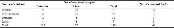
A total of 198 samples (75 liver samples and 123 intestinal samples) representing 40 flocks were collected from diseased chickens with clinical symptoms of necrotic enteritis (Table 1).
Table 1. Type and number of examined chicken samples from different flocks in El-Behera governorate
Bacterial isolation and identification
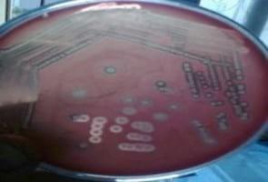
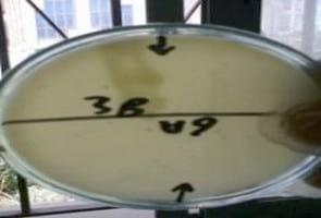
Intestinal and livers tissue samples were inoculated into tubes containing cooked meat medium (Willis, 1977) and incubated in anaerobic jar for 48 hrs at 37oC. Aliquots of 0.1 ml were streaked onto 5% sheep blood agar (with neomycin sulfate (200 ug/ml) (Carter and Cole, 1990); and Perfringens agar (Tryptose Sulphite Cycloserine TSC Agar, Oxoid) were incubated anaerobically at 37°C for 24 hrs. The colonies of C. Perfringens on blood agar appeared flat, olive color with characteristic double zone of hemolysis, the inner clear zone due to Beta toxin, and the outer zone due to alpha toxin as shown in (Fig. 1) (Vaikosen and Muller, 2001). On TSC agar colonies were black due to sulphite reduction ability of C. Perfringens. Microscopic examination revealed Gram positive non motile bacilli. Biochemical identification of C. Perfringens isolates showed catalase and indole negative, nitrate reduction positive, lecithinase (phospholipase C; α- toxin) activity on egg yolk agar with opalescence (Figure 2) (Cruickshank et al., 1975; Koneman et al., 1992 and Macfaddin, 2000).
Polymerase Chain Reaction (PCR)
Oligonucleotide primers (100 pmol) for the 3 toxin genes (alpha, beta, and epsilon) of C. Perfringens were selected according to Yoo et al. (1997) at concentration of 10 pmol, the sequence of the respective oligonucleotide primers were as follow:
C. Perfringens Alpha (CPA) for amplification of the alpha toxin gene (402bp) (Forward primer: 5`GTT GAT AGC GCA GGA CAT GTT AAG 3`; Reverse primer: 5` CAT GTA GTC ATC TGT TCC AGC ATC 3`(; C. Perfringens Beta (CPB) for amplification of the Beta toxin gene (236bp) (Forward primer: 5`ACTATACAGACAGATCATTCAACC3`; Reverse primer 5`TTAGGAGCAGTTAGAACTACAGAC 3`) and C. Perfringens Epsilon (CPE) for amplification of the Epsilon toxin gene (541bp) (Forward primer: 5` ACT GCA ACT ACTACT CAT ACT GTG 3 ; Reverse primer: 5` CTG GTG CCT TAA TAG AAA GAC TCC 3`(.
Extraction of DNA was done according to QIA amp DNA mini kit instructions, Batch No. (51304) and the amplification and cycling protocol for PCR were done using of PCR 1.1x Reddy Mix TM Master Mix (Thermo Scientific).
DNA samples were amplified in a total volume of 50 μl of the following reaction mixture: 25 μl of PCR master mix, 1μl of both of forward and reverse primer for each toxin gene (alpha, beta and epsilon), 14 μl of PCR grade water, and 5 μl of the template DNA (Effat et al., 2007 and Shanmugasamy and Rajeswar, 2012). Thermal cycling designed as initial denaturation for 5 min at 94°C, the samples were subjected to 30 cycles of denaturation at 94°C for 1 min, annealing at 55°C for 1 min and extension at 72°C for 1 min. After the last cycle, a final extension was performed for 3 min. at 72°C. The detection of amplified products were analyzed by electrophoresis on a 1% (w/v) agarose gel in the presence of 100-bp DNA ladder supplied from QIAGEN (Augustynowicz et al., 2000).
Antimicrobial susceptibility testing
Antimicrobials susceptibility testing of C. Perfringens isolates was performed using disc diffusion test (Oxoid, UK). The antimicrobials used were Ampicillin, Amoxicillin, Cefotaxime, Metronidazole, Florfenicol, Cephradine, Lincospectin, Lincomycin, Clindamycin, Norfloxacin, Ciprofloxacin and Enrofloxacin. All isolates were grown over night in 10% neomycin sheep blood agar, then cultures were suspended in 0.85% NaCl to an optical density equivalent to that of McFarland 0.5 standards; each isolate was then inoculated into Mueller Hinton agar medium (Oxoid, UK), 15 minutes later, antimicrobial discs were applied. Plates were incubated anaerobically at 37°C for 24 hours and the interpretation was performed according to the manufacturer (Martel et al., 2004).
Experimental induction of NE in broiler chickens
A total number of 140 broiler chicks at 1 day of age were reared for experimental study. They were divided into 14 groups, and birds in all groups were supplied with drinking water and commercial starter feed, ad-libitum. The feed constituents were modified to help stimulate Clostridium Perfringens proliferation. Mixture of wheat and barley were added to the feed at a concentration of 10%. Each group contained 10 birds. At day 12 of age, all chickens (except the control negative group) were orally inoculated with 100 ul of 1x10³ sporulated oocysts concentration of Eimeria tenella as a predisposing infection. Also, on the 14 day of age, all birds were vaccinated with an intermediate plus live Gumboro vaccine to enhance the incidence of NE.
Bacterial inoculum (Botlhoko, 2009)
At 19 days old; the birds were challenged via oral gavages with toxigenic strain of C. Perfringens type A by inoculation of 1ml of 6x10⁸cfu daily, for 3 consecutive days [19th-20th –and 21st day of age]. Three isolates were used for the experiment whereas each of them was used for infection of 4 subgroups. In addition to one group infected orally with E. tenella, and the 6th group was kept as control negative. Chickens in each of the three groups infected with each of C. Perfringens type A isolates were treated with 3 antibiotics that were effective in-vitro sensitivity (amoxicillin; metronidazole; florfenicol) and the treatments started on the 2nd day after infection and continued for 4 days.
Experimentally infected birds were observed for 2 weeks post infection.
Parameters used to evaluate the experimental infection and the efficacy of antibiotics treatment Body Weights (BW) on 19th day of age (on the same day of the experimental infection), 24th day and on the 33thday (at the end of the experiment).
Histopathological examination: samples were taken from intestine and liver 7 and 14 days PI (post infection) at the age of 26 and 33 days (3 and 10 days; respectively; after the last treatment dose) (Sivaseelanet al., 2013).
Determination of the effect of C. Perfringens on liver function enzymes: at the end of experiment (33th day of age, 10 days after the last treatment dose) using [Glutamic pyruvic transaminase; GPT (ALT) and Glutamate oxaloacetate transaminase; GOT (AST)] (Diamond Diagnostics Kits; Batch no: 30175 Hannover, Germany).
Resulte Sults and discussion
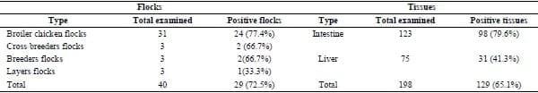
Necrotic enteritis (NE), costs the worldwide poultry industry $2 billion annually and also causes growth retardation and increased mortality (Cooper and Songer, 2009). In Egypt, a previous work reported the incidence of C. Perfringens in broiler chickens at a range between 45-83% and the most common type was A alpha toxin producing (Abd-El Gwad and Abd-El Kader, 2001; Osman et al., 2012 and Abd-El All and Maysa, 2014 ). In this study, C. Perfringens were isolated from 29 out of 40 chicken flocks (72.5%) (Table 2). Expressed as tissue samples, from 198 total tissue samples (123 intestine and 75 liver samples), intestinal and liver samples showed 79.6% and 41.3% positive, respectively (Table 2).
Table 2. Incidence of C.Perfringens among the examined chicken flocks and tissues during 2014 in El-Behera governorate.
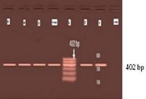
Molecular characterization proved that 5 of the examined field isolates were C. Perfringens type A (alpha toxin) only with a characteristic band at 402 pb (Fig. 3) and all isolates were negative for beta and epsilon toxins. This agreed with Effat et al. (2007) who reported that the same single amplicon, with the same size representing the alpha toxin encoding gene was amplified from all C. Perfringens field isolates causing outbreaks of severe NE during the winter of 2006 in Egypt.
The in-vitro sensitivity of C. Perfringens isolates to 12 different antimicrobials (Table 3). Revealed that these isolates were fairly sensitive to ampicillin, amoxicillin, metronidazole and florfenicol and this is supporting the fact that penicillin’s are known to be particularly active against C. Perfringens and resistance to penicillins and β-lactamase is very rare probably due to the ill-developed plasmid in this bacteria (Hafez, 2011). However C. Perfringens isolates showed intermediate sensitivity to cefotaxime, ciprofloxacin, norfloxacin and enrofloxacin but they recorded low sensitivity to cephradine, lincomycin, clindamycin and lincospectin. These results agreed with Abd-El Gawad and Abd-El Kader (2001) who said that C. Perfringens isolates were highly sensitive to ampicillin, ciprofloxacin, amoxicillin, colistine, lincomycin and moderately sensitive to enerofloxacin, chloramephenicol, erythromycin, oxytetracycline, nalidixicacid, while neomycin, gentamycin, streptomycin had no effect at all. Such data agree also with Llanco et al. (2012) who reported that 22 C. Perfringens type A isolates were susceptible to amoxicillin, amoxicillin-clavulanic acid, cefoxitin, chloramphenicol, enrofloxacin, metronidazole and penicillin- streptomycin combination. Most strains showed high rates of resistance to erythromycin, cephalexin and bacitracin and sulfaquinoaxlin. Also these results agree with Silva et al. (2014) who found that all isolated strains were susceptible to penicillin, metronidazole and vancomycin, whereas four(22.2%), five (27.8%) and 13(72.2%) strains were considered resistant to erythromycin, oxytetracycline and lincomycin respectively.
Table 3. In-vitro sensitivity of C. Perfringens isolates from the examined chicken flocks to different antimicrobial agents.
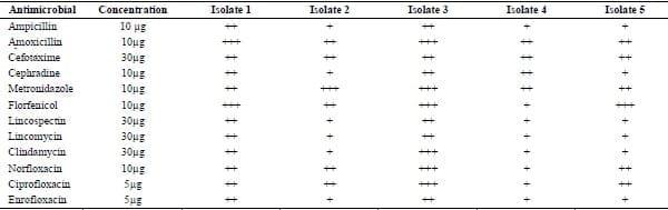
Antimicrobials sensitivity for C. Perfringenstype A isolates. (+) = Low sensitivity; (++) = Moderate sensitivity; (+++) = High sensitivity
Also, Gharaibeh et al. (2010) recorded that lincomycin, erythromycins and tilmicosin showed very high minimal inhibitory concentration (MIC50) of ≥256 μg/ml on C. Perfringens. However, tylosin, amoxicillin and ampicillin, penicillin, florfenicol, danofloxacin, enrofloxacin, chlortetracycline, doxycycline and oxytetracycline had variable MIC50 of 64, 0.5, 1, 1, 8, 4, 8, 4, 8 and 0.5 μg/ml; respectively. They suggested that florfenicol showed in-vitro activity due to fact that it is a fairly new antimicrobial in the field and no significant resistance has developed yet. However, its relatively high minimal inhibitory concentration doesn’t make it as a first choice of treatment of C. Perfringens infection. Metronidazole is a drug used only for the treatment of anaerobic infections in humans; although, because of sensitivity of the C. Perfringens strains isolated from chicken, its use could be a choice for controlling such infection in poultry (Llanco et al., 2012) but our results didn’t match with Hussein and Mostafa (1999) who stated that enrofloxacin was not effective. In the experimental infection, the clinical signs appeared 1 week post infection in the form of marked depression, ruffled feathers, in-appetence, soiled vent, and bloody stained diarrhea. The PM lesions were restricted to the small intestine in the form of enteritis which varied from focal necrosis with exudates in chickens treated with amoxicillin (score 1) to severe hemorrhages and necrosis in the intestinal tract in the form of darkly stained ingesta in chickens treated with florfenicol (score 4). Kidneys showed nephritis with area of hemorrhage and paleness due to C. Perfringens alpha toxin. Also, liver showed congestion and areas of whitish necrosis with distended gall bladder (cholangiohepatitis) in chickens treated with florfenicol.
There was no significant differences in the BW among each of the 3 isolates of C. Perfringens infected chickens. The BW was significantly higher in the control non infected group. Regarding the treatment groups, amoxicillin and metronidazole treated groups showed higher BW than florfenicol treated group. These results agree with Van Immerseel et al. (2004); Hamoda et al. (2011) and Opengart and Songer (2013) (Tables 4 and 5).
Table 4. Effect of experimental infection with C. Perfringens on body weight of infected chickens
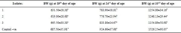
The means within the same column within the same isolate carry different superscripts are significantly different at (P<0.05).
Table 5. Effect of different treatments on the body weight of experimentally infected chickens
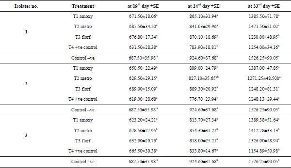
The means within the same column within the same isolate carry different superscripts are significantly different at (P<0.05). T1 amoxy: effect of amoxicillin, T2 metro: effect of metronidazole and T3 florf: effect of florfenicol.
The effect of C. Perfringens infection on liver function enzymes (GPT and GOT) were reported (Tables 6 and 7) as the 3 isolates of C. Perfringens increased the level of liver enzymes compared with control negative chickens with no significance difference among the 3 infected groups. All the antimicrobials decreased the values of liver enzymes compared with the infected non treated group, but amoxicillin caused the most marked decrease in the liver function enzymes. These results are in agreement with Atta et al. (2014). Our results also are in accordance with the note of Llancoet al. (2012) who suggested amoxicillin was effective against necrotic enteritis infection.
Table 6. Effect of experimental infection with C. Perfringens on liver function enzymes (GOT and GPT) of experimentally infected chickens.
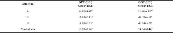
The means within the same column carry different superscripts are significantly different at (P<0.05).
Table 7. The effect of treatments with Amoxycillin, Metronidazol and Florfenicol on liver function enzymes (GOT and GPT) for each isolate of experimentally infected chickens.
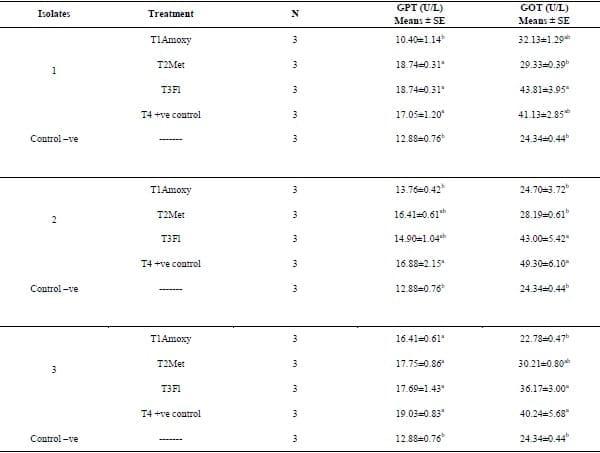
The means within the same column within the same isolate carry different superscripts are significantly different at (P<0.05). T1 amoxy: effect of amoxicillin on isolates, T2 Met: effect of metronidazole on isolates and T3Fl: effect of florfenicol on isolates.
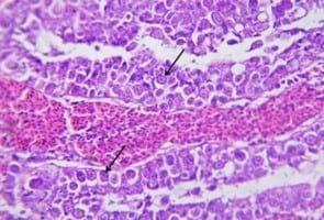
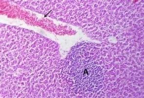
Histopathologically (Table 8), the examined intestine and liver of control negative group showed normal histological structure along the experiment, but in the coccidiosis infected group; the intestine exhibited hemorrhagic and necrotic enteritis due to replacement of the enterocytes with the developmental stage of E. tenella (Figure 4).
All the 3 isolates caused necrotic enteritis with inflammatory cells infiltrations and the presence of the developmental stages of E. tenella, while the liver showed necrotic hepatocytes with inflammatory cell infiltrations and congestion of blood vessel in comparison to the intestine and liver of control negative group (Fig. 5). These results were similar to those of Cooper and Songer (2009).
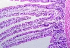
The intestine of chickens infected with each of the 3 isolates and treated with amoxicillin, metronidazol and florfenicol, 3 days after ending the antibiotic treatment, were still showing necrotic enteritis with infiltration of erythrocytes and inflammatory cells and the liver exhibited congestion of blood vessels and hepatic sinusoids. While a good regeneration of enterocytes was shown 10 days after stop of treatment especially in the amoxicillin treated group.
Table 8. Mortality rate and severity of histopathological lesions of intestine and liver of experimentally infected chickens.
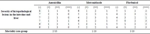
Number of chicken with lesions per total examined (10 chickens per group);Severity of lesions was graded by estimating the percentage area affected in the entire section; Lesion scoring: (-) absence of the lesion = 0%, (+) mild = 5–25%, (++) moderate = 26–50% and (+++) severe > 50% of the examined tissue sections.
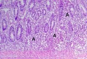
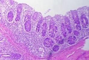
The intestine of infected chickens and treated with florfenicol and metronidazol on the 4th day after ending the treatment showed necrotic enteritis with infiltration of erythrocytes and inflammatory cells: The liver exhibited extravasations of erythrocytes which replaced necrotic hepatocytes besides congestion of blood vessel (Fig. 6 and Fig. 7). The microscopic pictures of the intestine of infected chickens and treated with amoxicillin during 4 days after ending the treatment were necrotic enteritis with mild inflammatory cells infiltration (Fig. 8). We could conclude that the intestine of the infected and treated chickens of all groups explored a good regeneration of enterocytes 11 days after treatment ended especially in the amoxicillin treated groups.
In this work, there was no correlation between the in-vitro and the in-vivo sensitivity of clostridial isolates to the different antibacterials. This can be attributed to the fact that in the gut there are a lot of interfering factors that may affect the antibacterial pharmacodynamics of the antibiotics; like un-healthy gut mucosa due to mycotoxins or viruses like rota or reovirus, artificial infection with coccidiosis, in addition to the un-healthy liver tissue that is unable to metabolize the antibiotics.
Finally, we could conclude that C. Perfringens type A was the most predominant etiology of necrotic enteritis in the examined chicken flocks in El-Behera governorate. The isolated strains of C. Perfringens were mostly in-vitro sensitive to amoxicillin, ampicillin, metronidazole, florfenicol and norfloxacin and they were very weak sensitive to cephradine, lincomycin, clindamycin and lincospectin. There were no differences in the pathogenicity among the examined C. Perfringens isolates and according to the lesion scoring and histopathological results, amoxicillin was the most effective antibiotic on all the 3 isolates of C. Perfringens infected chickens.
Acknowledgement
Grateful thanks to poultry and fish diseases department, faculty of veterinary medicine, Damanhour university for performing laboratory work and experimental infection and also to animal health Research Institute, Dokki, Giza, Egypt for conducting the PCR.
References
1. Abd-El All AM and Maysa A (2014). Toxin genotyping of Clostridium Perfringens isolated from broiler and layer farms and their workers in Egypt. Revue Médecine Vétérinaire, 165, 9-10, 272-279.
2. Abd-El Gwad AM and Abd El-Kader HA (2001). The occurrence of Clostridium Perfringens in the intestine of broiler chickens Assiut governorate. Assiut University Bulletin Environmental Research:4, (2): 13- 22.
3. Atta AH, Shalaby MA and Saifan HY (2014). Efficacy of commiphoramolmol extract against Clostridium Perfringens experimental infection in chickens. World Journal of Pharmacy and Pharmaceutical Sciences, 3 :(12): 365-380.
4. Augustynowicz E, Gzyl A and Slusarczyk J (2000). Molecular epidemiology survey of toxinogenic Clostridium Perfringens strain types by multiplex PCR. Scandinavian Journal of Infectious Diseases, 32(6): 637–641.
5. Botlhoko TD (2009). Performance of Clostridium Perfringens challenged broilers inoculated with effective microorganisms. MSc Agric. (Animal Science), Fac. Of natural and agricultural science, Pretoria Univ.
6. Carter GR and Cole JR (1990). Diagnostic procedures in veterinary bacteriology and mycology.” 5th Ed., Academic Press, Harcourt, BoaceJov. Publisher, New York, Boston, Tokyo, Toronto.
7. Cruickshank R, Duguid JP, Marmion BR and Swain RHA (1975). “Medical Microbiology”, 12t Ed., Living s tone, London, NewYork: , pp: 812-825.
8. Cooper KK, Trinh HT and Songer JG (2009). Immunization with recombinant alpha toxin partially protects broiler chicks against experimental challenge with Clostridium Perfringens. Veterinary Microbiology: ,133: 92–97.
9. Effat MM, Abdallah YA, Soheir MF and Rady MM (2007).Characterization of Clostridium Perfringens field isolates implicated in necrotic enteritis outbreaks on private broiler farms in Cairo, by multiplex PCR. African Journal of Microbiology Research:, pp: 29-32.
10. Elwinger K, Schneitz C, Berndtson E, Fossum O,Teglof B and Engstom B (1992). Factors affecting the incidence of necrotic enteritis, caecal carriage of Clostridium Perfringens and bird performance in broiler chicks. Acta Veterinaria Scandinavia: , 33:369–378.
11. Hafez MH (2011). Enteric Diseases of Poultry with Special Attention to Clostridium Perfringens. Pakistan Veterinary Journal, 31(3): 175-184.
12.Gharaibeh S, AlRifai R and Al-Majali A (2010). Molecular typing and antimicrobial susceptibility of Clostridium Perfringens from broiler chickens. Anaerobe:, 16: 586- 589.
13. Hamouda AS, Amer MM, Mohamed AA, Sherin SA and Merati R (2011). Experimental induction of subclinical necrotic enteritis using Clostridium Perfringens field isolate in male layer chickens. Giza. Veterinary Medical Journal, 59(3): 363-383.
14. Hussein SZ and Mostfa FA (1999). Necrotic enteritis in broiler chicken in Assiut Governorate. Assiut Veterinary Medical Journal, 41 (82): 239-246.
15. Kaldhusdal M and Løvland A (2000). The economical impact of Clostridium Perfringens is greater than anticipated. World Poultry, 16, 50-51.
16. KonemanEW, Allen SD, Dowell VR and Summers HW (1992). “Colour atlas and text book of diagnostic m microbiology.” 4Ed.J.B.LippinCott, New York, London.
17. Llanco LA, Viviane N, Ferreira AJ and Avilacampos MJ (2012). Toxinotyping and antimicrobial susceptibility of Clostridium Perfringensisolated from broiler chickens with necrotic enteritis. International Journal of Microbiology Research, 4: 290-294.
18. MacFaddin JF (2000). Biochemical tests for identification of medical bacteria. Baltimore: Lippincott Williams and Wilkins; 1-450. 39 pp. 1781–1791.
19. Martel A,Devriese LA, CauwertsK,De-Gussem K, Decostere A and Haesebrouck F (2004).Susceptibility of Clostridium PerfringensllS strains from broiler chickens to antibiotics and anticoccidials. Avian Pathology: ,31: 3-7.
20. Opengart K and Songer JS (2013). Necrotic Enteritis Diseases of Poultry. In diseases of poultry. 13th ed.: Swayne,D. E., Glisson JR, McDougald LR,Nolan Lisa K., Suarez, D. L. and Venugopal Nair. Ames, IA, USA: Iowa State Press; 2013. pp. 949-953.
21. Osman KM, Soliman YA, Amin ZMS and Aly MAK (2012). Prevalence of Clostridium Perfringens type A isolates in commercial broiler chickens and parent broiler breeder hens in Egypt. Revue Scientifique et Technique (International office of Epizootics): , 31: (3): 931-941.
22. Shanmugasamy M and Rajeswar J (2012). Alpha toxin specific PCR for detection of toxigenic strains of Clostridium Perfringensin Poultry. Veterinary World: 5, (6): 365- 368.
23. Silva OS, Francisco CF, Marcus VR, Carlos AO, Nelson R and Francisco CF (2014). Genotyping and antimicrobial susceptibility of Clostridium Perfringens isolated from Tinamidae, Cracidae and Ramphastidae species in Brazil. Ciência Rural:, (44): 486-491.
24. Sivaseelan S, Vijayakumar S, Malmarugan S, Balachandran P and Balasubramaniam GA (2013). Assessment of predisposing effect of coccidiosis to necrotic enteritis in experimentally induced broiler chicken. Veterinary arhive: ,83: 653-664.
25. Vaikosen ES and Muller W (2001). Evaluating biochemical tests for isolation and identification of Clostridium Perfringens in fecal samples of small ruminants in Nigeria. Bulletin Animal Health and production in Africa, 49(4): 244-248.
26. Van Immersee IF, De Buck J, Pasmans F, Huyghebaert G, Haesebrouck F and Ducatelle R (2004).Clostridium Perfringens in poultry: an emerging threat for animal and public health. Avian Patholology: ,33:537–549.
27. Willis AT (1977). Characteristics of the pathogenic and related Clostridia. Anaerobic Bacteriology. Clinical laboratory practice3rd Ed.
28. Yoo HS, Lee SU, Park KY and Park YH (1997) Molecular typing and epidemiological survey of prevalence of Clostridium Perfringens types by multiplex PCR. Journal of Clinical Microbiology: ,35: 228–232.


.jpg&w=3840&q=75)