Explore
Communities in English
Advertise on Engormix

Usefulness of ASF diagnostic techniques in the prevention and control of the disease
Published: October 15, 2021
By: Carmina Gallardo, Raquel Nieto, Alejandro Soler, Jovita Fernández-Pinero, Marisa Arias / European Union Reference Laboratory for African Swine Fever (EURL), Centro de Investigación en Sanidad Animal, INIA-CISA, Valdeolmos 28130, Madrid, Spain.
Introduction
Although African swine fever (ASF) was first described almost a century ago, controlling the disease has proven to be a challenge, in particular because no vaccine or treatment are available. Spread of ASF can only be prevented by early detection and the application of strict compliance of classical disease control methods, including surveillance, epidemiological investigation, tracing of pigs, stamping out in infected holdings, strict quarantine and biosecurity measures and animal movement control. Epidemiology of ASF is very complex by the existence of different virus circulating which induce different clinical forms, different reservoirs and scenarios depending on the geographical localization, and the on-going spread of the disease through Europe and Asia. Survivor pigs can remain persistently infected for months, which may contribute to virus transmission and thus the spread and maintenance of the disease, thereby complicating attempts to control it (Arias et al., 2018). 
Over the last decade, ASF has spread to several European and Asian countries and is now one of the major threats to profitable pig production world-wide (FAO 2020). From the genetic point of view, all the ASFV strains circulating in Europe (except in Sardinia) and in Asia belong to the p72 genotype II and shows high genetic stability with a homology of more than 99.9% (Bao et al., 2019; Forth et al., 2019; Gallardo et al., 2014; Garigliany et al., 2019; Ge et al., 2018; Kim et al., 2020; Malogolovkin et al., 2012; Mazur‑Panasiuk et al., 2019a, b; Le et al., 2019; Rowlands et al., 2008). From the clinical point of view, most of the genotype II isolates of the “Georgia 2007 type” that are currently circulating in Eastern and Central Europe and, now in Asia, are highly virulent and cause very high mortality rates of 91–100% (Pikalo et al., 2019; Zhao et al., 2019). However, experimental and field data provide evidence of the natural evolution of the genotype II ASFVs in Central-Eastern Europe from virulent to attenuated strains able to induce different ASF clinical forms from acute to subclinical infections. These different clinical forms are coexisting in the field, in more or less proportion (Gallardo et al., 2019a, b; Nurmoja et al., 2017; Sargsyan et al., 2018; Zani et al., 2018). 
Given the demonstrated clinical evolution of the disease in some affected areas in Europe, mainly in those areas where the ASFV persist within the wild boar population, every country should update the contingency plan and the early warning system in place to prevent the ASFV entry in free areas, its spread, and subsequent maintenance. Any delay in outbreak response and implementation of control measures can result in greater viral contamination of the environment and promote disease spread (Bellini et al., 2016). Highly virulent ASFV isolates are associated to evident clinical forms and should therefore be easier to detect by passive surveillance. However, the demonstrated presence of animals that survive from sub-acute infections or even are subclinical infected, makes the passive surveillance not sufficient for early disease detection in the case of infection with moderately virulent or attenuated ASFV isolates. This is particularly important in high-risk areas. In case of wild life infected animals a combination of passive surveillance of dead wild boar and active surveillance in areas at highest risk should be considered (Arias et al., 2018). The active surveillance will also provide very valuable data on the evolution of the disease and guidance on the assessment of the effectiveness of the control measures. A surveillance system, to be successful, must have adequate laboratory support for a rapid diagnosis, being a key step to design effective control and eradication programs.
Available ASF diagnostic tests
Availability of reliable and accurate diagnostic assays is a prerequisite for an efficient disease control, as any clinical suspicion of ASF in domestic pigs and wild boar has to be verified by laboratory diagnostic methods. On the international level, laboratory methods, as well as sampling and shipping guidelines, can be found in the OIE Manual of Diagnostic Tests and Vaccines for Terrestrial Animals (Chapter 3.8.1, 2019 edition) and the respective EU Diagnostic Manual (European Commission Decision EC 2003/422/EC). 
The starting point for any laboratory investigation of ASF is sample collection. An important consideration is the purpose of the investigation, for example disease diagnosis, disease surveillance, or health certification. Which animals to sample will depend on the objective of the sampling. For example, when investigating an outbreak (passive surveillance), sick and dead animals should be targeted, while the oldest animals should be sampled when checking if animals have been exposed to the disease (active surveillance) (Beltrán-Alcrudo et al 2017). To be effective, proper samples combined with the selection of diagnostic methods, is of fundamental importance in order to make a rapid and reliable diagnosis. Samples collected from live pigs should include anticoagulated whole blood for the detection of virus or viral nucleic acid and serum for the detection of antibodies, whereas samples collected from dead pigs should comprise tissues from virus detection using. Target organs are spleen, lymph nodes, liver, tonsil, heart, lung, and kidney. Of these, spleen and lymph nodes are the most important as they usually contain the highest amounts of virus. Bone marrow is also useful in incidents involving dead wild animals, as it might be the only tissue that is comparatively well preserved if an animal has been dead for some time. Intra-articular tissues of joints can be examined to check for the presence of low virulent isolates. Tissue fluids are also useful for serological investigations (Gallardo et al., 2015a, 2019b).
In certain situations, sampling can be the bottleneck of swine fever diagnosis, especially in the case of wild boar, but also in remote areas. For this reason, alternative sampling strategies and sample matrices (oral fluids, dried blood-spots on filter papers and swabs) have been tested for ASF (often combined with Classical swine fever sampling) especially for wildlife specimens and under rural conditions. However, most of them are not yet in routine use and need further validation (Blome et al., 2014; Braae et al., 2015; Davies et al., 2017; Grau et al., 2015; Michaud et al., 2007; Mur et al., 2013; Petrov et al., 2014; Randriamparany et al., 2016). 
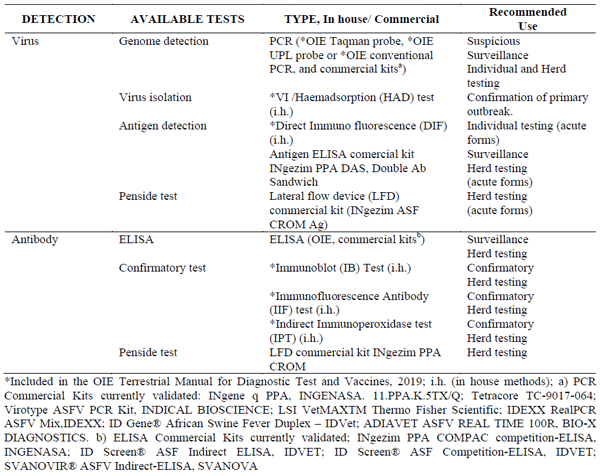
For agent identification, nucleic acid detection tests (real-time PCR, gel-based RT-PCR), virus isolation and haemadsorbing (HAD) assay or antigen detection tests like direct fluorescent antibody test (DIF) on fixed cryosections of organ material and enzyme-linked immunosorbent assays (ELISAs) detecting p72 antigen are available (OIE 2019) (see table 1). Antigen ELISA is a rapid method which can be fully automated; however, its sensitivity is rather low (Gallardo et al., 2015a; 2019b; Oura et al., 2013); therefore, it is not to be used for testing individual animals. ASF antigen detection by DIF yields quick results, and is a highly sensitive test for cases of peracute and acute ASF. It is a robust test, but has been largely replaced by PCR and reagents are no longer widely available. It is important to note that in subacute and chronic disease, where antibodies are present, the sensitivity of this method is also limited (40%) and interpretation of test results is difficult and requires well-trained and experienced laboratory staff. 
Virus isolation (VI) and identification by HAD tests, a characteristic feature of the ASFV-infected cells, are recommended as a reference confirmatory tests in the event of a primary outbreak or a case of ASF (EC 2003). In theory, all of the ASFVs collected from natural outbreaks can be isolated in susceptible primary leukocyte cultures of swine origin, either from blood or lung (alveolar) monocytes or from macrophages cells. However, growing ASFV isolates is a critical step for diagnosis at the national reference laboratories, since it is more expensive than other techniques, requires both specialized facilities and training, is time consuming and cannot be adapted to high throughput. In addition, attempts to isolate infectious virus from field-derived samples provide irregular results. The reason lies in the poor state of samples received, which affects the virus viability, especially on samples obtained from dead or hunted animals, such as wild boar (Gallardo et al., 2015a, 2019b). Despite these constraints, virus isolation is essential to obtain virus stocks for future molecular and biological characterization studies. The use of established cell lines, such as COS-1, IPAM or wild boar lung cells (WSL), can overcome the difficulty in obtaining the primary cells but they are not always suitable for the ASFV isolation from field samples without a little apparent adaptation (Carrascosa et al., 2011; Gallardo et al., 2013). Therefore, further evaluation studies are required for the potential use of established cell lines in routinely diagnosis.
Among the above listed tests the PCR is by far the most sensitive method for the detection of the agent and should be regarded as the method of choice for first-line laboratory diagnosis. A variety of PCR tests, including both conventional and real time (rPCR), have been developed and validated to detect a wide range of ASF isolates belonging to different known virus genotypes, non-HAD strains, and diverse virulence (Agüero et al, 2003; Fernández-Pinero et al, 2013; King et al, 2003; Tignon et al, 2011; Zsak et al, 2005). All of them have been designed in the VP72-coding region, a highly conserved gene coding the major viral protein, assuring the (potential) detection of any ASFV isolate (Oura et al., 2013; Gallardo et al., 2015a). The OIE rPCRs developed by King et al. (2003; OIE 2019) and the OIE Universal probe library (UPL) rPCR developed by Fernández-Pinero et al., (2013; OIE 2019) are the most widely used for routine diagnosis at the EU´s national reference laboratories (NRLs) level (Nieto R., personal communication 2018). Both methods are able to provide a confident ASF diagnosis, although the UPL-PCR has greater diagnostic sensitivity for detecting survivors and allows earlier detection of the disease even when the typical clinical signs are not yet evident (Fernández-Pinero et al., 2013; Gallardo et al., 2015a). Finally, the number of commercial kits for ASFV genome detection based on published rPCRs, has greatly increased over recent years (table 1). These represent an alternative that can guarantee a certain homogeneity in results, which is important in establishing testing procedures to be adopted by many laboratories. Each of the new ASF-commercial assays must to be evaluated and validated following international guidance to ensure they are specific, sensitive, reproducible, precise, robust and accurate. In summary, the PCR is a basic diagnostic tool for surveillance considering the long-term viremia and high viral load that exhibits in the infected animals suffering acute or subacute clinical courses. It is quick and can be used for individual as well as pooled samples. However, the occurrence of low virulent strains in recent years within the EU has made the diagnosis more problematic as some of these strains show only a short period of viremia (Gallardo et al., 2015c; 2018; 2019a). 
Table 1: African swine fever validated ASFV and antibody detection tests.
Whenever the suspicion is raised that ASFV is circulating in a pig population, serological assays must also be used for the diagnosis of disease. Moreover, serology is applied for surveillance purposes and is a valuable tool for further epidemiological investigations, for example, for determining the time point of agent introduction into a pig herd or into a wild boar population. Anti-ASFV antibodies appear soon after infection and persist for up to several months or even years (Arias and Sánchez-Vizcaino 2002, 2012; Arias et al., 2018; Gallardo et al., 2015a). Additionally, no vaccine is available against ASFV, which means that the presence of anti-ASFV antibodies always indicates infection. Antibody-based surveillance is therefore essential for the detection of surviving animals, to elucidate the epidemiological characteristics of the epidemics, i.e., time since the virus introduction into a farm, and for detecting incursions involving low virulence ASFV isolates (Arias et al., 2018; Gallardo et al., 2015a, b, 2019b; Laddomada et al., 2019). The use of antibody detection assays was also crucial for successful eradication programs in the past (Arias and Sánchez-Vizcaíno 2002, 2012). Current ASFV antibody-based tests approved by the OIE involve the use of an ELISA for antibody screening, backed up by Immunoblotting (IB), Indirect Immunofluorescence (IIF) or the Indirect immunoperoxidase tests (IPT) as confirmatory tests (OIE 2019). The IPT has been proved as the best test for ASF serological diagnosis due its superior sensitivity, but moreover, its performance to test any kind of porcine material such as blood, exudate tissues or body fluids (Gallardo et al., 2015a; 2019b). This is particularly relevant for wild boar surveillance and control programs, where poor sample quality can result in false-positive or false-negative reactions in ELISA test kits, a problem which is in particular observed in wild boar specimen. 
The use of the pen-side tests offers a first-line diagnosis that can be useful for rapid application in case of sanitary emergency. The time elapsed between the clinical suspicion and laboratory confirmation used to be relatively long due to the logistics of sending samples to official laboratories. On the other hand, in most cases, regional laboratories do not have the expertise, equipment and/or facilities to diagnose exotic diseases such as ASF. This can be sorted out by the use of pen-side tests for a first front-line diagnosis under field conditions, giving real-time data on the animal’s infection status. Two different lateral flow devices (LFDs) for the detection of antibodies or the viral antigen in blood are commercially available by INGENASA so far, and it is expected several others come in the near future. However, from the published data, the LFDs should not be used alone due to a very limited sensitivity compared to the gold standard methods, overall in the case of the LFD for the detection of antigens (Gallardo et al., 2019b). It is important to point out antibody LFD penside tests are not useful for the detection of acute forms of the disease. They need to be accompanied by virus/antigen detection techniques, due to antibodies cannot be detected before 12-14 days post infection using these tests. The analysis of suspicious samples by both, virus and antibody detection techniques/penside tests will give us a complete picture of what is going on. Test sensitivity needs to be high, whereas specificity is less critical, since any positive result will need to be verified by the competent National reference laboratory. Now, the possible contribution of pen-side tests to the control of notifiable diseases is still discussed controversially and their application may be limited due to national and international regulations. In general, there are a number of commercial diagnostic test kits and reagents on the market and routinely used in the laboratories; however, the quality of the products can vary considerably and in particular, in case of an outbreak scenario, availability of certain test kits on short notice can be problematic.
Taken together, sensitive, specific and robust laboratory diagnostic assays are available; nevertheless, continuous improvement of diagnostic tests in terms of sensitivity, specificity, costs, practicability and robustness is necessary. For the control of the disease, it is important to push the worldwide implementation of already available modern diagnostic techniques in the laboratories involved in ASF diagnosis further ahead. Proficiency laboratory tests, organized by the EU reference laboratory for ASF on a yearly basis, regularly evaluate the status quo of the currently performed diagnostic assays in the EU member states and third countries, thus representing a valuable tool for identification of general gaps in laboratory diagnosis of ASF as well as individual laboratory deviations. 
All newly developed diagnostic tests need to be validated appropriately prior to implementation in routine diagnostics. However, while there are general guidelines for validation of diagnostic tests (e.g., OIE), there are no standardized procedures for validation of specific tests, as the respective criteria have not yet been defined. In this context, the development of standardized reference material (e.g., non-infectious molecular standards for nucleic acid detection assays and reference serum standards for ELISA batch testing) could help to increase the comparability of test validation between laboratories. Development of new tests, improvement of existing ones or even maintenance of fully validated kits currently on the market are time- and labour-consuming tasks; therefore, cooperation between laboratories engaged in diagnostics and research with expertise in diagnostic techniques and commercial companies with expertise in licensing and marketing such products is very useful. 
The ASF diagnostic interpretation
As in any other disease, there is not a single test being 100% reliable (sensitive and specific). For this reason, final diagnosis should be based on the interpretation of the results derived from the use of a number of validated tests, in combination with the information coming from disease epidemiology, scenario, and the clinical signs. A detailed understanding of the time course of viremia and antibody seroconversion during the ASFV infection is a prerequisite to obtain relevant information about the dynamic of the infection in affected areas and to support control and eradication programs. An appropriate diagnosis therefore should involve the detection and identification of ASFV-specific antigens or DNA and antibodies. 
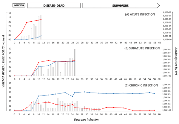
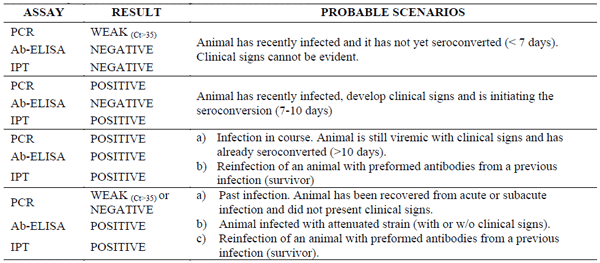
On the basis of experimental data gained at the EURL, upon infection with virulent ASFV strains weak viremia (Cycle threshold Ct >35) can be detectable by real time PCR at on average of 3.75 ± 1.4 days, two days before the onset of the clinical signs. No antibodies are developed. Therefore, a weak PCR positive result on a field blood sample in absence of antibodies could be an indicator of an early phase of the infection (< 1week). Spleen, lymph nodes, liver, and lungs are the sites of secondary viral growth and after 24-30 hours of the primary viremia all tissues contained the virus, reaching the maximum titres around 7-8 days to being infected. The ASFV is therefore easily detected in any kind of porcine sample by real time PCR and even using the antigen detection techniques (DIF or ELISA). A weak antibody response can be detected as early as 7-8 days by IPT in sera and in exudate tissues, mainly spleen and lung, 2-3 days before than with the ELISA (in sera), although with the latter it is rarely detected. The mortality reachs up 92% to 100% within 4 (peracute form) to 12 (acute form) days after the infection.  
Since acute forms are predominant at the beginning of outbreaks, the measures taken in free areas bordering infected areas is based on a risk assessment and on enhanced passive surveillance and PCR testing (SANTE/7113/2015 – Rev 11). However, false positive PCR results, although rare, can occur (e.g., due to lab contamination or other factors). It is unlikely that a primary outbreak (or case) of ASF would be made on the basis of a PCR positive result alone. The most conclusive evidence of infection is isolation of ASFV, but there will be situation in which this is not possible since is time-consuming, requires specialized laboratory and staff and, in comparison with the PCR has limited sensitivity, particularly on samples obtained from dead or hunted wild boar, or in weak positive PCR samples (Gallardo et al., 2015a, 2019b). The occurrence of ASF virus infection can be confirmed if clinical signs or lesions of disease have been detected in the pigs in question and at least two distinct virus or antibody detection tests have given a positive result on samples taken from the same-suspected pig. In wild boar, if virus isolation is not possible, a primary case of ASF can be confirmed when at least two virus or antibody detection tests have given a positive result (EC 2003).
Where ASF becomes endemic, increased numbers of subacute, chronic, and subclinical infections occur and that mortality rates decline over time (Gallardo et al., 2015a, 2015c, 2018, 2019a, b). In clinical terms, subacute ASF develops over a 10-20-day period and the mortality rate ranges from 30 to 70% after 20 days post infection (Arias and Sánchez-Vizcaino, 2012; Beltrán-Alcrudo et al., 2017). Viremia can be detected by real time PCR on average of 8.5 ± 3.6 days and antibodies by ELISA and IPT from 10 days, reaching mean antibody titres of 1:20,000 from the third week. All tissues obtained from animals that succumb within the first month to the infection are positive by PCR and in IPT (Gallardo et al., 2018). The presence of ASF is therefore easily confirmed combined both virus and antibody detection tests, in either blood, sera or tissue samples. 
In recovered pigs surviving acute or subacute infections, the viremia, clearly detectable within the first month, decline over the time and only weak PCR results (Ct >35) can be sporadically detected for up to 78 days (Gallardo, et al., 2018). The common feature in these survivors is the presence of high antibody titer in either blood, sera or tissue samples for the entire life of the animal. Nevertheless, the detection of antibodies in a field sample that resulted PCR weak or negative, is not only an indicator of the presence of survivors from acute or subacute infections. Animals infected with non-HAD and low virulence strains seroconvert after the first week of the infection even in absence of clinical signs or viremia. The antibodies are easily detected by IPT and ELISA, reaching antibody levels >1:160,000 after one month which are maintained over the time (Gallardo et al., 2015c, 2019a; Leitao et al., 2001; Sánchez-Cordon et al., 2017; Sánchez-Vizcaíno et al., 2015). The relative low percentage of non- HAD viruses isolated within the EU could come to the fact that these non-HAD viruses are more difficult to isolate than HAD viruses since the viraemia they cause is sporadic and virus has mostly been isolated in small amounts from the organs. Infection of wild boar with these attenuated non-HAD isolates may account for the seropositive animals detected in the field in absence of clinical signs and viremia. These data emphasize the fact that early detection based only on clinical signs and ASFV genome detection is not an efficient approach for the control of ASF in the current epidemiological situation in Europe. It is likely that the European wild boar is getting endemically infected in certain regions within the EU becoming a recurrent source of infection to other wild boar but also, to domestic pigs. Since each animal could be at a different stage of the disease, both virus and antibody detection tests, for confirming transient viremia and the presence of anti-ASFV specific antibodies, could make sub-clinically ASFV infected wild boar or domestic pigs detectable. A positive test for the presence of the virus indicates that the tested animal was undergoing infection at the time of sampling. On the other hand, a positive ASFV antibody test indicates an ongoing or past infection, where the animals have recovered (and may remain seropositive for life; Tab.2, Fig. 1). 
Table 2. Interpretation of the ASF diagnostic results.
Interpretation of the ASF diagnostic results.
Figure 1. Viremia (measured by real-time PCR) and antibody response (determined by IPT) over time and in relation to the stage of ASF virus infection, as observed in European domestic pigs infected with genotype II ASFV isolates circulating in the EU (2014-2019). Clinical score, expressed in bars, overlapped with viremia and antibody response. 
Viremia (measured by real-time PCR) and antibody response (determined by IPT) over time and in relation to the stage of ASF virus infection, as observed in European domestic pigs infected with genotype II ASFV isolates circulating in the EU (2014-2019). Clinical score, expressed in bars, overlapped with viremia and antibody response.
Final remarks
In conclusion, control-eradication programs in areas with a clear endemic tendency should be reviewed and updated and include parallel routine laboratory monitoring, together with the regular clinical inspection. The use of the most fitting diagnostic tools combining both ASF virus and antibody detection will improve the efficacy of disease control measures, regardless of the nature of the circulating ASFV strains (Arias and Sánchez-Vizcaíno 2002, 2012; Gallardo et al., 2015a, 2019b). An accurate evaluation of the results of the serological and virological tests must be carried out, taking into account all the clinical and epidemiological findings, in the framework of the enquiry to be carried out in case of suspicion or confirmation of ASF.
Published in the proceedings of the International Pig Veterinary Society Congress – IPVS2020. For information on the event, past and future editions, check out https://ipvs2022.com/en.

Agüero M, Fernández J, Romero L, Sánchez Mascaraque C, Arias M, Sánchez-Vizcaíno JM. Highly sensitive PCR assay for routine diagnosis of African swine fever virus in clinical samples. J Clin Microbiol. Sep;41(9):4431-4, 2003. 

Arias M, Jurado C, Gallardo C, Fernández-Pinero J, Sánchez-Vizcaíno JM. Gaps in African swine fever: Analysis and priorities (2018). Transbound Emerg Dis. 2018 May;65 Suppl 1:235-247, 2018. 

Arias M, Sánchez-Vizcaíno JM. African swine fever eradication: The Spanish model. In: Morilla A, Jin K, Zimmerman J, editors. Trends in Emerging Viral Infections of Swine. 1. Ames, IA, USA: Iowa State University Press; pp. 133-139, 2002. 

Arias M, Sánchez-Vizcaíno JM. African swine fever In: Zimmerman, J., Karriker, L.A., Ramirez, A., Schwartz, K.J, Stevenson, G.W., Diseases of swine, 10th Edition. pp. 396-404. Editors: John Wiley and Sons, United States of America, 2012. 

Bao J, Wang Q, Lin P, Liu C, Li L, Wu X, Chi T, Xu T, Ge S, Liu Y, Li J, Wang S, Qu H, Jin T, Wang Z. Genome comparison of African swine fever virus China/2018/AnhuiXCGQ strain and related European p72 Genotype II strains. Transbound Emerg Dis. May;66(3):1167-1176, 2019.

Bellini S, Rutili D, Guberti V. Preventive measures aimed at minimizing the risk of African swine fever virus spread in pig farming systems. Acta Vet Scand. 2016 Nov 29;58(1):82. Review. 

Beltrán-Alcrudo, D, Arias M, Gallardo C, Kramer S, Penrith, ML. African swine fever: detection and diagnosis – A manual for veterinarians. FAO Animal Production and Health Manual No. 19. Rome. Food and Agriculture Organization of the United Nations (FAO). 88 page. Available at http://www.fao.org/3/a-i7228e.pdf. 2017. 

Blome S, Goller KV, Petrov A, Dräger C, Pietschmann J, Beer M. Alternative sampling strategies for passive classical and African swine fever surveillance in wild boar--extension towards African swine fever virus antibody detection. Vet Microbiol. 2014 Dec 5;174(3-4):607-608. 2014. 

Braae UC, Johansen MV, Ngowi HA, Rasmussen TB, Nielsen J, Uttenthal Å. Detection of African swine fever virus DNA in blood samples stored on FTA cards from asymptomatic pigs in Mbeya region, Tanzania. Transbound Emerg Dis. 2015 Feb;62(1):87-90, 2015. 

Carrascosa AL, Bustos MJ, de Leon P. Methods for growing and titrating African swine fever virus: field and laboratory samples. Curr Protoc Cell Biol Chapter 26, Unit 26 14, 2011. 

Cappai S, Rolesu S, Coccollone A, Laddomada A, Loi F. Evaluation of biological and socio-economic factors related to persistence of African swine fever in Sardinia. Prev Vet Med. 2018 Apr 1;152:1-11, 2018. 

Davies K, Goatley LC, Guinat C, Netherton CL, Gubbins S, Dixon LK, Reis AL. Survival of African Swine Fever Virus in Excretions from Pigs Experimentally Infected with the Georgia 2007/1 Isolate. Transbound Emerg Dis. 2017 Apr;64(2):425-431, 2017. 

European Commission (EC). African swine fever diagnostic manual (notified under document number C (2003) 1696) 2003/422/EC). Available at https://eur-lex.europa.eu/legal-content/EN/TXT/PDF/?uri=CELEX:32003D0422& from=EN, 2003.

Fernandez-Pinero J, Gallardo C, Elizalde M, Robles A, Gomez C, Bishop R, Heath L, Couacy-Hymann E, Fasina FO, Pelayo V, Soler A, Arias M. Molecular diagnosis of African Swine Fever by a new real-time PCR using universal probe library. Transbound Emerg Dis 60(1), 48-58, 2013. 

Food and Agriculture Organization of Animal Health (FAO). Emergency Prevention System for Animal Health (EMPRES-AH): ASF situation in Asia update. Available from http://www.fao.org/ag/againfo/programmes/en/empres/ASF/situation_update.html (Accessed: February 05, 2020), 2020. 

Forth JH, Tignon M, Cay AB, Forth LF, Höper D, Blome S, Beer M. Comparative Analysis of Whole-Genome Sequence of African Swine Fever Virus Belgium 2018/1. Emerg Infect Dis. Jun;25(6):1249-1252, 2019. 

Gallardo C, Soler A, Nieto R, Carrascosa AL, De Mia GM, Bishop RP, Martins C, Fasina FO, Couacy-Hymman E, Heath L, Pelayo V, Martín E, Simón A, Martín R, Okurut AR, Lekolol I, Okoth E, Arias M. Comparative evaluation of novel African swine fever virus (ASF) antibody detection techniques derived from specific ASF viral genotypes with the OIE internationally prescribed serological tests. Vet Microbiol. 2013 Feb 22;162(1):32-43, 2013. 

Gallardo C, Fernandez-Pinero J, Pelayo V, Gazaev I, Markowska-Daniel I, Pridotkas G, Nieto, R, Fernandez-Pacheco P, Bokhan S, Nevolko O, Drozhzhe Z, Perez C, Soler A, Kolvasov D, Arias M. Genetic variation among African swine fever genotype II viruses, eastern and central Europe. Emerg Infect Dis 20(9), 1544-7, 2014. 

Gallardo C, Nieto R, Soler A, Pelayo V, Fernández-Pinero J, Markowska-Daniel I, Pridotkas G, Nurmoja, I, Granta R, Simón A, Pérez C, Martín E, Fernández-Pacheco P, Arias M. Assessment of African Swine Fever Diagnostic Techniques as a Response to the Epidemic Outbreaks in Eastern European Union Countries: How To Improve Surveillance and Control Programs. J Clin Microbiol. 2015 Aug;53(8):2555-65, 2015a. 

Gallardo MC, Reoyo AT, Fernández-Pinero J, Iglesias I, Muñoz MJ, Arias ML. African swine fever: a global view of the current challenge. Porcine Health Manag. Dec 23;1:21, 2015b. 

Gallardo C, Soler A, Nieto, R, Sánchez MA, Martins C, Pelayo V, Carrascosa A, Revilla Y, Simón A, Briones V, Sánchez-Vizcaíno JM, Arias M Experimental Transmission of African Swine Fever (ASF) Low Virulent Isolate NH/P68 by Surviving Pigs. Transbound Emerg Dis. Dec;62(6):612-22, 2015c. 

Gallardo C, Nurmoja I, Soler A, Delicado V, Simón A, Martin E, Perez C, Nieto R and Arias M. Evolution in Europe of African swine fever genotype II viruses from highly to moderately virulent. Vet Microbiol. 2018 Jun;219:70-79. doi: 10.1016/j.vetmic.2018.04.001. Epub Apr 7, 2018.

Gallardo C, Soler A, Rodze I, Nieto R, Cano-Gómez C, Fernandez-Pinero J, Arias M. Attenuated and nonhaemadsorbing (non-HAD) genotype II African swine fever virus (ASFV) isolated in Europe, Latvia 2017. Transbound Emerg Dis. 2019 May;66(3):1399-1404, 2019a. 

Gallardo C, Fernández-Pinero J, Arias M. African swine fever (ASF) diagnosis, an essential tool in the epidemiological investigation.(2019b). Virus Res. 2019 Oct 2;271:197676. doi: 10.1016/j.virusres.2019.197676. Epub 2019 Jul 27. Review.PubMed PMID: 31362027, 2019b. 

Garigliany M, Desmecht D, Tignon M, Cassart D, Lesenfant C, Paternostre J, Volpe R, Cay AB, van den Berg T, Linden A. Phylogeographic Analysis of African Swine Fever Virus, Western Europe, 2018. Emerg Infect Dis. Jan;25(1):184-186, 2019.

Ge S, Li J, Fan X, Liu F, Li L, Wang Q, Ren W, Bao J, Liu C, Wang H, Liu Y, Zhang Y, Xu T, Wu X, Wang Z. Molecular Characterization of African Swine Fever Virus, China, 2018. Emerg Infect Dis. Nov;24(11):2131-2133, 2018. 

Grau FR, Schroeder ME, Mulhern EL, McIntosh MT, Bounpheng MA. Detection of African swine fever, classical swine fever, and foot-and-mouth disease viruses in swine oral fluids by multiplex reverse transcription real-time polymerase chain reaction. J Vet Diagn Invest. 2015 Mar;27(2):140-9, 2015. 

Kim HJ, Cho KH, Lee SK, Kim DY, Nah JJ, Kim HJ, Kim HJ, Hwang JY, Sohn HJ, Choi JG, Kang HE, Kim YJ. Outbreak of African swine fever in South Korea, 2019. Transbound Emerg Dis. 2020 Jan 19. doi: 10.1111/tbed.13483. [Epub ahead of print] PubMed PMID: 31955520, 2019. 

King DP, Reid SM, Hutchings GH, Grierson SS, Wilkinson PJ, Dixon LK, Bastos AD, Drew TW. Development of a TaqMan PCR assay with internal amplification control for the detection of African swine fever virus. J Virol Methods. 2003 Jan;107(1):53-61, 2003. 

Laddomada A, Rolesu S, Loi F, Cappai S, Oggiano A, Madrau MP, Sanna ML, Pilo G, Bandino E, Brundu D, Cherchi S, Masala S, Marongiu D, Bitti G, Desini P,Floris V, Mundula L, Carboni G, Pittau M, Feliziani F, Sanchez-Vizcaino JM, Jurado C, Guberti V, Chessa M, Muzzeddu M, Sardo D, Borrello S, Mulas D, Salis G, Zinzula P, Piredda S, De Martini A, Sgarangella F. Surveillance and control of African Swine Fever in free-ranging pigs in Sardinia. Transbound Emerg Dis. 2019 May;66(3):1114-1119, 2019. 

Le VP, Jeong DG, Yoon SW, Kwon HM, Trinh TBN, Nguyen TL, Bui TTN, Oh J, Kim JB, Cheong KM, Van Tuyen N, Bae E, Vu TTH, Yeom M, Na W, Song D. Outbreak of African Swine Fever, Vietnam, 2019. Emerg Infect Dis. 2019 Jul;25(7):1433-1435. doi: 10.3201/eid2507.190303. Epub 2019 Jul 17. PubMed PMID: 31075078; PubMed Central PMCID: PMC6590755. 

Leitão A, Cartaxeiro C, Coelho R, Cruz B, Parkhouse RM, Portugal F, Vigário JD, Martins CL. The nonhaemadsorbing African swine fever virus isolate ASFV/NH/P68 provides a model for defining the protective anti-virus immune response. J Gen Virol. Mar;82(Pt 3):513-23, 2001 

Malogolovkin A, Yelsukova A, Gallardo C, Tsybanov S, Kolbasov D. Molecular characterization of African swine fever virus isolates originating from outbreaks in the Russian Federation between 2007 and 2011. Vet Microbiol 158(3-4), 415-9, 2012. 

Mazur-Panasiuk N, Woźniakowski G. The unique genetic variation within the O174L gene of Polish strains of African swine fever virus facilitates tracking virus origin. Arch Virol. Jun;164(6):1667-1672, 2019a. 

Mazur-Panasiuk N, Woźniakowski G, Niemczuk K. The first complete genomic sequences of African swine fever virus isolated in Poland. Sci Rep. 2019b Mar 14;9(1):4556, 2019b. 

Michaud V Gil P, Kwiatek O, Prome S, Dixon L, Romero L, Le Potier MF, Arias M, Couacy-Hymann E, Roger F, Libeau G, Albina E. Long-term storage at tropical temperature of dried-blood filter papers for detection and genotyping of RNA and DNA viruses by direct PCR. J Virol Methods. 2007 Dec;146(1-2):257-65, 2007. 

Mur L, Gallardo C, Soler A, Zimmermman J, Pelayo V, Nieto R, Sánchez-Vizcaíno JM, Arias M. Potential use of oral fluid samples for serological diagnosis of African swine fever. Vet Microbiol. 2013 Jul 26;165(1-2):135-9, 2013. 

Nurmoja I, Petrov A, Breidenstein C, Zani L, Forth J H, Beer M, Kristian M, Viltrop A, Blome S. Biological characterization of African swine fever virus genotype II strains from north-eastern Estonia in European wild boar. Transbound Emerg Dis. Jan 24, 2017. 

Oura CA, Edwards L, Batten CA. Virological diagnosis of African swine fever--comparative study of available tests. Virus Res. Apr;173(1):150-8. Review, 2013. 

Petrov A, Schotte U, Pietschmann J, Dräger C, Beer M, Anheyer-Behmenburg H, Goller KV, Blome S. Alternative sampling strategies for passive classical and African swine fever surveillance in wild boar. Vet Microbiol. 2014 Oct 10;173(3-4):360-5, 2014. 

Pikalo J, Zani L, Hühr J, Beer M, Blome S. Pathogenesis of African swine fever in domestic pigs and European wild boar - lessons learned from recent animal trials. Virus Res. 2019 Apr 3, 2019. 

Randriamparany T, Kouakou KV, Michaud V, Fernández-Pinero J, Gallardo C, Le Potier MF, Rabenarivahiny R, Couacy-Hymann E, Raherimandimby M, Albina E. African Swine Fever Diagnosis Adapted to Tropical Conditions by the Use of Dried-blood Filter Papers. Transbound Emerg Dis. 2016 Aug;63(4):379-88, 2016. 

Rowlands RJ, Michaud V, Heath L, Hutchings G, Oura C, Vosloo W, Dwarka R, Onashvili T, Albina E, Dixon L K. African swine fever virus isolate, Georgia, 2007. Emerg Infect Dis 14(12), 1870-4, 2008. 

Sánchez-Cordón PJ, Chapman D, Jabbar T, Reis AL, Goatley L, Netherton CL, Taylor G, Montoya M, Dixon L. Different routes and doses influence protection in pigs immunised with the naturally attenuated African swine fever virus isolate OURT88/3. Antiviral Res. Feb;138:1-8, 2017.

Sánchez-Vizcaíno JM, Mur L, Gomez-Villamandos, JC, Carrasco, L. An update on the epidemiology and pathology of African swine fever. J Comp Pathol. Jan;152(1):9-21, 2015.

Sargsyan MA, Voskanyan HE, Karalova EM, Hakobyan LH, Karalyan ZA. Third wave of African swine fever infection in Armenia: Virus demonstrates the reduction of pathogenicity. Vet World. 2018 Jan;11(1):5-9, 2018.

Tignon M, Gallardo C, Iscaro C, Hutet E, Van der Stede Y, Kolbasov D, De Mia GM, Le Potier MF, Bishop RP, Arias M, Koenen F. Development and inter-laboratory validation study of an improved new real-time PCR assay with internal control for detection and laboratory diagnosis of African swine fever virus. J Virol Methods. 2011 Dec;178(1-2):161-70, 2011.

World Organisation for Animal Health (OIE). African swine fever. In: Manual of diagnostic tests and vaccines for terrestrial animals 2019; Vol 2, Chapter 3.8.1. http://www.oie.int/fileadmin/Home/eng/Health_standards/tahm/3.08.01_ASF.pdf, 2019. 

Zani L, Forth JH, Forth L, Nurmoja I, Leidenberger S, Henke J, Carlson J, Breidenstein C, Viltrop A, Höper D, Sauter-Louis C, Beer M, Blome S. Deletion at the 5'-end of Estonian ASFV strains associated with an attenuated phenotype. Sci Rep. Apr 25;8(1):6510, 2018. 

Zhao D, Liu R, Zhang X, Li F, Wang J, Zhang J Liu X, Wang L, Zhang J, Wu X, Guan Y, Chen W, Wang X, He X, Bu Z. (2019). Replication and virulence in pigs of the first African swine fever virus isolated in China. Emerg Microbes Infect.;8(1):438-447, 2019. 

Zsak L, Borca MV, Risatti GR, Zsak A, French RA, Lu Z, Kutish GF, Neilan JG, Callahan JD, Nelson WM, Rock DL (2005). Preclinical diagnosis of African swine fever in contact-exposed swine by a real-time PCR assay. J Clin Microbiol. Jan;43(1):112-9, 2005.

Related topics
Authors:
Carmina Gallardo
INIA España
Follow
Join to be able to comment.
Once you join Engormix, you will be able to participate in all content and forums.
* Required information
Would you like to discuss another topic? Create a new post to engage with experts in the community.
Create a post
Join Engormix and be part of the largest agribusiness social network in the world.
LoginRegister