Explore
Communities in English
Advertise on Engormix

Recent Advances in the Diagnosis of Classical Swine Fever and Future Perspectives

Published: December 14, 2020
By: Lihua Wang 1, Rachel Madera 1, Yuzhen Li 1, David Scott McVey 2, Barbara S. Drolet 2 and Jishu Shi 1. / 1 Department of Anatomy and Physiology, College of Veterinary Medicine, Kansas State University, Manhattan, KS 66506, USA; 2 United States Department of Agriculture, Arthropod-Borne Animal Diseases Research Unit, Center for Grain and Animal Health Research, Manhattan, KS 66502, USA.
Summary

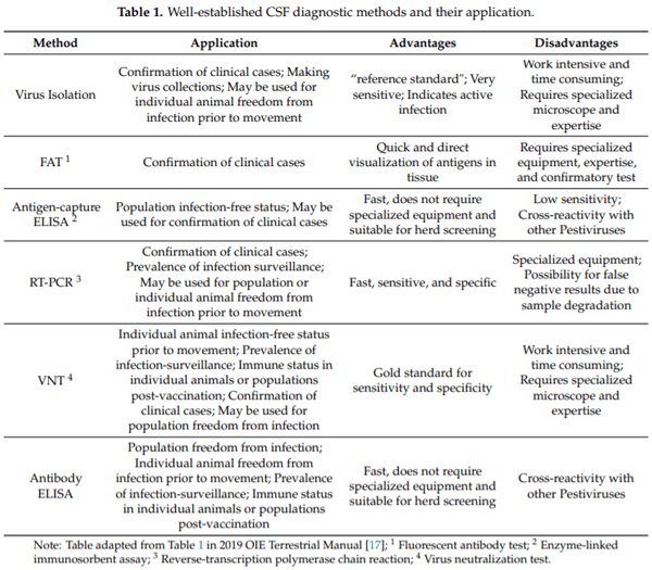
Classical swine fever (CSF) is a highly contagious viral disease of pigs, including wild boar. It is regarded as one of the major problems in the pig industry as it is still endemic in many regions of the world and has the potential to cause devastating epidemics, particularly in countries free of the disease. Rapid and reliable diagnosis is of utmost importance in the control of CSF. Since clinical presentations of CSF are highly variable and may be confused with other viral diseases in pigs, laboratory diagnosis is indispensable for an unambiguous diagnosis. On an international level, well-established diagnostic tests of CSF such as virus isolation, fluorescent antibody test (FAT), antigen capture antibody enzyme-linked immunosorbent assay (ELISA), reverse-transcription polymerase chain reaction (RT-PCR), virus neutralization test (VNT), and antibody ELISA have been described in detail in the OIE Terrestrial Manual. However, improved CSF diagnostic methods or alternatives based on modern technologies have been developed in recent years. This review thus presents recent advances in the diagnosis of CSF and future perspectives.

Keywords: classical swine fever; laboratory diagnosis; technologies; future perspectives.

1. Introduction
Classical swine fever (CSF), a list-A disease classified by the World Organization for Animal Health (OIE), is considered as a transboundary animal disease by the Food and Agriculture Organization of the United Nations (FAO) [1]. The disease causes high morbidity and mortality in both feral and domestic pigs and can result in significant economic losses to the swine industry worldwide [2]. Currently, it is present in many countries in Asia, the Caribbean islands, Africa, and South and Central America (Figure 1). It is most likely to be introduced to CSF-free countries through inadvertent or deliberate importation of classical swine fever virus (CSFV) infected animals, animal products, and animal feed [2,3].
Classical swine fever virus (CSFV) is the etiologic agent of CSF and belongs to the genus Pestivirus in the Flaviviridae family [4]. The genome of CSFV is a positive single-strand RNA of about 12.3 kb. It contains untranslated regions at 5′ and 3′ ends and a single large open reading frame (ORF). The ORF codes four structural (C, Erns, E1, and E2) and eight nonstructural viral proteins (Npro, p7, NS2, NS3, NS4A, NS4B, NS5A, and NS5B) [5,6]. Based on the nucleotide sequences of 5′-non-translated region (5′-NTR) and glycoprotein E2, CSFVs are divided into three genotypes and 11 sub-genotypes (1.1–1.4, 2.1–2.3, and 3.1–3.4) [7–9]. As reported, CSFV genotype 2.1 and genotype 2.3 caused the more recent outbreaks in Europe [10]. Sub-genotypes 1.1, 2.1, 2.2, and 2.3 are prevalent in Asia [11], while sub-genotypes 3.1-3.4 are distributed in other separated geographic regions [1,12–14].
Recent Advances in the Diagnosis of Classical Swine Fever and Future Perspectives - Image 1
Traditional diagnostics for CSF include clinical signs, pathological findings, and antigen and antibody detection [15]. Although unique clinical and pathological observations such as “button” ulcers in the cecum and large intestine mucosa may be found exclusively in CSF, other clinical signs and pathological findings in pigs infected with CSFV are highly variable and are often similar to that of other viral diseases of pigs, such as African swine fever, pseudorabies, porcine dermatitis, and nephropathy syndrome (PDNS), post-weaning multisystemic wasting syndrome (PMWS), thrombocytopenic purpura, and various septicemic conditions [16]. Thus, laboratory diagnosis of CSF for detection of the specific CSFV antigen and antibody is indispensable [15,16]. The well-established diagnostic methods of CSF such as virus isolation, fluorescent antibody test (FAT), antigen capture antibody enzyme-linked immunosorbent assay (ELISA), reverse-transcription polymerase chain reaction (RT-PCR), virus neutralization test (VNT), and antibody ELISA (Table 1) have been widely used and well described in the OIE Terrestrial Manual [17]. Recently developed techniques and alternatives have made significant improvements in several key components of CSF diagnosis, including less sample and reagents required, less effort and time needed, increased detection efficiency (multiplexing), ease of performing and disposal, automation, and point of care (POC). This review provides an updated overview on laboratory diagnosis of CSF and future perspectives.
Recent Advances in the Diagnosis of Classical Swine Fever and Future Perspectives - Image 2
2. Antigen Detection
2.1. Virus Isolation
Virus isolation in cell culture is the oldest laboratory technique for detecting CSFV. Porcine kidney cell lines (PK-15 and SK-6) are often used for isolation of CSFV [17]. However, the use of other porcine cells including swine primary cells (pulmonary alveolar macrophages and peripheral blood mononuclear cells) may enhance the chances of obtaining different CSFVs with different growth characteristics. Since CSFV does not cause a cytopathic effect (CPE), the growth of CSFV in the cells is usually visualized by using immunological technologies with fluorescent or horseradish peroxidase (HRF)-conjugated antibodies [17–19].
The cell culture, virus propagation, and staining are labor intensive and time-consuming (weeks). In addition, skilled and experienced personnel and adequate facilities are needed for cell culture, handling CSFVs and accurate interpretation of the CPE. These disadvantages make virus isolation less attractive for mass surveillance or rapid diagnosis. However, virus isolation is still considered the “gold standard” for confirming CSF clinical cases and the only method for making virus collections (Table 1).
2.2. Fluorescence Antibody Test (FAT)
FAT is the commonly used staining method for CSFV detection. It utilizes fluorescein isothiocyanate (FITC) labeled antibodies to detect CSFV proteins in the slices of cryostat (frozen) tissues or fixed cells. Anti-CSFV gamma-globulins prepared from specific pathogen-free pigs are recommended to be used. These globulins can ensure that most variant CSFVs will be captured. The differentiation of CSFV from other pestiviruses in FAT positive samples, especially bovine viral diarrhea virus (BVDV) and border disease virus (BDV), can be done using RT-PCR with genetic typing or virus isolation in cell culture with specific monoclonal antibody (mAb) typing [17,19].
The main advantages of FAT are that it is relatively easy and rapid to perform and allows direct visualization of the CSFVs in stained tissues. Therefore, it is useful for a first laboratory investigation in suspected clinical cases (Table 1). Several FITC conjugated anti-CSFV antibodies (polyclonal or monoclonal) for FAT are commercially available for research purposes, such as those from Creative Diagnostics, Bioss Inc., Biorbyt LLC, and so on. However, FAT requires highly specialized equipment (i.e., fluorescent microscope) and immunohistochemical staining expertise. It is only recommended to be used in laboratories that have the expertise of performing this technique. The novel ViewRNA in situ hybridization method can detect CSFV RNA directly in infected cells [20]. Using RNA in an in situ hybridization method and specific probes of CSFV RNA, the relative location of CSFV RNA can be visualized in PK15 cells. The sensitivity of this method was three to four orders of magnitude higher than that of FAT. The specificity experiment showed that it was highly specific for CSFV (sub-genotypes 1.1, 2.1, 2.2, and 2.3) and without cross-reaction with other pestiviruses including BVDV, porcine parvovirus (PPV), porcine pseudorabies virus (PRV), and porcine circovirus II (PCV-2). This assay has the potential to be used for testing for CSFV in cells. However, it remains to be determined whether this method can be used to detect CSFV in swine tissues and it is still expensive and is not commercially available yet.
2.3. Antigen-Capture ELISA
Antigen-capture ELISA uses anti-CSFV antibodies on an ELISA plate to capture the CSFV proteins [21]. It has been developed for the rapid screening of large numbers of pigs with clinical suspicion of CSFV infection [15,17,21–23]. Commercial antigen-capture ELISA kits are available from several commercial vendors including IDEXX Laboratories, Thermo Fisher Scientific, MEDIAN Diagnostics, and so on. These kits are double-antibody-sandwich (DAS)-based ELISA for detecting CSFV E2 or Erns protein in serum, blood, plasma, or tissue extracts (Table 2).
Antigen-capture ELISA is fast (provides results within 4 h), easy to perform, and does not require specialized equipment. It can be applied at a herd level for confirmation of clinical cases or determining infection-free population status (Table 1). However, its sensitivity and specificity are lower than most of the other diagnostics, especially the real-time RT-PCR. It is not recommended for testing individual animals and has been increasingly discouraged in recent years.
Recent Advances in the Diagnosis of Classical Swine Fever and Future Perspectives - Image 3
2.4. Real-Time Reverse Transcription Polymerase Chain Reaction (Real-Time RT-PCR)
Real-time RT-PCR has now replaced the traditional RT-PCR and has become an essential tool in the routine diagnosis of CSFV [24–26]. It is a suitable approach for confirmation of clinical cases and prevalence of infection surveillance for CSF (Table 1). Several commercial real-time RT-PCR kits are available for rapid and specific detection of CSFV RNA, including IDEXX RealPCR CSFV RNA Mix, virotypefi CSFV RT-PCR Kit, CSFV dtec-RT-qPCR Test, ADIAVET CSF REAL TIME, CSFV genesigfi Advanced and standard kits, and so on (Table 3). These kits use either SYBR green or TaqMan probe to detect the accumulation of amplicon during the exponential phase of the reaction, which can specifically and sensitively test the CSFV in serum, blood, plasma, viral culture, tissue, or swabs.
The disadvantages of real-time RT-PCR are its high cost and complexity due to simultaneous thermal cycling and fluorescence detection, false positives caused by laboratory contamination from polluted specimens or equipment, and false negatives caused by PCR inhibitors in the sample or degraded RNA [27]. The improved real-time RT-PCRs and advanced alternatives have been designed to help resolve these issues. One-step and automated RT-PCRs can reduce the risk of contamination [27–29]. The primer-probe energy transfer RT-PCR assay provides a higher specificity by analyzing the melting curve following PCR amplification [30,31]. The loop-mediated isothermal amplification (LAMP) assay can accumulate the CSFV amplicon under isothermal conditions [32–34]. The functionalized gold nanoparticles were developed as nanoflare probes for rapid detection of CSFV without nucleic acid amplification [35].
Multiplex real-time RT-PCR as a powerful technique has expanded exponentially in the diagnosis of CSF in recent years. It is quite useful and convenient for quick and accurate detection of different pathogens in mixed infections, which is common in swine production systems. Multiplex RT-PCR assays for rapid detection and genotyping of CSFVs [36,37], simultaneous detection, and differentiation of common swine viruses [38–41] have been developed. Additionally, multiplex combined high-throughput molecular diagnostic platform, user-friendly electronic microarray, magnetoelastic sensor, and microfluidic detection systems were developed as potential alternatives for detection and surveillance of CSFV infection [42–46]. These assays can save considerable time and e ort without compromising robustness and sensitivity and can reduce the sample and reagent requirement as well.
Recent Advances in the Diagnosis of Classical Swine Fever and Future Perspectives - Image 4
2.5. Next Generation Sequencing (NGS)
Next Generation Sequencing (NGS) is a highly sensitive method for generating sequence data and exploring the genetic characters of infectious agents. It has been extensively applied to metagenomics and whole-genome sequencing of infectious viral diseases of livestock [47,48]. By using NGS, researchers found that there might be a long-term persistence of genotype 2.3 CSFV strains in wild boar in Germany [49]. By analyzing NGS data of CSFV isolates of varying virulence in infected pigs, higher quasispecies diversity and more nucleotide variability were found in viral samples from pigs infected with the highly virulent isolates compared to samples of pigs infected with low and moderately virulent isolates [50]. Evolutionary changes in virus populations following the challenge of naïve and vaccinated pigs with the highly virulent CSFV strain were studied using the NGS technology and this study found that vaccination imposes a strong selective pressure on CSF viruses that subsequently replicate within the vaccinated animals [51].
The complete genome sequences obtained from NGS can provide detailed genetic information for construction of reliable phylogenetic relationships of CSFVs for monitoring the evolution and transmission patterns during field outbreaks or epidemics of CSF. One phylogenetic analysis using 58 CSFV complete genome sequences from different Asian countries indicated that the circulating Indian CSFV strains belong to different branches of the 1.1 sub-genotype [52]. These data combined those obtained from other different diagnostic tests can be used for meta-analysis of CSF prevalence, which is important for the investigation of CSF prevalence in different regions [52].
Currently, most of the NGS platforms are expensive to establish and require highly skilled molecular biologists and bioinformaticians. The implementation of NGS is still a challenge and cannot be used as a routine test for disease diagnosis due to cost and the time required [47,48]. However, with the novel and emerging sequencing technologies, cost-effective, user-friendly, and portable NGS will be developed and will act as an effective tool for CSF control and prevention.
3. Antibody Detection
3.1. Virus Neutralization Test (VNT)
VNT is the gold standard for sensitivity and specificity of antibody detection methods. It can be used for confirmation of clinical cases, prevalence of infection surveillance, evaluation of the immune status post-vaccination, and the efficacy of CSF vaccines (Table 1) [53–55]. However, VNT is a work-intensive and time-consuming procedure that requires cell culture and a high-containment laboratory that can handle infectious CSF virus. In addition, it cannot be automated, thus it is not suitable for mass analysis of samples [14,26,53–55].
More recently, alternatives have been developed to overcome the disadvantages of VNT. A neutralizing mAb-based competitive ELISA (cELISA) with emphasis on the replacement of VNT for C-strain post–vaccination monitoring was developed in our group. The test principle of this cELISA is that the neutralizing mAb can compete with C-strain vaccine induced neutralizing antibodies in pig serum to bind the capture antigen C-strain E2 protein. The established cELISA showed 100% sensitivity (95% confidence interval: 94.87 to 100%) and 100% specificity (95% confidence interval: 100 to 100%) when testing C-strain VNT negative pig sera (n = 445) and C-strain VNT positive pig sera (n = 70) and showed excellent agreement (Kappa = 0.957) with VNT when testing the pig sera (n = 139) in parallel. The inhibition rate of serum samples in the cELISA is highly correlated with their titers in VNT (r2= 0.903, p < 0.001). The C-strain antibody can be tested in pigs as early as 7 days post vaccination with the cELISA. This cELISA is a reliable, rapid, simple, safe, and cost-effective tool for sero-monitoring of C-strain vaccination at a population level [56]. In addition, another group developed a high-throughput VNT by using the recombinant CSFV possessing a small report tag and luciferase system. As reported, the VNT titers of the serum can be determined tentatively at 2 days post-infection (dpi) and are comparable to those obtained by conventional VNTs at 3 or 4 dpi. This system allows CSF virus growth to be easily and rapidly monitored and enabled the rapid and easy determination of the VNT titer using a luminometer, which could be a powerful tool to replace the conventional VNT as a high-throughput antibody test for CSFV infections [57].
3.2. Antibody ELISA
Antibody ELISA is the quickest, easiest, and most widely used technique for serological diagnosis and epidemiological investigation of CSF. It is suitable for herd or individual animal CSFV infection screening, prevalence of infection surveillance, and immune status checking in individual animals or populations post-vaccination (Table 1). The E2 protein is crucial for inducing an immune response in the host following CSFV infection [58]. Detection of E2 antibodies in the serum of animals is an easy and reliable method for monitoring CSFV infection during and after outbreaks and for testing coverage of immunization after vaccination [59–61].
Several commercial CSF antibody ELISA kits are available including those from Biocheck, Boehringer Ingelheim, Cusabio Technology LLC, IDEXX Laboratories, ID VET, Indical Bioscience, iNtRON Biotechnology, Median Diagnostics Inc., Thermo Fisher Scientific, and so on. Most of the commercial kits are indirect, competitive, or blocking ELISAs based on the detection of envelop glycoprotein E2 specific antibodies (Table 4). Limitations of these antibody ELISAs are lower specificity (i.e., cross-reactions with BVDV, BDV, and other pestiviruses) and inability to discriminate animals vaccinated with conventional attenuated vaccines or E2-based subunit vaccines.
Recent Advances in the Diagnosis of Classical Swine Fever and Future Perspectives - Image 5
4. Differentiation of Infected from Vaccinated Animals (DIVA) Diagnostic Methods
4.1. Genetic DIVA
The rationale of genetic DIVA (differentiation of infected from vaccinated animals) is the identification of genetic differences between vaccine strains and wild-type CSFVs. Both traditional RT-PCR and real-time RT-PCR (single-plex or multiplex)-based CSF genetic DIVA systems have been developed and evaluated [62–69]. Multiplex nested RT-PCR and real-time RT-PCR assays have been developed for differential detection of wild-type virus from C-strain vaccine [62–68]. A one-step RT-PCR using TaqMan minor-groove-binding (MGB) probes was developed to distinguish between attenuated Korean LOM and wild-type strains of CSFV in Korea [69]. A simple RT-PCR based on the T-rich insertions in CSFV genome was developed for rapid differentiation of wild-type and at least three attenuated lapinized vaccine strains [70]. The modified genotype 1.1 (including C-strain) real-time RT-PCR assay with a real-time RT-PCR assay that detects all known CSFV strains has been successfully used to distinguish C-strain vaccine from the circulating field strains that do not belong to genotype 1 [71].
The genetic DIVA approach facilitates a rapid and reliable differentiation of field virus infected from live attenuated virus vaccinated domestic pigs and wild boars. It is especially useful for detection of the infected animals that are incompletely protected by vaccination and will play a critical role for making decisions prior to and during cessation of a control strategy that employs vaccination with CSF live vaccines.
4.2. Serological DIVA
The ideal serological DIVA test has the ability to discriminate antibodies induced by CSFV infection from the vaccine-derived antibodies, so it can rule out CSFV infected pigs from vaccinated pigs [72]. This can be obtained by detection of specific antibodies against antigens or epitopes that are modified or lacking in a subunit or marker vaccine. It has been shown that antibodies to Erns can be used as an indicator of CSFV infection in pigs and the Erns-based ELISA can be used as a companion diagnostic test to identify CSFV-infected pigs vaccinated with the E2-based subunit or marker vaccines [73–76]. Currently, two Erns ELISAs are commercially available and have been evaluated as accompanying DIVA diagnostic tools for E2 subunit vaccines, CP7_E2alf, or similar chimeric vaccines. One is prioCHECK CSFV Erns (Thermo Fisher Scientific, Waltham, MA, USA); the other is pigtype CSFV Erns Ab (Indical Bioscience, GMBH, Leipzig, Germany) (Table 4). Published data on their evaluations showed that prioCHECK CSFV Erns has a sensitivity of 90–98% with sera from CSFV infected domestic pigs and a specificity of 89–96% with sera from vaccinated domestic pigs [77]. In combination with the marker vaccine “CP7_E2alf”, pigtype CSFV Erns Ab has a sensitivity of 90.2% and a specificity of 93.8% [78]. However, cross-reactivity with antibodies against other pestiviruses was observed for these two Erns ELISAs [77,78]. Depending on the represented data, these two Erns ELISAs are recommended to be used on a herd basis and not for diagnostic analysis on samples of single animals.
Additional approaches or alternatives are undergoing development or further optimization. These include the multiplex microsphere immunoassay [79], which is capable of discrimination within epitope-specific antibody populations [80] and the indirect Erns antibody ELISA with Pichia pastoris-expressed Erns [81]. Recently, our research group successfully generated a mAb against Erns, which can specifically recognize C-strain, but not react with wild-type CSFVs or other viruses in the genus Pestivirus. A cELISA was developed in our group based on the strategy that the C-strain-specific mAb will compete with the C-strain vaccine-induced antibodies in pig serum to bind the capture antigen (C-strain Erns) [unpublished data]. Different from the CSFV neutralizing monoclonal anti-E2 antibody based cELISA for sero-monitoring of C-strain vaccination at a population level [56], this novel anti-ErnsmAb-based cELISA is a valuable tool for measuring and differentiating immune responses to C-strain vaccination and/or infection in pigs. The data about the establishment and validation of this C-strain specific cELISA will be published separately at a later date. In brief, suitable tools for serological DIVA are available. However, there is room for improvement, especially with respect to cross-reactivity issues.
5. Point-of-Care (POC) Diagnostics
User-friendly, cost-effective, rapid, and reliable POC diagnostics (i.e., diagnosis of diseases directly on-site) are indispensable tools for immediate decisions of effective and evidence-based disease control strategies [82]. For example, dipstick tests are designed to use thin paper/plastic strips coated with specific antiviral antibodies to detect viral antigens in serum and other body fluids. Lateral flow assays (LFA) and microfluidic devices are two different and yet more complex technologies that are also based on the biochemical interaction of antigen–antibody. The principles for these three immunochromatographic assays are the same as sandwich ELISA and the major difference between them is that the immunological reaction is carried out on different platforms for different assays. POC studies in animal health management are rare compared to human and companion animal medicine. The immunochromatographic assay-based kits including Antigen Rapid Test Kit (Ring Biotechnology Co., Ltd., Beijing, China), LiliF CSFV antibody rapid test kit (iNtRON Biotechnology, Inc., Gyeonggi, South Korea), and CSFV Antibodies Rapid Test Kit (Antibodies-online Inc., Limerick, PA, USA) are commercially available for rapid testing of CSFV antigen or antibodies in the field. Laboratory-based assays, including the loop-mediated isothermal amplification, combined with a lateral flow dipstick assay [83], the immunochromatographic strip [84], and duplex lateral flow assay [85] have been investigated as potential CSF POC tools as well.
POC diagnostics showed advantages in rapidity and portability, which are the most important parameters considered by farmers and veterinarians [86]. It is foreseeable that as interests and needs of stakeholders increase and new portable POC technologies emerge, novel and applicable POC diagnostics will be developed for detection, control, and prevention of CSF in the field in the near future.
6. Future Perspectives
Although commercial and in-house diagnostics (antigen detection and antibody detection) of CSF are available, there is still room for improvement. The authors suggest that the following aspects should be considered: (i) Continuously improving the sensitivity, specificity, costs, speed, automation, and POC is necessary; (ii) Reference materials (serum bank, virus bank, and non-infectious molecular standards) should be produced and be accessible for validation of the developed CSF diagnostics; (iii) The development of DIVA diagnostics without cross-reaction with antibodies induced by other pestiviruses is critical.
The other emerging infectious diseases, such as the spreading of African swine fever in Asia and the ongoing pandemic of Coronavirus disease 2019 (COVID-19), may shift focus away from the CSF [87,88]. However, as long as CSF exists, it will remain a continuous threat to the pig industry worldwide. Therefore, international cooperation on surveillance and control of CSF becomes even more crucial, both currently and in the future. Researchers should continue to work on developing novel rapid and reliable diagnostics to facilitate the surveillance and control of CSF.
Author Contributions: Conceptualization: J.S., L.W., D.S.M. and B.S.D.; Data curation: L.W., R.M. and Y.L.;Investigation: L.W., R.M. and Y.L.; Supervision: J.S.; Writing–original draft: L.W.; Writing–review & editing: J.S., D.S.M. and B.S.D. All authors have read and agreed to the published version of the manuscript.
Funding: This research is supported by awards from the National Bio and Agro-Defense Facility TransitionFund, the USDA National Institute of Food and Agriculture, Hatch-Multistate project, grant number [1021491]; USDA ARS Non-Assistance Cooperative Agreements, grant numbers [58-8064-8-011, 58-8064-9-007, 58-3020-9-020, 59-0208-9-222]; National Pork Board Grant, grant number [18-059]. The funders had no role in the study design, data collection and analysis, decision to publish, or preparation of the manuscript.
Conflicts of Interest: The authors declare no conflict of interest.
This article was originally published in Pathogens 2020, 9, 658; doi:10.3390/pathogens9080658. This is an Open Access article distributed under the terms and conditions of the Creative Commons Attribution (CC BY) license (http://creativecommons.org/licenses/by/4.0/).
  1. Blome, S.; Staubach, C.; Henke, J.; Carlson, J.; Beer, M. Classical swine fever-an updated review. Viruses 2017, 9, 86. [CrossRef]
  2. Brown, V.R.; Bevins, S.N. A review of classical swine fever virus and routes of introduction into the United States and the potential for virus establishment. Front. Vet. Sci. 2018, 5, 31. [CrossRef] [PubMed]
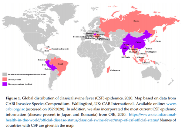
  3. CABI. Invasive Species Compendium; CAB International: Wallingford, UK, 2020; Available online: www.cabi.org/isc (accessed on 29 May 2020).
  4. Schirrmeier, H.; Strebelow, G.; Depner, K.; Ho mann, B.; Beer, M. Genetic and antigenic characterization of an atypical pestivirus isolate, a putative member of a novel pestivirus species. J. Gen. Virol. 2004, 85, 3647–3652. [CrossRef] [PubMed]
  5. Thiel, H.J.; Stark, R.; Weiland, E.; Rumenapf, T.; Meyers, G. Hog cholera virus: Molecular composition of virions from a pestivirus. J. Virol. 1991, 65, 4705–4712. [CrossRef] [PubMed]
  6. Meyers, G.; Thiel, H.J.; Rümenapf, T. Classical swine fever virus: Recovery of infectious viruses from cDNA constructs and generation of recombinant cytopathogenic defective interfering particles. J. Virol. 1996, 70, 1588–1595. [CrossRef]
  7. Lowings, P.; Ibata, G.; Needham, J.; Paton, D. Classical swine fever virus diversity and evolution. J. Gen. Virol. 1996, 77, 1311–1321. [CrossRef]
  8. Paton, D.J.; Mcgoldrick, A.; Greiser-Wilke, I.; Parchariyanon, S.; Song, J.Y.; Liou, P.P.; Stadejek, T.; Lowings, J.P.; Björklund, H.; Belák, S. Genetic typing of classical swine fever virus. Vet. Microbiol. 2000, 73, 137–157. [CrossRef]
  9. Deng, M.C.; Huang, C.C.; Huang, T.S.; Chang, C.Y.; Lin, Y.J.; Chien, M.S.; Jong, M.H. Phylogenetic analysis of classical swine fever virus isolated from Taiwan. Vet. Microbiol. 2005, 106, 187–193. [CrossRef]
  10. Postel, A.; Moennig, V.; Becher, P. Classical swine fever in Europe-the current situation. Berl. Munch. Tierarztl. Wochenschr. 2013, 126, 468–475.
  11. Postel, A.; Nishi, T.; Kameyama, K.I.; Meyer, D.; Suckstor , O.; Fukai, K.; Becher, P. Reemergence of Classical Swine Fever, Japan, 2018. Emerg. Infect. Dis. 2019, 25, 1228–1231. [CrossRef]
  12. Gomez-Villamandos, J.C.; Carrasco, L.; Bautista, M.J.; Sierra, M.A.; Quezada, M.; Hervas, J.; Chacón, M.F.; Ruiz-Villamor, E.; Salguero, F.J.; Sónchez-Cordón, P.J.; et al. African swine fever and classical swine fever: A review of the pathogenesis. Dtsch. Tierarztl. Wochenschr. 2003, 110, 165–169. [PubMed]
  13. Paton, D.J.; Greiser-Wilke, I. Classical swine fever: An update. Res. Vet. Sci. 2003, 75, 169–178. [CrossRef]
  14. Zhou, B. Classical swine fever in China—An update Minireview. Front. Vet. Sci. 2019, 13, 187. [CrossRef] [PubMed]
  15. Greiser-Wilke, I.; Blome, S.; Moennig, V. Diagnostic methods for detection of Classical swine fever virus—Status quo and new developments. Vaccine 2007, 25, 5524–5530. [CrossRef] [PubMed]
  16. Moennig, V.; Floegel-Niesmann, G.; Greiser-Wilke, I. Clinical signs and epidemiology of classical swine fever: A review of new knowledge. Vet. J. 2003, 165, 11–20. [CrossRef]
  17. OIE Terrestrial Manual 2019. Available online: https://www.oie.int/fileadmin/Home/eng/Health_standards/tahm/3.08.03_CSF.pdf (accessed on 29 May 2020).
  18. Grummer, B.; Fischer, S.; Depner, K.; Riebe, R.; Blome, S.; Greiser-Wilke, I. Replication of classical swine fever virus strains and isolates in di erent porcine cell lines. Dtsch. Tierarztl. Wochenschr. 2006, 113, 138–142. [PubMed]
  19. Chander, V.; Nandi, S.; Ravishankar, C.; Upmanyu, V.; Verma, R. Classical swine fever in pigs: Recent developments and future perspectives. Anim. Health Res. Rev. 2014, 15, 87–101. [CrossRef]
  20. Zhang, Q.; Xu, L.; Zhang, Y.; Wang, T.; Zou, X.; Zhu, Y.; Zhao, C.; Chen, K.; Sun, Y.; Sun, J.; et al. A novel ViewRNA in situ hybridization method for the detection of the dynamic distribution of Classical Swine Fever Virus RNA in PK15 cells. Virol. J. 2017, 14, 81. [CrossRef]
  21. Shannon, A.D.; Morrissy, C.; Mackintosh, S.G.; Westbury, H.A. Detection of hog cholera virus antigens in experimentally-infected pigs using an antigen-capture ELISA. Vet. Microbiol. 1993, 34, 233–248. [CrossRef]
  22. Penrith, M.L.; Vosloo, W.; Mather, C. Classical swine fever (hog cholera): Review of aspects relevant to control. Transbound. Emerg. Dis. 2011, 58, 187–196. [CrossRef]
  23. Moennig, V.; Becher, P. Pestivirus control programs: How far have we come and where are we going? Anim. Health Res. Rev. 2015, 16, 83–87. [CrossRef] [PubMed]
  24. Dewulf, J.; Koenen, F.; Mintiens, K.; Denis, P.; Ribbens, S.; de Kruif, A. Analytical performance of several classical swine fever laboratory diagnostic techniques on live animals for detection of infection. J. Virol. Methods 2004, 119, 137–143. [CrossRef] [PubMed]
  25. Depner, K.; Ho mann, B.; Beer, M. Evaluation of real-time RT-PCR assay for the routine intra vitam diagnosis of classical swine fever. Vet. Microbiol. 2007, 121, 338–343. [CrossRef] [PubMed]
  26. Postel, A.; Austermann-Busch, S.; Petrov, A.; Moennig, V.; Becher, P. Epidemiology, diagnosis and control of classical swine fever: Recent developments and future challenges. Transbound. Emerg. Dis. 2018, 65, 248–261. [CrossRef]
  27. Espy, M.J.; Uhl, J.R.; Sloan, L.M.; Buckwalter, S.P.; Jones, M.F.; Vetter, E.A.; Yao, J.D.; Wengenack, N.L.; Rosenblatt, J.E.; Cockerill, F.R., 3rd; et al. Real-time PCR in clinical microbiology: Applications for routine laboratory testing. Clin. Microbiol. Rev. 2006, 19, 165–256. [CrossRef] [PubMed]
  28. Lung, O.; Pasick, J.; Fisher, M.; Buchanan, C.; Erickson, A.; Ambagala, A. Insulated isothermal reverse transcriptase PCR (iiRT-PCR) for rapid and sensitive detection of classical swine fever virus. Transbound. Emerg. Dis. 2016, 63, e395–e402. [CrossRef]
  29. Lung, O.; Fisher, M.; Erickson, A.; Nfon, C.; Ambagala, A. Fully automated and integrated multiplex detection of high consequence livestock viral genomes on a microfluidic platform. Transbound. Emerg. Dis. 2019, 66, 144–155. [CrossRef]
  30. Liu, L.; Xia, H.; Belák, S.; Widén, F. Development of a primer-probe energy transfer real-time PCR assay for improved detection of classical swine fever virus. J. Virol. Methods 2009, 160, 69–73. [CrossRef]
  31. Zhang, X.J.; Xia, H.; Everett, H.; Sosan, O.; Crooke, H.; Belák, S.; Widén, F.; Qiu, H.J.; Liu, L. Evaluation of a primer-probe energy transfer real-time PCR assay for detection of classical swine fever virus. J. Virol. Methods 2010, 168, 259–261. [CrossRef]
  32. Chen, H.T.; Zhang, J.; Ma, L.N.; Ma, Y.P.; Ding, Y.Z.; Liu, X.T.; Chen, L.; Ma, L.Q.; Zhang, Y.G.; Liu, Y.S. Rapid pre-clinical detection of classical swine fever by reverse transcription loop-mediated isothermal amplification. Mol. Cell. Probes 2009, 23, 71–74. [CrossRef]
  33. Yin, S.; Shang, Y.; Zhou, G.; Tian, H.; Liu, Y.; Cai, X.; Liu, X. Development and evaluation of rapid detection of classical swine fever virus by reverse transcription loop-mediated isothermal amplification (RT-LAMP). J. Biotechnol. 2010, 146, 147–150. [CrossRef] [PubMed]
  34. Zhang, X.J.; Sun, Y.; Liu, L.; Belák, S.; Qiu, H.J. Validation of a loop-mediated isothermal amplification assay for visualised detection of wild-type classical swine fever virus. J. Virol. Methods 2010, 167, 74–78. [CrossRef] [PubMed]
  35. Ning, P.; Wu, Z.; Li, X.; Zhou, Y.; Hu, A.; Gong, X.; He, J.; Xia, Y.; Guo, K.; Zhang, R.; et al. Development of functionalized gold nanoparticles as nanoflare probes for rapid detection of classical swine fever virus. Colloids Surf. B Biointerfaces 2018, 171, 110–114. [CrossRef] [PubMed]
  36. Huang, Y.L.; Pang, V.F.; Pan, C.H.; Chen, T.H.; Jong, M.H.; Huang, T.S.; Jeng, C.R. Development of a reverse transcription multiplex real-time PCR for the detection and genotyping of classical swine fever virus. J. Virol. Methods 2009, 160, 111–118. [CrossRef]
  37. Zheng, H.H.; Zhang, S.J.; Cui, J.T.; Zhang, J.; Wang, L.; Liu, F.; Chen, H.Y. Simultaneous detection of classical swine fever virus and porcine circovirus 3 by SYBR green I-based duplex real-time fluorescence quantitative PCR. Mol. Cell. Probes 2020, 50, 101524. [CrossRef]
  38. Díaz de Arce, H.; Pérez, L.J.; Frías, M.T.; Rosell, R.; Tarradas, J.; Núñez, J.I.; Ganges, L. A multiplex RT-PCR assay for the rapid and di erential diagnosis of classical swine fever and other pestivirus infections. Vet. Microbiol. 2009, 139, 245–252. [CrossRef]
  39. Haines, F.J.; Hofmann, M.A.; King, D.P.; Drew, T.W.; Crooke, H.R. Development and validation of a multiplex, real-time RT PCR assay for the simultaneous detection of classical and African swine fever viruses. PLoS ONE 2013, 8, e71019. [CrossRef]
  40. Shi, X.; Liu, X.; Wang, Q.; Das, A.; Ma, G.; Xu, L.; Sun, Q.; Peddireddi, L.; Jia, W.; Liu, Y.; et al. A multiplex real-time PCR panel assay for simultaneous detection and di erentiation of 12 common swine viruses. J. Virol. Methods 2016, 236, 258–265. [CrossRef]
  41. Zhao, Y.; Liu, F.; Li, Q.; Wu, M.; Lei, L.; Pan, Z. A multiplex RT-PCR assay for rapid and simultaneous detection of four RNA viruses in swine. J. Virol. Methods 2019, 269, 38–42. [CrossRef]
  42. Xiao, L.; Wang, Y.; Kang, R.; Wu, X.; Lin, H.; Ye, Y.; Yu, J.; Ye, J.; Xie, J.; Cao, Y.; et al. Development and application of a novel Bio-Plex suspension array system for high-throughput multiplexed nucleic acid detection of seven respiratory and reproductive pathogens in swine. J. Virol. Methods 2018, 261, 104–111. [CrossRef]
  43. Deregt, D.; Gilbert, S.A.; Dudas, S.; Pasick, J.; Baxi, S.; Burton, K.M.; Baxi, M.K. A multiplex DNA suspension microarray for simultaneous detection and di erentiation of classical swine fever virus and other pestiviruses. J. Virol. Methods 2006, 136, 17–23. [CrossRef] [PubMed]
  44. Erickson, A.; Fisher, M.; Furukawa-Sto er, T.; Ambagala, A.; Hodko, D.; Pasick, J.; King, D.P.; Nfon, C.; Ortega Polo, R.; Lung, O. A multiplex reverse transcription PCR and automated electronic microarray assay for detection and di erentiation of seven viruses a ecting swine. Transbound. Emerg. Dis. 2018, 65, e272–e283. [CrossRef] [PubMed]
  45. Guo, X.; Gao, S.; Sang, S.; Jian, A.; Duan, Q.; Ji, J.; Zhang, W. Detection system based on magnetoelastic sensor for classical swine fever virus. Biosens. Bioelectron. 2016, 82, 127–131. [CrossRef] [PubMed]
  46. Fu, Y.; Li, W.; Dai, B.; Zheng, L.; Zhang, Z.; Qi, D.; Cheng, X.; Zhang, D.; Zhuang, D. Diagnosis of mixed infections with swine viruses using an integrated microfluidic platform. Sens. Actuators B Chem. 2020, 312, 128005. [CrossRef]
  47. Van Borm, S.; Belák, S.; Freimanis, G.; Fusaro, A.; Granberg, F.; Höper, D.; King, D.P.; Monne, I.; Orton, R.; Rosseel, T. Next-generation sequencing in veterinary medicine: How can the massive amount of information arising from high-throughput technologies improve diagnosis, control, and management of infectious diseases? Methods Mol. Biol. 2015, 1247, 415–436.
  48. Kumar, D.; Rao, P.P.; Hegde, N.R. Next-generation sequencing as diagnostic tool in veterinary research. J. Anim. Res. 2019, 9, 797–806. [CrossRef]
  49. Leifer, I.; Ho mann, B.; Höper, D.; Bruun Rasmussen, T.; Blome, S.; Strebelow, G.; Höreth-Böntgen, D.; Staubach, C.; Beer, M. Molecular epidemiology of current classical swine fever virus isolates of wild boar in Germany. J. Gen. Virol. 2010, 91, 2687–2697. [CrossRef]
  50. Töpfer, A.; Höper, D.; Blome, S.; Beer, M.; Beerenwinkel, N.; Ruggli, N.; Leifer, I. Sequencing approach to analyze the role of quasispecies for classical swine fever. Virology 2013, 438, 14–19. [CrossRef]
  51. Fahnøe, U.; Pedersen, A.G.; Johnston, C.M.; Orton, R.J.; Höper, D.; Beer, M.; Bukh, J.; Belsham, G.J.; Rasmussen, T.B. Virus adaptation and selection following challenge of animals vaccinated against classical swine fever virus. Viruses 2019, 11, 932. [CrossRef]
  52. Malik, Y.S.; Bhat, S.; Kumar, O.R.V.; Yadav, A.K.; Sircar, S.; Ansari, M.I.; Sarma, D.K.; Rajkhowa, T.K.; Ghosh, S.; Dhama, K. Classical swine fever virus biology, clinicopathology, diagnosis, vaccines and a meta-analysis of prevalence: A review from the Indian Perspective. Pathogens 2020, 9, 500. [CrossRef]
  53. Madera, R.; Gong, W.; Wang, L.; Burakova, Y.; Lleellish, K.; Galliher-Beckley, A.; Nietfeld, J.; Henningson, J.; Jia, K.; Li, P.; et al. Pigs immunized with a novel E2 subunit vaccine are protected from heterologous classical swine fever virus challenge. BMC Vet. Res. 2016, 12, 197. [CrossRef]
  54. Madera, R.; Wang, L.; Gong, W.; Burakova, Y.; Buist, S.; Nietfeld, J.; Henningson, J.; Ozuna, A.G.C.; Tu, C.; Shi, J. Towards the development of a one-dose classical swine fever subunit vaccine: Antigen titration, onset and duration of immunity. J. Vet. Sci. 2018, 19, 393–405. [CrossRef] [PubMed]
  55. Laughlin, R.C.; Madera, R.; Peres, Y.; Berquist, B.R.; Wang, L.; Buist, S.; Burakova, Y.; Palle, S.; Chung, C.J.; Rasmussen, M.V.; et al. Plant-made E2 glycoprotein single-dose vaccine protects pigs against classical swine fever. Plant. Biotechnol. J. 2019, 17, 410–420. [CrossRef] [PubMed]
  56. Wang, L.; Mi, S.; Madera, R.; Ganges, L.; Borca, M.V.; Ren, J.; Cunningham, C.; Cino-Ozuna, A.G.; Li, H.; Tu, C.; et al. A neutralizing monoclonal antibody-based competitive ELISA for classical swine fever C-strain post-vaccination monitoring. BMC Vet. Res. 2020, 16, 14. [CrossRef]
  57. Tetsuo, M.; Matsuno, K.; Tamura, T.; Fukuhara, T.; Kim, T.; Okamatsu, M.; Tautz, N.; Matsuura, Y.; Sakoda, Y. Development of a high-throughput serum neutralization test using recombinant pestiviruses possessing a small reporter tag. Pathogens 2020, 9, 188. [CrossRef] [PubMed]
  58. Moser, C.; Ruggli, N.; Tratschin, J.D.; Hofmann, M.A. Detection of antibodies against classical swine fever virus in swine sera by indirect ELISA using recombinant envelope glycoprotein E2. Vet. Microbiol. 1996, 51, 41. [CrossRef]
  59. Cheng, T.C.; Pan, C.H.; Chen, C.S.; Chuang, K.H.; Chuang, C.H.; Huang, C.C.; Chu, Y.Y.; Yang, Y.C.; Chu, P.Y.; Kao, C.H.; et al. Direct coating of culture medium from cells secreting classical swine fever virus E2 antigen on ELISA plates for detection of E2-specific antibodies. Vet. J. 2015, 205, 107–109. [CrossRef]
  60. Clavijo, A.; Lin, M.; Riva, J.; Mallory, M.; Lin, F.; Zhou, E.M. Development of a competitive ELISA using a truncated E2 recombinant protein as antigen for detection of antibodies to classical swine fever virus. Res. Vet. Sci. 2001, 70, 1–7. [CrossRef]
  61. Kumar, R.; Barman, N.N.; Khatoon, E.; Kumar, S. Development of single dilution immunoassay to detect E2 protein specific classical swine fever virus antibody. Vet. Immunol. Immunopathol. 2016, 172, 50–54. [CrossRef]
  62. Li, Y.; Zhao, J.J.; Li, N.; Shi, Z.; Cheng, D.; Zhu, Q.H.; Tu, C.; Tong, G.Z.; Qiu, H.J. A multiplex nested RT-PCR for the detection and di erentiation of wild-type viruses from C-strain vaccine of classical swine fever virus. J. Virol. Methods 2007, 143, 16–22. [CrossRef]
  63. Zhao, J.J.; Cheng, D.; Li, N.; Sun, Y.; Shi, Z.; Zhu, Q.H.; Tu, C.; Tong, G.Z.; Qiu, H.J. Evaluation of a multiplex real-time RT-PCR for quantitative and di erential detection of wild-type viruses and C-strain vaccine of Classical swine fever virus. Vet. Microbiol. 2008, 126, 1–10. [CrossRef] [PubMed]
  64. Leifer, I.; Depner, K.; Blome, S.; Le Potier, M.F.; Le Dimna, M.; Beer, M.; Ho mann, B. Di erentiation of C-strain “Riems” or CP7_E2alf vaccinated animals from animals infected by classical swine fever virus field strains using real-time RT-PCR. J. Virol. Methods 2009, 158, 114–122. [CrossRef] [PubMed]
  65. Liu, L.; Xia, H.; Everett, H.; Sosan, O.; Crooke, H.; Meindl-Böhmer, A.; Qiu, H.; Moennig, V.; Belák, S.; Widén, F. A generic real-time TaqMan assay for specific detection of lapinized Chinese vaccines against classical swine fever. J. Virol. Methods 2011, 175, 170–174. [CrossRef] [PubMed]
  66. Zhang, X.J.; Han, Q.Y.; Sun, Y.; Zhang, X.; Qiu, H.J. Development of a triplex TaqMan real-time RT-PCR assay for di erential detection of wild-type and HCLV vaccine strains of classical swine fever virus and bovine viral diarrhea virus 1. Res. Vet. Sci. 2012, 92, 512–518. [CrossRef] [PubMed]
  67. Everett, H.E.; Crudgington, B.S.; Sosan-Soulé, O.; Crooke, H.R. Di erential detection of classical swine fever virus challenge strains in C-strain vaccinated pigs. BMC Vet. Res. 2014, 10, 281. [CrossRef]
  68. Widén, F.; Everett, H.; Blome, S.; Fernandez Pinero, J.; Uttenthal, A.; Cortey, M.; von Rosen, T.; Tignon, M.; Liu, L. Comparison of two real-time RT-PCR assays for di erentiation of C-strain vaccinated from classical swine fever infected pigs and wild boars. Res. Vet. Sci. 2014, 97, 455–457. [CrossRef]
  69. Cho, H.S.; Park, S.J.; Park, N.Y. Development of a reverse-transcription polymerase chain reaction assay with fluorogenic probes to discriminate Korean wild-type and vaccine isolates of Classical swine fever virus. Can. J. Vet. Res. 2006, 70, 226–229.
  70. Pan, C.H.; Jong, M.H.; Huang, Y.L.; Huang, T.S.; Chao, P.H.; Lai, S.S. Rapid detection and di erentiation of wild-type and three attenuated lapinized vaccine strains of classical swine fever virus by reverse transcription polymerase chain reaction. J. Vet. Diagn. Investig. 2008, 20, 448–456. [CrossRef]
  71. Blome, S.; Gabriel, C.; Staubach, C.; Leifer, I.; Strebelow, G.; Beer, M. Genetic di erentiation of infected from vaccinated animals after implementation of an emergency vaccination strategy against classical swine fever in wild boar. Vet. Microbiol. 2011, 153, 373–376. [CrossRef]
  72. Schroeder, S.; von Rosen, T.; Blome, S.; Loe en, W.; Haegeman, A.; Koenen, F.; Uttenthal, A. Evaluation of classical swine fever virus antibody detection assays with an emphasis on the di erentiation of infected from vaccinated animals. Rev. Sci. Tech. 2012, 31, 997–1010. [CrossRef]
  73. Lin, M.; Trottier, E.; Pasick, J. Antibody responses of pigs to defined Erns fragments after infection with classical swine fever virus. Clin. Diagn. Lab. Immunol. 2005, 12, 180–186. [CrossRef]
  74. Moormann, R.J.; Bouma, A.; Kramps, J.A.; Terpstra, C.; De Smit, H.J. Development of a classical swine fever subunit marker vaccine and companion diagnostic test. Vet. Microbiol. 2000, 73, 209–219. [CrossRef]
  75. Langedijk, J.P.; Middel, W.G.; Meloen, R.H.; Kramps, J.A.; de Smit, J.A. Enzyme-linked immunosorbent assay using a virus type-specific peptide based on a subdomain of envelope protein Erns for serologic diagnosis of pestivirus infections in swine. J. Clin. Microbiol. 2001, 39, 906–912. [CrossRef] [PubMed]
  76. de Smit, A.J. Laboratory diagnosis, epizootiology, and e cacy of marker vaccines in classical swine fever: A review. Vet. Q. 2000, 22, 182–188. [CrossRef] [PubMed]
  77. Pannhorst, K.; Fröhlich, A.; Staubach, C.; Meyer, D.; Blome, S.; Becher, P. Evaluation of an Erns-based enzyme-linked immunosorbent assay to distinguish Classical swine fever virus-infected pigs from pigs vaccinated with CP7_E2alf. J. Vet. Diagn. Investig. 2015, 27, 449–460. [CrossRef] [PubMed]
  78. Meyer, D.; Fritsche, S.; Luo, Y.; Engemann, C.; Blome, S.; Beyerbach, M.; Chang, C.Y.; Qiu, H.J.; Becher, P.; Postel, A. The double-antigen ELISA concept for early detection of Erns -specific classical swine fever virus antibodies and application as an accompanying test for di erentiation of infected from marker vaccinated animals. Transbound. Emerg. Dis. 2017, 64, 2013–2022. [CrossRef]
  79. Xia, H.; Harimoorthy, R.; Vijayaraghavan, B.; Blome, S.; Widén, F.; Beer, M.; Belák, S.; Liu, L. Di erentiation of classical swine fever virus infection from CP7_E2alf marker vaccination by a multiplex microsphere immunoassay. Clin. Vaccine Immunol. 2015, 22, 65–71. [CrossRef]
  80. Bruderer, U.; van de Velde, J.; Frantzen, I.; De Bortoli, F. Discrimination within epitope specific antibody populations against classical swine fever virus is a new means of di erentiating infection from vaccination. J. Immunol. Methods 2015, 420, 18–23. [CrossRef]
  81. Luo, Y.; Li, L.; Austermann-Busch, S.; Dong, M.; Xu, J.; Shao, L.; Lei, J.; Li, N.; He, W.R.; Zhao, B.; et al. Enhanced expression of the Erns protein of classical swine fever virus in yeast and its application in an indirect enzyme-linked immunosorbent assay for antibody di erentiation of infected from vaccinated animals. J. Virol. Methods 2015, 222, 22–27. [CrossRef]
  82. Manessis, G.; Gelasakis, A.I.; Bossis, I. The challenge of introducing point of care diagnostics in farm animal health management. Biomed. J. Sci. Tech. Res. 2019, 14, 002601.
  83. Chowdry, V.K.; Luo, Y.; Widén, F.; Qiu, H.J.; Shan, H.; Belák, S.; Liu, L. Development of a loop-mediated isothermal amplification assay combined with a lateral flow dipstick for rapid and simple detection of classical swine fever virus in the field. J. Virol. Methods 2014, 197, 14–18. [CrossRef] [PubMed]
  84. Li, X.; Wang, L.; Shi, X.; Zhao, D.; Yang, J.; Yang, S.; Zhang, G. Development of an immunochromatographic strip for rapid detection of antibodies against classical swine fever virus. J. Virol. Methods 2012, 180, 32–37. [CrossRef] [PubMed]
  85. Sastre, P.; Pérez, T.; Costa, S.; Yang, X.; Räber, A.; Blome, S.; Goller, K.V.; Gallardo, C.; Tapia, I.; García, J.; et al. Development of a duplex lateral flow assay for simultaneous detection of antibodies against African and Classical swine fever viruses. J. Vet. Diagn. Investig. 2016, 28, 543–549. [CrossRef]
  86. Nannucci, L.; Barattini, P.; Bossis, I.; Wo´zniakowski, G.; Balka, G.; Pugliese, C. Point-of-service diagnostic technology for detection of swine viral diseases. J. Vet. Res. 2020, 64, 15–23. [CrossRef] [PubMed]
  87. Dixon, L.K.; Sun, H.; Roberts, H. African swine fever. Antivir. Res. 2019, 165, 34–41. [CrossRef]
  88. Arshad Ali, S.; Baloch, M.; Ahmed, N.; Arshad Ali, A.; Iqbal, A. The outbreak of coronavirus disease 2019 (COVID-19)-An emerging global health threat. J. Infect. Public Health 2020, 13, 644–646. [CrossRef]
Related topics
Authors:
Lihua Wang
Kansas State University
Follow
Join to be able to comment.
Once you join Engormix, you will be able to participate in all content and forums.
* Required information
Would you like to discuss another topic? Create a new post to engage with experts in the community.
Create a post
David MUPENZI
5 de marzo de 2022
WE WISH TO BUY D Screen• Classical Swine Fever Kit content: Test kit components: *Coated Microtiter Plates *Uncoated Microtiter plates * Diluting and Washing Solution Concentrated * Conjugate *Chromogen Solution *Using instructions QTY 2
Join Engormix and be part of the largest agribusiness social network in the world.
LoginRegister