Explore
Communities in English
Advertise on Engormix

Intimate Attachment of Escherichia coli O157:H7 to Urinary Bladder Epithelium in the Gnotobiotic Piglet Model

Published: August 7, 2020
By: Rodney A. Moxley 1, Tom W. Bargar 2, Stephen D. Kachman 3, Diane R. Baker 4 and David H. Francis 4. / 1 School of Veterinary Medicine and Biomedical Sciences, University of Nebraska-Lincoln, Lincoln, NE, USA; 2 Electron Microscopy Core Facility, University of Nebraska Medical Center, Omaha, NE, USA; 3 Department of Statistics, University of Nebraska-Lincoln, Lincoln, NE, USA; 4 Department of Veterinary and Biomedical Sciences, South Dakota State University, Brookings, SD, USA.
Summary

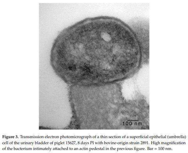
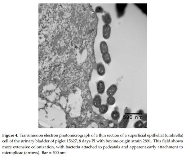
Enterohemorrhagic Escherichia coli (EHEC), a pathogenic subset of Shiga toxin-producing E. coli (STEC), is an important cause of hemorrhagic colitis and hemolytic–uremic syndrome (HUS), and a rare cause of urinary tract infections (UTIs) with associated HUS. EHEC strains attach intimately to intestinal epithelium with formation of actin pedestals (attaching-effacing (A/E) lesions); however, the mechanism of EHEC attachment to the uroepithelium is unknown. We conducted a retrospective study on archived urinary bladder specimens from gnotobiotic piglets that naturally developed cystitis associated with EHEC O157:H7 infection following oral inoculation and fecal shedding. Paraffin-embedded bladder tissues from three piglets with cystitis and immunohistochemical evidence of EHEC O157:H7 adherence to the uroepithelium were processed for and examined by transmission electron microscopy. EHEC O157:H7 bacteria were found in one of three piglets, intimately attached to pedestals on the apical surfaces of the superficial urothelium (umbrella cells). Cystitis was significantly associated with the length of survival of the piglets post-inoculation (p = 0.0339; estimated odds ratio = 2.6652). This is the first report of E. coli causing A/E-like lesions in the uroepithelium, and also evidence of the utility of the gnotobiotic piglet as a model for studies of the pathogenesis of EHEC UTIs.

Keywords: Shiga toxin-producing E. coli; enterohemorrhagic E. coli; attaching-effacing; pedestals; urinary tract infection; cystitis; pigs; gnotobiotic piglets

1. Introduction
Shiga toxin-producing Escherichia coli (STEC) is an important cause of foodborne illness in many countries of the world [1]. STEC infections often cause hemorrhagic colitis and subsequent hemolytic–uremic syndrome (HUS) [2,3]. E. coli O157:H7 was the first STEC serotype recognized to cause sporadic cases and outbreaks of these illnesses [4–6]. Subsequently, non-O157 STEC were recognized as important causes of hemorrhagic colitis and HUS [7]. In the U.S., non-O157 outnumber O157 foodborne STEC infections, but the latter are approximately 10 times more likely to lead to a diagnosis of HUS (1% versus 11%, respectively) [8–10]. STEC strains isolated from human patients with hemorrhagic colitis or HUS, and those isolated from any source that carry genes that encode for virulence factors that allow them to colonize the intestine, e.g., eae (intimin), are known as enterohemorrhagic E. coli (EHEC) [11].
Although HUS commonly occurs with EHEC enteric infection, it occurs rarely in association with E. coli urinary tract infection (UTI). A recent systematic review of the literature found 28 individually documented cases of HUS preceded by an E. coli UTI in patients ranging from 2 days to 75 years of age [12]. In 19 of these cases, Shiga toxin testing of the E. coli isolates was done, and 15 of the isolates tested positive, i.e., they were confirmed to be EHEC. Hence, EHEC is a rare but established cause of HUS in children and adults.
Adherence to epithelial cells is a critical early step in the pathogenesis of many bacterial pathogens, including E. coli [13]. With regard to the urinary tract, the most common cause of uncomplicated infections is uropathogenic E. coli (UPEC), which accounts for approximately 90% of the cases [13–15]. Numerous studies have addressed the mechanisms of adherence of UPEC to the urothelium, and these findings, among other facets of the pathogenesis, have been summarized in many different reviews [13,14,16–21]. Although multiple bacterial virulence and host factors are involved, UPEC are thought to adhere to the superficial uroepithelial (umbrella) cells primarily via several different fimbria and other cell-surface adhesins, and subsequently invade these cells with the establishment of intracellular bacterial communities [13–22].
Although much is known about the pathogenesis of UPEC adherence and colonization of the uroepithelium, and also the pathogenesis of EHEC adherence to intestinal epithelium with the formation of attaching-effacing (A/E) lesions [23,24], the mechanism of adherence of EHEC to the uroepithelium is unknown. In a previous study, we reported the finding of cystitis in gnotobiotic piglets inoculated with EHEC O157:H7 strains [25]. Adherence of EHEC O157:H7 bacterial cells to the uroepithelium of the bladder as seen by light microscopy was reported in that publication [25]. In order to further investigate adherence, we conducted a retrospective study utilizing transmission electron microscopy (TEM) on blocks of formalin-fixed bladder tissues from these same piglets. EHEC O157:H7 intimately adhered to pedestals on the apical surfaces of the superficial urothelium in a manner similar to classical A/E lesions in the intestine. We also retrospectively conducted statistical analyses on clinical data of the piglets and found that the incidence of cystitis was significantly associated with the length of survival post-inoculation (PI).
2. Materials and Methods
2.1. Bacterial Strains and Gnotobiotic Piglet Studies
The bacterial strains, inoculum preparation, gnotobiotic piglet challenge studies, histological examinations, Vero cell cytotoxicity assays, and immunohistochemistries were previously described [25]. Animal experiments were approved by the South Dakota State University, Institutional Animal Care and Use Committee. All strains were EHEC O157:H7 and positive for stx1, stx2 and eae. The strains included EDL933 as a positive control, and 10 different strains each that were of human or bovine origin. Gnotobiotic piglets were orally inoculated 24 to 30 h after birth with ~3 × 10⁹ CFU, and observed for clinical signs of illness for a maximum of 8 d PI (Table 1). Ten piglets were inoculated with EDL933 and 5 per strain inoculated with either a human- or bovine-origin strain [25]. One litter containing control piglets that did not die or become moribund within 8 d after challenge with EDL933 was not included in the comparison of the virulence of human- and bovine-origin strains in the published study, but since urinary bladders had been collected from piglets in this litter, they were examined for cystitis [25]. With inclusion of this litter, the study included 126 piglets, with 105 having the bladder examined histologically. Individual piglets with cystitis, and their corresponding litters of origin and inoculum strains, which is information that had not been listed in the previous publication [25], is shown in Table 1.
Intimate Attachment of Escherichia coli O157:H7 to Urinary Bladder Epithelium in the Gnotobiotic Piglet Model - Image 1
2.2. Transmission Electron Microscopy
Three piglets with cystitis that had the greatest number of bacteria adherent to the mucosal epithelium visible in tissue sections (11090, 15627, and 15637) [25] (Table 1) were selected for TEM. Areas corresponding to sites of bacterial adherence were dissected from paraffin blocks, 1 to 2 mm2 in size. Samples were deparaffinized at 65°C for 2 h to remove the majority of the paraffin. Following heat treatment, the pieces were passed through 4 exchanges of 100% xylene, soaking for 1 h during each exchange to remove any remaining paraffin. Samples were then placed in 2% glutaraldehyde, 2% paraformaldehyde in 0.1 M Sorensen’s phosphate buffer (SPB), pH 7.2 and stored overnight at 4°C. The following day, samples were washed 3 times in 0.1 M SPB, soaking for 15 min in each wash. After washing, samples were post-fixed in 1% osmium tetroxide in water for 1 h, and then washed in SPB 3 times, soaking 15 min in each wash. Samples were dehydrated through a graded ethanol series 50%, 70%, 90%, 95%, 100% x 3, and 100% propylene oxide x 3, at 15 min per step. Samples were placed in a 1:1 mixture of 100% propylene oxide and Araldite 502 embedding medium (Electron Microscopy Sciences, Hatfield, PA, USA) and left overnight in a fume hood. The following day, samples were soaked in fresh Araldite 502 for 4 h, followed by final embedding in a flat silicon embedding mold in fresh Araldite 502, and placed in an oven set at 65°C overnight for polymerization of the blocks. Sections 1 µm thick were cut from the polymerized blocks using a Diatome diamond knife (Diatome USA, Electron Microscopy Sciences) on a UC6 Ultramicrotome (Leica Microsystems, Wetzlar, Germany), then stained with toluidine blue and examined by light microscopy to locate areas of bacterial adherence to the mucosal surface. If bacteria were seen, thin sections, 60–90 nm in thickness, were then cut using the same ultramicrotome and diamond knife, and placed on 200-mesh copper grids. Sections on grids were stained with 1% uranyl acetate for 5 min, washed in water, and stained with Reynolds lead citrate for 5 min and washed. Sections were then examined on a Philips 410 transmission electron microscope operated at 80 Kv (ThermoFisher FEI, Hillsboro, OR). Images were acquired with an AMT digital camera system (AMT, Danvers, MA, USA).
2.3. Statistical Analyses
Cystitis was analyzed using logistic regression models. Each of the models included litter as a blocking factor. Models looking at the effect of PI days of survival included days of survival as a covariate, and models looking at the effect of bacterial strain origin (control, human or bovine) included origin as a classification factor. All analyses were carried out using the SAS® software’s GLIMMIX procedure.
3. Results and Discussion
As noted previously, 14 of 126 (13.3%) piglets orally inoculated with EHEC O157:H7 strains developed mild to moderate purulent cystitis within 8 d PI [25] (Table 1). Further, 8 of 14 piglets with cystitis had coccobacillary bacteria attached to the apical surfaces of superficial urothelial cells, and in all eight piglets, adherent bacteria stained positive immunohistochemically for E. coli O157 antigen [25] (Table 1).
In 1-µm thick Araldite sections of urinary bladder prepared from tissues originally contained within the paraffin blocks [25], bacteria were detected only in piglet 15627 (Figure 1). In this piglet, in which bladder sections were oriented such that all three cell layers (basal, intermediate and superficial or umbrella) of the urothelium [26–29] were visible, bacteria, corresponding to bovine-origin strain 2891, were diffusely adherent to the apical surfaces of umbrella cells. By TEM, bacterial cells were found intimately attached to pedestals (Figures 2–4). Some bacterial cells appeared to be attached to microplicae, preceding pedestal formation (Figure 4).
Intimate Attachment of Escherichia coli O157:H7 to Urinary Bladder Epithelium in the Gnotobiotic Piglet Model - Image 2
 
Intimate Attachment of Escherichia coli O157:H7 to Urinary Bladder Epithelium in the Gnotobiotic Piglet Model - Image 3
 
Intimate Attachment of Escherichia coli O157:H7 to Urinary Bladder Epithelium in the Gnotobiotic Piglet Model - Image 4
 
Intimate Attachment of Escherichia coli O157:H7 to Urinary Bladder Epithelium in the Gnotobiotic Piglet Model - Image 5
Morphologically, the pedestals to which bacteria in the urinary bladder of piglet 15627 were intimately attached were consistent with actin pedestals induced by EHEC and enteropathogenic E. coli (EPEC) in intestinal epithelium [24]. To our knowledge, this is the first report of intimate bacterial adherence and actin pedestals in the uroepithelium in any species. Staley et al. [30] first reported these lesions in 1969, describing them as attachment and microvillous exfoliation in ileal enterocytes of newborn, cesarean-derived piglets intragastrically inoculated with an E. coli strain belonging to a classical EPEC serotype, O55:H7. Takeuchi et al. [31] later described these lesions as occurring in rabbits inoculated with RDEC-1, a rabbit-origin O15:NM E. coli later classified as an EPEC [23]. Soon thereafter, the lesions were recognized in human infants with EPEC infection [32,33]. Moon et al. coined the term “attaching and effacing” to describe intimate attachment and effacement of microvilli in the intestinal epithelium of piglets and rabbits by EPEC [23]. Knutton et al. [34] first determined that the electron-dense material underlying the bacteria within the pedestals was filamentous actin. Over the past 50 years since the initial report by Staley et al. [30], numerous studies, many at the molecular level, have elucidated key bacterial and host factors involved in the pathogenesis of intimate attachment and pedestal formation [29].
The urinary tracts of the piglets in our study were infected naturally following exposure to the respective E. coli O157:H7 inoculum strains through fecal shedding and environmental contact (i.e., feed bowls and surfaces within the isolator units). Similarly, in humans, UTIs occur naturally most often through exposure to fecal microbiota [17,20,35]. Hence, we hypothesized that piglets that survived longer PI would have a higher incidence of cystitis since they would have had longer exposure to fecal and environmental bacteria in the isolator units. Indeed, the incidence of cystitis was significantly associated with the length of survival of the piglets PI (p = 0.0339; estimated odds ratio = 2.6652), but not the origin (control, human, or bovine) of the strain (p = 0.4435). In humans, UTIs are more common in females, in part due to the greater proximity of the urethral opening to the decreased length of the urethra [17,20,35]. The same anatomical predisposition is true for females of other mammalian species, including pigs. Unfortunately, the sex of the piglets in our study had not been recorded [25]; hence, we were not able to address sex as a risk factor.
We are aware of only one other study in the literature that had addressed EHEC bacterial adherence to urothelium [36]. This was an in vitro study involving T24 human transitional cell carcinoma cells. EHEC O157:H7 bacteria were found to invade T24 cells, but there was no report of intimate adherence or pedestal formation [36].
The urothelium of the pig shares many features structurally and functionally with that of humans [26–28,37]. Since EHEC is an established, albeit rare, cause of UTIs in humans, the gnotobiotic piglet, which we have herein shown to be susceptible to spontaneous UTI following enteric infection with EHEC, may be a useful UTI model for further studies. Research questions addressed by the model could include, e.g., those aimed at the identification of fimbria or other EHEC adhesins important for uroepithelial adherence and colonization, or the testing of preventive or treatment strategies.
This article was originally published in Microorganisms 2020, 8, 263; doi:10.3390/microorganisms8020263. This is an Open Access article distributed under the terms and conditions of the Creative Commons Attribution (CC BY) license (http://creativecommons.org/licenses/by/4.0/).

1. Majowicz, S.E.; Scallan, E.; Jones-Bitton, A.; Sargeant, J.M.; Stapleton, J.; Angulo, F.J.; Yeung, D.H.; Kirk, M.D. Global incidence of human Shiga toxin-producing Escherichia coli infections and deaths: A systematic review and knowledge synthesis. Foodborne Pathog. Dis. 2014, 11, 447–455. [CrossRef] [PubMed]

2. Tarr, P.I.; Gordon, C.A.; Chandler, W.L. Shiga-toxin-producing Escherichia coli and haemolytic uraemic syndrome. Lancet 2005, 365, 1073–1086. [CrossRef]

3. Kavanagh, D.; Raman, S.; Sheerin, N.S. Management of hemolytic uremic syndrome. F1000Prime Rep. 2014, 6, 119. [CrossRef] [PubMed]

4. Centers for Disease Control. Isolation of E. coli O157:H7 from sporadic cases of hemorrhagic colitis—United States. Morb. Mortal. Wkly. Rep. 1982, 31, 580–585.

5. Riley, L.W.; Remis, R.S.; Helgerson, S.D.; McGee, H.B.; Wells, J.G.; Davis, B.R.; Hebert, R.J.; Olcott, E.S.; Johnson, L.M.; Hargrett, N.T.; et al. Hemorrhagic colitis associated with a rare Escherichia coli serotype. N. Engl. J. Med. 1983, 308, 681–685. [CrossRef]

6. Remis, R.S.; MacDonald, K.L.; Riley, L.W.; Puhr, N.D.; Wells, J.G.; Davis, B.R.; Blake, P.A.; Cohen, M.L. Sporadic cases of hemorrhagic colitis associated with Escherichia coli O157:H7. Ann. Intern. Med. 1984, 101, 624–626. [CrossRef]

7. Brooks, J.T.; Sowers, E.G.; Wells, J.G.; Greene, K.D.; Griffin, P.M.; Hoekstra, R.M.; Strockbine, N.A. Non-O157 Shiga toxin-producing Escherichia coli infections in the United States, 1983–2002. J. Infect. Dis. 2005, 192, 1422–1429. [CrossRef]

8. Scallan, E.; Hoekstra, R.M.; Angulo, F.J.; Tauxe, R.V.; Widdowson, M.A.; Roy, S.L.; Jones, J.L.; Griffin, P.M. Foodborne illness acquired in the United States—Major pathogens. Emerg. Infect. Dis. 2011, 17, 7–15. [CrossRef]

9. Gould, L.H.; Mody, R.K.; Ong, K.L.; Clogher, P.; Cronquist, A.B.; Garman, K.N.; Lathrop, S.; Medus, C.; Spina, N.L.; Webb, T.H.; et al. Increased recognition of non-O157 Shiga toxin-producing Escherichia coli infections in the United States during 2000–2010: Epidemiologic features and comparison with E. coli O157 infections. Foodborne Pathog. Dis. 2013, 10, 453–460. [CrossRef]

10. Luna-Gierke, R.E.; Griffin, P.M.; Gould, L.H.; Herman, K.; Bopp, C.A.; Strockbine, N.; Mody, R.K. Outbreaks of non-O157 Shiga toxin-producing Escherichia coli infection: USA. Epidemiol. Infect. 2014, 142, 2270–2280. [CrossRef]

11. Croxen, M.A.; Law, R.J.; Scholz, R.; Keeney, K.M.; Wlodarska, M.; Finlay, B.B. Recent advances in understanding enteric pathogenic Escherichia coli. Clin. Microbiol. Rev. 2013, 26, 822–880. [CrossRef] [PubMed]

12. Lavrek, D.; Lava, S.A.G.; Milani, G.P.; Simonetti, G.D.; Bianchetti, M.G.; Giannini, O. Hemolytic-uremic syndrome after Escherichia coli urinary tract infection in humans: systematic review of the literature. J. Nephrol. 2018, 31, 919–924. [CrossRef] [PubMed]

13. Kalita, A.; Hu, J.; Torres, A.G. Recent advances in adherence and invasion of pathogenic Escherichia coli. Curr. Opin. Infect. Dis. 2014, 27, 459–464. [CrossRef] [PubMed]

14. Nielubowicz, G.R.; Mobley, H.L.T. Host-pathogen interactions in urinary tract infection. Nat. Rev. Urol. 2010, 7, 430–441. [CrossRef] [PubMed]

15. Robino, L.; Scavone, P.; Araujo, L.; Algorta, G.; Zunino, P.; Pírez, M.C. Intracellular bacteria in the pathogenesis of Escherichia coli urinary tract infection in children. Clin. Infect. Dis. 2014, 59, e158–e164. [CrossRef] [PubMed]

16. Mulvey, M.A.; Schilling, J.D.; Martinez, J.J.; Hultgren, S.J. Bad bugs and beleaguered bladders: interplay between uropathogenic Escherichia coli and innate host defenses. Proc. Natl. Acad. Sci. USA 2000, 97, 8829–8835. [CrossRef]

17. Mulvey, M.A. Adhesion and entry of uropathogenic Escherichia coli. Cell. Microbiol. 2002, 4, 257–271. [CrossRef]

18. Rosen, D.A.; Hooton, T.M.; Stamm, W.E.; Humphrey, P.A.; Hultgren, S.J. Detection of intracellular bacterial communities in human urinary tract infection. PLoS Med. 2007, 4, e329. [CrossRef]

19. Wiles, T.J.; Kulesus, R.R.; Mulvey, M.A. Origins and virulence mechanisms of uropathogenic Escherichia coli. Exp. Mol. Pathol. 2008, 85, 11–19. [CrossRef]

20. Mobley, H.L.; Donnenberg, M.S.; Hagan, E.C. Uropathogenic Escherichia coli. EcoSal Plus 2013. [CrossRef]

21. Tamadonfar, K.O.; Omattage, N.S.; Spaulding, C.N.; Hulgren, S.J. Reaching the end of the line: urinary tract infections. Microbiol. Spectrum 2019, 7, BAI-0014-2019. [CrossRef]

22. Eberly, A.R.; Beebout, C.J.; Tong, C.M.C.; Van Horn, G.T.; Green, H.D.; Fitzgerald, M.J.; De, S.; Apple, E.K.; Schrimpe-Rutledge, A.C.; Codreanu, S.G.; et al. Defining a molecular signature for uropathogenic versus urocolonizing Escherichia coli: The status of the field and new clinical opportunities. J. Mol. Biol. 2019. [CrossRef]

23. Moon, H.W.; Whipp, S.C.; Argenzio, R.A.; Levine, M.M.; Giannella, R.A. Attaching and effacing activities of rabbit and human enteropathogenic Escherichia coli in pig and rabbit intestines. Infect. Immun. 1983, 41, 1340–1351. [CrossRef] [PubMed]

24. Lai, Y.; Rosenshine, I.; Leong, J.M.; Frankel, G. Intimate host attachment: Enteropathogenic and enterohaemorrhagic Escherichia coli. Cell. Microbiol. 2013, 15, 1796–1808. [PubMed]

25. Baker, D.R.; Moxley, R.A.; Steele, M.B.; LeJeune, J.T.; Christopher-Hennings, J.; Chen, D.-G.; Hardwidge, P.R.; Francis, D.H. Differences in virulence among Escherichia coli O157:H7 strains isolated from humans during disease outbreaks and from healthy cattle. Appl. Environ. Microbiol. 2007, 73, 7338–7346. [CrossRef] [PubMed]

26. Scheidegger, G. Der aufbau des übergangsepithels der harnblase bei schwein, schaf, ratte und spitzmaus (Structure of the transitional epithelium in the urinary bladder of the pig, sheep, rat and shrew). Acta Anat. 1980, 107, 268–275. [CrossRef] [PubMed]

27. Liebhold, M.; Wendt, M.; Kaup, F.-J.; Drommer, W. Licht- und elektronenmikroskopische studien zur struktur des normalen blasenepithels beim weiblichen schwein (Light- and electron-microscope study of the structure of the normal bladder epithelium in female pigs). Anat. Histol. Embryol. 1995, 24, 47–52. [CrossRef]

28. Khandelwal, P.; Abraham, S.N.; Apodaca, G. Cell biology and physiology of the uroepithelium. Am. J. Physiol. Renal Physiol. 2009, 297, F1477–F1501. [CrossRef]

29. Hicks, R.M. The mammalian urinary bladder: An accommodating organ. Biol. Rev. Camb. Phil. Soc. 1975, 50, 215–246. [CrossRef]

30. Staley, T.E.; Jones, E.W.; Corley, L.D. Attachment and penetration of Escherichia coli into intestinal epithelium of the ileum in newborn pigs. Am. J. Pathol. 1969, 56, 371–392.

31. Takeuchi, A.; Inman, L.R.; O’Hanley, P.D.; Cantey, J.R.; Lushbaugh, W.B. Scanning and transmission electron microscopic study of Escherichia coli O15 (RDEC-1) enteric infection in rabbits. Infect. Immun. 1978, 19, 686–694. [CrossRef] [PubMed]

32. Ulshen, M.H.; Rollo, J.L. Pathogenesis of Escherichia coli gastroenteritis in man—Another mechanism. N. Engl. J. Med. 1980, 302, 99–101. [CrossRef] [PubMed]

33. Rothbaum, R.; McAdams, A.J.; Giannella, R.; Partin, J.C. A clinicopathologic study of enterocyte-adherent Escherichia coli: A cause of protracted diarrhea in infants. Gastroenterology 1982, 83, 441–454. [CrossRef]

34. Knutton, S.; Baldwin, T.; Williams, P.H.; McNeish, A.S. Actin accumulation at sites of bacterial adhesion to tissue culture cells: Basis of a new diagnostic test for enteropathogenic and enterohemorrhagic Escherichia coli. Infect. Immun. 1989, 57, 1290–1298. [CrossRef] [PubMed]

35. Geerlings, S.E. Clinical presentations and epidemiology of urinary tract infections. Microbiol. Spectrum 2016, 4, UTI-0002-2012. [CrossRef] [PubMed] 36. Oelschlaeger, T.A.; Barrett, T.J.; Kopecko, D.J. Some structures and processes of human epithelial cells involved in uptake of enterohemorrhagic Escherichia coli O157:H7 strains. Infect. Immun. 1994, 62, 5142–5150. [CrossRef]

37. Turner, A.M.; Subramaniam, R.; Thomas, D.F.M.; Southgate, J. Generation of a functional, differentiated porcine urothelial tissue in vitro. Eur. Urol. 2008, 54, 1423–1432. [CrossRef]

Related topics
Authors:
Rodney Moxley
University of Nebraska-Lincoln
Recommend
Comment
Share
Profile picture
Would you like to discuss another topic? Create a new post to engage with experts in the community.
Join Engormix and be part of the largest agribusiness social network in the world.