Explore
Communities in English
Advertise on Engormix

Detection of antibodies against pathogens in feral and domestic pigs (Sus scrofa) at the Sierra La Laguna Biosphere Reserve, Mexico

Published: June 10, 2019
By: Claudia Mariana Pérez-Rivera 1, Mauro Sanvicente López 2, Gustavo Arnaud Franco 3, Rosalba Carreón Nápoles 4.
Summary

Several diseases that were believed to be controlled or eradicated have reappeared and have had catastrophic effects on humans and on domestic and wild animals. Approximately 60 % of recently registered disease outbreaks are caused by zoonotic agents, and 72 % of these originated in a wild species. Swine (Sus scrofa) is a species that favors the propagation of pathogens, and they can be a reservoir of many diseases. Thus, the objective of this study was to detect the presence of viral and bacterial diseases that could impair the health of wild animals and humans in both feral and domesticated pigs at the Sierra La Laguna Biosphere Reserve. Diagnosis was performed with serological tests on 70 animals to detect antibodies against swine influenza virus (SIV), porcine respiratory reproductive syndrome virus (PRRSV), Aujeszky’s disease virus (ADV), leptospirosis (Lp), salmonellosis (Sal), and brucellosis (Br).

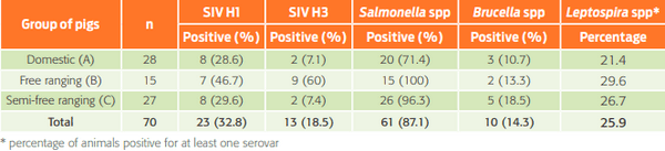
No antibodies were detected against PRRS or AD, whereas the seropositivity was 30.7 % for SI, 25.9 % for Lp, 87.1 % for Sal, and 14.3 % for Br. This evidence supports the presence of these pathogens in Sierra Laguna, and implies that swine could be an important factor for transmission to other wild species, as well as to people who have had contact with or consumed these animals. Thus, developing management and epidemiological surveillance plans for the animals inhabiting the region is of paramount importance.

Keywords: Feral swine; zoonosis; antibodies; Mexico.

Introduction
Globalization has played an important role in the dissemination of diseases because it allows the mobilization of people, animals and products from one place to another in a short time1. In addition, population growth, deforestation, the introduction of exotic species, changes in land use, habitat fragmentation and alternative tourism have increased the possibility of contact with wild animals, and their consumption by human2. Interactions within the human-animal environment are thus fundamental for the appearance of zoonoses2,3, and greater attention has been given to diseases of domestic animals because of their interaction with wild species. However, the difficulty of studying diseases in wildlife has led to wild animals receiving less attention, despite acknowledging their important role in the transmission of pathogens2. In these terms, feral pigs (Sus scrofa) play an important role in the transmission of pathogens because they are carriers of numerous viral, bacterial, and parasitic diseases that can potentially be passed onto humans and to other wild and domestic animals4-6. In addition, they are considered as a major worldwide invasive species, and can also participate in the propagation of diverse exotic diseases4,7,8
The Sierra La Laguna Biosphere Reserve (REBIOSLA is the acronym in Spanish) is a site that has a great biodiversity of mammals9 and a geological history that gave rise to a unique ecosystem in Mexico10. Approximately 70 % of the total population of terrestrial and aerial mammals that live in the state of Baja California Sur are found here11. Small communities of farmers live within the limits of REBIOSLA, and domestic pigs were introduced during the 1940s11. Due to production practices, some of these pigs have escaped from confinement and have established a feral population. The abundance of the feral pigs varies due to climatological factors, such as droughts and hurricanes, which have an impact on the food supply and tend to reduce the population12. The size of the feral population is influenced by the high mortality rate (51 %) of suckling pigs, as well as by hunting and by some management practices, such as the castration of males13. In addition, feral pigs have become part of the trophic chain because they are prey for the mountain lion (Felis concolor improcera)11 and are the second most important food source for the coyote (Canis latrans peninsulae)12. Some inhabitants of the region have stated that foxes (Urocyon cinereoargenteus peninsularis) and bobcats (Lynx rufus peninsularis) also prey on feral pigs, particularly on the young.
This study was aimed at detecting the presence of antibodies to viral and bacterial infections in feral and domestic pigs, by using serological tests. The literature indicates that swine influenza virus (SIV) could harm humans, as well as wild and domestic animals14,15. Swine play an important role in the ecology of disease because several subtypes of SIV can infect simultaneously15. Aujeszky’s disease is another viral infection in which swine are the exclusive natural host and are mainly carriers, but it can affect other mammals, such as ruminants, carnivores and rodents, as well as wildlife animals such as raccoons, skunks, foxes and big cats14,16,17. Additionally, antibodies to leptospirosis, salmonellosis, and brucellosis infection were determined. All of these diseases have a high zoonotic potential and could affect a wide range of animals as well as humans.
Materials and methods
Study location
The Sierra La Laguna Biosphere Reserve is located in the Northwest of Mexico at the southern end of the Baja California Peninsula in the state of Baja California Sur (BCS), between 23°42´ - 23°20’ S and 109°46’ - 110°11’ W10,18.
Animals
Thirty-two domestic swine farms were previously studied within the limits of the Reserve and in neighboring areas by the Grupo de Ecología Animal of the Centro de Investigaciones Biológicas del Noroeste, A.C. According to the compiled data of previous studies, 3 groups of swine were established as sampling subjects12,13. The criteria for their classification included management practices and whether the animals were roaming freely. The 3 groups were:
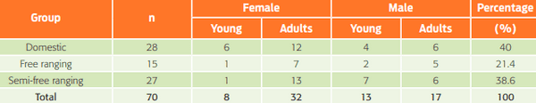
  • Domestic pigs (Group A); pigs kept inside a barn or another facility and fed by their owners.
  • Free ranging pigs (Group B); pigs roaming mostly free that obtained food by themselves. They reproduced naturally, without any type of handling.
  • Semi-free ranging pigs (Group C); feral pigs that had been used for consumption and were fed by farmers. Seventy animals were used for the study. The sample size was based on an expected prevalence of 5 % with a statistical error of 5 %19. The sex and age of the pigs are shown in Table 1.
Table I. Number and percentage of animals sampled in each group.
Detection of antibodies against pathogens in feral and domestic pigs (Sus scrofa) at the Sierra La Laguna Biosphere Reserve, Mexico - Image 1
Sampling and sample processing
Blood was sampled from each animal following the technique described by Straw et al. 20. Sterile Vacutainer® needles were used (21 G × 32 mm, green cap). Vacutainer® tubes without anticoagulant were used to collect 12 mL of blood, in 2 tubes per animal. Once the samples were obtained, they were labeled with a permanent marker and were recorded with the sex of each animal, the production purpose and the color. Samples were kept in a fresh spot for approximately 30 min; afterwards, they were placed in an icebox with refrigerants for their transport to the laboratory21.
The blood samples were centrifuged at 1500 rpm for 10 min to separate the serum from the other cell components. The serum was collected with a sterile micropipette, placed in a 2-mL Eppendorf tube and stored at -20°C until processing, according to protocols below, established in the diagnostic laboratories of the Facultad de Medicina Veterinaria y Zootecnia (Universidad Nacional Autónoma de México) and Centro Nacional de Investigación Disciplinaria-Microbiología (CENID-Microbiología, Instituto Nacional de Investigaciones Agrícolas, Forestales y Pecuarias).
Diagnostic techniques
  • The hemagglutination inhibition test (HI) was used for the diagnosis of swine influenza following the protocol in Beltrán (2009)22, which is considered the standard test for this disease by the World Organization for Animal Health23 (OIE for its acronym in French, http://www.oie.int/es/ retrieved 24/06/2012). The viruses A/swine/NewJersey/11/76 (H1N1) and A/swine/Minnesota/9088-2/98 (H3N2) were used as the antigens, and sera that showed sedimentation in a dilution greater than or equal to 1:80 were considered positive.
  • ELISA tests were used for the diagnosis of Aujeszky’s disease (Hipra CIVTEST SUIS ADVgE No. CAE.12, Spain); PRRS: an indirect ELISA test was used (Hipra CIVTEST SUIS PRRS A/S No. 40ND, Spain); and for Salmonellosis, Idexx was used (Idexx - SWINE SALMONELLA HERD CHEK No. 44100 T 161, USA). Idexx detects antibodies for the most common Salmonella serotypes and indicates the exposure of the herd to these bacteria. The manufacturer’s protocols were followed to perform and interpret each assay.
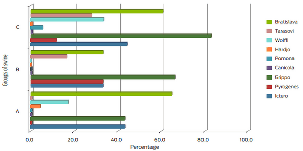
  • Microscopic microagglutination (MAT) was used to diagnose leptospirosis. It is the reference test given by OIE, and antigens representative of the area inhabited by the animals were used23,24. Nine Leptospira interrogans serovarieties from the collection of CENID-Microbiología were used. This test was scored as positive starting at a 1:100 dilution based on the minimum significant titer of the OIE.
  • Card Test (3 %), an indirect diagnostic test also known as Rose Bengal, was used for the serological diagnosis of brucellosis. The antigen Brucella abortus strain 1119-3 at 8 % was used for the specific diagnosis of brucellosis in swine. It was stained with Rose Bengal from the Productora Nacional de Biológicos Veterinarios (PRONABIVE) which detects B. abortus, B. suis, and B. melitensis. This test was performed following the manufacturer’s instructions.
Statistical analysis
The results are presented using descriptive statistics and graphs generated with the Sigma plot © version 10.0 software. To compare the proportion of positives among the three groups for the diseases that were positive, a contingency analysis was performed using the chi-square distribution via the JMP 8.0 statistical program.
Results and discussion
Specific antibodies related to four infections were found: influenza (viral) and leptospirosis, salmonellosis, and brucellosis (bacterial). More animals in the free feral pig group had antibodies than either of the other 2 groups, likely because this group of animals is widely distributed at the site with a probable home range of 1.1 to 5.32 km2 25. Because feral pigs are free, they can roam at will and thus come into contact with wildlife. They can carry and share infectious agents26.
Influenza
The free ranging swine group (B) had the highest percentage of both viral subtypes (H1N1 46.7 %, H3N2 60 %), while the other two groups (A and C) tended to be similar, higher for H1N1 and lower for H3N2, but had a lower percentage of seropositivity than group B (Table 2). In general, a greater percentage of animals was positive for H1N1 than H3N2, except for group B. The ranges of antibody titers for H1N1 were from 1:80 to 1:320 and were higher for H3N2, from 1:80 to 1:1280. No significant differences were found among the three pig groups for H1N1 (P = 0.437), but the differences were significant for H3N2 (P < 0.001).
Detection of influenza antibodies in REBIOSLA swine suggests exposure to this agent. Seropositivity found in this study was double that found by Hall et al.15, in which the highest prevalence found in feral swine in the USA was 14 % for subtype H3N2. Only a 4 % prevalence for subtype H1N1 was found in a study performed in Spain that analyzed 78 feral swine samples with the same test as this study27. There was no vaccination history in this study’s region.
Groups of swine in captivity at REBIOSLA (A and C) showed similar positivity percentages, which could be because both groups live in similar captivity conditions independently from their origin, hence the circulation of the virus with other possible hosts or factors that trigger the disease is similar. On the other hand, the free ranging swine group had the highest percentage of positivity to both subtypes (53.1 %), which could suggest that the circulation of the virus with other cohabiting species is possible, since swine are exposed to a natural exchange of this virus with other species28.
Table 2. Percentage and number of animals by group
Detection of antibodies against pathogens in feral and domestic pigs (Sus scrofa) at the Sierra La Laguna Biosphere Reserve, Mexico - Image 2
Leptospirosis
Antibodies were higher in the free ranging swine (group B) with 29 % positivity to one or more of the nine serovarieties used for the diagnosis, whereas the group with the lowest percentage of antibodies was domestic swine (21 %). The range of antibody titers for L. interrogans was 1:100 to 1:800.
Swine are very important as reservoirs of leptospirosis and excrete the bacteria intermittently, even in swine in which the circulation of specific antibodies occurs4. Therefore, the 25.9 % seropositivity in REBIOSLA swine is relevant. These animals interact closely with other both domestic and wild species at shared hydration sites, and can be reservoirs and infection sources for other productive species and humans. A statistically significant difference between the percentages of positive animals in the study groups was found for the serovars pyrogenes, pomona and wolffi (P = 0.005, 0.048, 0.578, respectively, P < 0.05).
The most frequently found serovarieties in the three groups were bratislava and grippotyphosa. No antibodies were detected for canicola in any group (Figure 1). Similar studies in different countries have shown different prevalence levels. Vicente et al.27 found a prevalence of 12 % for L. pomona in Spain, while Montagnaro et al.29 in Italy examined 342 sera showing 2.6 % positivity for the serovars L. copenagheni, L. bratislava and tarassovi. In the studies mentioned above and others, the focus was on only one or three serovars, while we evaluated 9 serovars in this study.
Detection of antibodies against pathogens in feral and domestic pigs (Sus scrofa) at the Sierra La Laguna Biosphere Reserve, Mexico - Image 3
Figure 1. Percentage of positivity per serovariety and sampling group
Salmonellosis
The free ranging pigs (group B) had 100 % seropositivity, and the captive feral swine group had 96.3 % seropositivity. The ELISA used in this study detected different serovarieties, and among them, S. typhimurium, S. choleraesuis, and S. derby stand out. A significant difference between the prevalence in the groups studied was noted.
The ELISA revealed the presence of antibodies to Salmonella spp. in 61 of 70 swine (87.14 %). These seroprevalence values exceed those reported for feral swine populations in Italy (19.3 %)29, Spain (4 %)27, and the USA (5 %)30. A study in Slovenia on 178 samples obtained during the wild boar hunting season used the same serological test as this study and reported a prevalence of 47 %31. It is worthwhile to emphasize that our results are in a wild population, suggesting that the presence of this infectious agent is undeniable and alarming, especially due to its relevance as a zoonotic pathogen.
Brucellosis
The results obtained for antibodies to Brucella spp. are shown in Table 1. Group C had the highest seropositivity (18.5 %), and group A had the lowest (10.7 %). However, no significant differences between the three groups of pigs were found (P = 0.062). These results are similar to results from the USA and Italy by Wyckoff et al.32 and Montaganaro et al.29, respectively.
It is important to mention that the Card test does not specifically detect antibodies to B. suis; it also detects antibodies to B. abortus and B. melitensis, but it does indicate the presence of the B. suis antigen in the population. It must also be mentioned that cattle can be naturally infected with B. suis, and can potentially transmit it to swine, via fluids excreted while giving birth, as well as via aborted fetuses33. This indicates that the contact of swine with domestic bovines is relevant for the transmission of diseases like brucellosis. Such contact is very common in the REBIOSLA on and off the farm. This can also occur among other species inhabiting REBIOSLA. In addition, it should be mentioned that due to a national campaign against brucellosis in animals in Mexico, the role played by wild species as carriers of this disease becomes very important by compromising the eradication of this pathogen from domestic populations32.
No antibodies against Aujeszky’s disease or PRRS were detected in the 3 groups, likely because PRRS is predisposed to large populations where overcrowding, direct contact, and other factors are critical for the occurrence and dissemination of the disease. This situation is not relevant to the REBIOSLA swine, although circulation of these agents in the REBIOSLA cannot be discounted. Despite the small sample size, which may limit the power of the detection of these pathogens, the absence of antibodies against Aujeszky’s etiologic agent is in accord with to the sanitary conditions in the area, which is considered free of this disease.
Diseases can alter the population dynamics of wild fauna28, that is, changes in the structure of these populations can be affected in terms of the size, age groups, sex ratios and the physical dimensions of their members, among other factors. It is very important to know that diverse epidemiologically relevant pathogens are present in the REBIOSLA, and can affect other mammals such as coyotes, foxes, bobcats, deer, raccoons, and also humans who consume their meat without the necessary hygienic measures34
Conclusions
Of the six studied diseases, antibodies were detected only for influenza (30.7 %), leptospirosis (25.9 %), salmonellosis (87.1 %) and brucellosis (14.3 %). These results and a lack of reports of clinical disease show that the population is probably not affected by these pathogens. Nevertheless, the pathogens were evident in this study. This is important because the coexistence of local and feral species such as pigs and cattle is narrow, and they are considered healthy and important food sources.
The study of pathogens in wild animals can provide an efficient epidemiological overview that allows an evaluation of the risk of infection with a zoonotic agent, and the propagation of disease in a free-roaming population, as well as in domestic animals and even in humans35. It is important to mention the type of wildlife studies that involve distant work sites, as well as the difficulty in capturing animals, sampling, and even the tests available for wildlife36. However, further studies in these populations are imperative.
No reports have emerged in Mexico about the epidemiological situation of the feral swine population. The data obtained in this study was compared with other similar studies, and shows that even though the REBIOSLA population is small, the presence of these infectious agents can be relevant. The presence of these animals and their possible role in the dynamics of infectious disease in this geographical area are significant because of the biological relevance of this ecosystem and the number of species that inhabit it. More studies are needed to provide current data and compare it with other sources.
Funding
We acknowledge CONACYT-Mexico for the fellowship (CVU 364439).
Acknowledgements
We thank Dr Ricardo Flores Castro and Dr José Francisco Morales Álvarez from Centro Nacional de Investigación Disciplinaria en Microbiología Animal del Instituto Nacional de Investigaciones Forestales, Agrícolas y Pecuarias (Cenid-Microbiología), for their support in analyzing the samples. To Dr. Roberto Martínez Gamba for his help in the statistical analysis, and to MSc. Francisco Cota Castro (technician of the Ecology Laboratory) and the inhabitants of Sierra La Laguna for their support during field work. We also thank the Department of Swine Medicine and Zootechnics of the FMVZ UNAM, a Conacyt scholarship (CVU 364439), and Dr Renate Marie Thummler Blum for her support in translating the manuscript.
Conflicts of interest
The authors have no conflict of interest in this study.
Author contributions
CMPR participated in the collection and analyses of samples, as well as in the data analysis of the results and wrote the manuscript.
MSV designed the experiment and participated in the analysis of the results. GA participated in the experiments and sample design. RCN performed the laboratory tests, participated in the design and helped to write the paper.
This article was originally published in Veterinaria México OA. 2017;4(1). doi:10.21753/vmoa.4.1.378. This is an Open Access article distributed under a Creative Commons Attribution License CC-BY 4.0.

1. Petersen EA. Emerging infectious disease. Arch Intern Med. 1996;156:124. doi: 10.1001/archinte.1996.00440020010001.

2. Daszak P. Emerging infectious diseases of wildlife-- Threats to biodiversity and human health. Science. 2000;287:443-9. doi: 10.1126/science.287.5452.443.

3. FAO-OIE-WHO. Influenza and other emerging zoonotic diseases at the human-animal interface. Food and Agriculture Organisation of the United Nations - World Organisation for Animal Health - World Health Organisation ed. Verona, Italy2010 2010.

4. Meng XJ, Lindsay DS, Sriranganathan N. Wild boars as sources for infectious diseases in livestock and humans. Phil Trans R Soc B: Biol Sci. 2009;364:2697- 707. doi: 10.1098/rstb.2009.0086.

5. Seward N, VerCauteren K, Witmer G, Engeman R. Feral swine impacts on agriculture and the environment. Sheep Goat Res J. 2004;19:34-40.

6. Gibbs EPJ. The public health risks associated with wild and feral swine. Rev Sci Tech - Off Int Epizoot. 1997;16:594-8. doi: 10.20506/rst.16.2.1052.

7. Vitousek P, D´Antonio C, Loope L, Rejmánek M, Westbrooks R. Introduced species: a significant component of human-caused global chance. N Z J Ecol. 1997;21:1-16.

8. Hutton T, DeLiberto T, Owen S, Morrison B. Disease risks associated with increasing feral swine numbers and distribution in the United States. USA: Midwest Association of Fish and Wildlife Agencies; 2006 7 Nov.

9. Ortega-Rubio A, Lagunas-Vázques M, Beltrán-Morales LF. Evaluación biológica y ecológica de la Reserva de la Biósfera Sierra la Laguna, Baja California Sur: Avances y retos. Ortega-Rubio A, Lagunas-Vázques M, Beltrán-Morales LF, editors. La Paz, BCS: Centro de Investigaciones Biológicas del Noroeste, SC; 2012. 422 p.

10. CONABIO. Programa de manejo de la Reserva de la Biósfera Sierra La Laguna. Comisión Nacional de Áreas Naturales Protegidas DGdMplC, editor. México: Comisión Nacional de Áreas Naturales Protegidas; 2003. 208 p.

11. Arnaud G, Álvarez S, Cortés P. Mamíferos de la Reserva de la Biósfera Sierra la Laguna. In: Ortega-Rubio A, Lagunas-Vázques M, Beltrán-Morales LF, editors. Evaluación de la Reserva de la Biósfera Sierra la Laguna, Baja California Sur: Avances y Retos. La Paz: Centro de Investigaciones Biológicas de Baja California Sur A.C.; 2012. p. 412.

12. Breceda A, Arnaud G, Álvarez S, Galina P, Montes J. Evaluación de la población de cerdos asilvestrados (Sus scrofa) y su impacto en la Reserva de la Biosfera Sierra La Laguna , Baja California Sur , México. Trop Conserv Sci. 2009;2:173-88.

13. Montes-Sánchez J, León de la Luz JL, Buntinx-Dios S, Aguilar-Marcelino L, Blázquez-Moreno MC. Dieta, crecimiento y reproducción del cerdo asilvestrado Sus scrofa en la Reserva de la Biósfera Sierra la Laguna. In: Ortega-Rubio A, Lagunas- Vázques M, Beltrán-Morales LF, editors. Evaluación de la Reserva de la Biósfera Sierra la Laguna, Baja California Sur: Avances y Retos. La Paz: Centro de Investigaciones Biológicas del Noroeste S.C.; 2012. p. 183-204.

14. Glass CM, McLean RG, Katz JB, Maehr DS, Cropp CB, Kirk LJ, et al. Isolation of pseudorabies (Aujeszky’s disease) virus from a florida panther. J Wildlife Dis. 1994;30:180-4. doi: 10.7589/0090-3558-30.2.180.

15. Hall JS, Minnis RB, Campbell TA, Barras S, DeYoung RW, Pabilonia K, et al. Influenza exposure in United States feral swine populations. J Wildlife Dis. 2008;44:362-8. doi: 10.7589/0090-3558-44.2.362.

16. Raymond JT, Gillespie RG, Woodruff M, Janovitz EB. Pseudorabies in captive Coyotes. J Wildlife Dis. 1997;33:916-8. doi: 10.7589/0090-3558-33.4.916.

17. Kirkpatrick CM, Kanitz CL, McCrocklin SM. Possible role of wild mammals in transmission of pseudorabies to swine. J Wildlife Dis. 1980;16:601-14.

18. Arriaga L, Ortega A. La Sierra de la Laguna de Baja California Sur. Arriaga L, Ortega A, editors. La Paz: Centro de Investigaciones Biológicas de Baja California Sur A.C.; 1989. 237 p.

19. Cortés F. Tamaño de muestra y análisis de asociación. Rev Mex Sociol. 1982;44(4):1381-411. doi: 10.2307/3540134.

20. Straw BE, Meuten DJ, Thacker BJ. Physical examination. In: Straw B, D’Allaire S, Mengeling W, Taylor D, editors. Diseases of swine. 8 ed. Iowa, USA: Iowa State University Press/ Ames; 1999. p. 15-7.

21. Segalés J, Martínez J, Catellà J, Darwich L, Domingo M, Mateu E, et al. Manual de diagnóstico laboratorial porcino. MSD SA, editor. Navarra, España: Servet; 2013. 120 p.

22. Beltrán Figueroa R. Identificación del virus de influenza porcina subtipos H1N1 y H3N2 mediante RT-PCR [Tesis de licenciatura]. Ciudad de México: Universidad Nacional Autónoma de México; 2009.

23. OIE. Manual de las pruebas de diagnóstico y de las vacunas para los animales terrestres. 7 ed. Paris, France: Office International des Epizooties; 2012. 1404 p.

24. Olsen S. Porcine brucellosis. OIE Manual of diagnostic tests and vaccines for terrestrial animals. 2. 6 ed. Paris, France: Office International des Epizooties; 2008. p. 1343.

25. Gaston W, Armstrong JB, Arjo W, Stribling HL. Home range and habitat use of feral hogs (Sus scrofa) on Lowndes Country WMA, Alabama. National conference on feral hogs April 13-15; MO, USA2008. p. 6.

26. Ruiz-Fons F, Vidal D, Vicente J, Acevedo P, Fernández-de-Mera IG, Montoro V, et al. Epidemiological risk factors of Aujeszky’s disease in wild boars (Sus scrofa) and domestic pigs in Spain. Eur J Wildl Res. 2008;54:549-55. doi: 10.1007/ s10344-008-0179-6.

27. Vicente J, León-Vizcaíno L, Gortázar C, Cubero MJ, González M, MartínAtance P. Antibodies to selected viral and bacterial pathogens in european wild boars from Southcentral Spain. J Wildlife Dis. 2002;38:649-52. doi: 10.7589/0090-3558-38.3.649.

28. Ruiz-Fons F. Riesgos sanitarios asociados a la producción cinegética del jabalí: la enfermedad de Aujeszky [Tesis de doctorado]. Ciudad Real, España: CSICUCLM - Instituto de Investigación en Recursos Cinegéticos (IREC) Universidad de Castilla-La Mancha; 2006.

29. Montagnaro S, Sasso S, De Martino L, Longo M, Iovane V, Ghiurmino G, et al. Prevalence of antibodies to selected viral and bacterial pathogens in wild boar (Sus scrofa) in Campania region, Italy. J Wildlife Dis. 2010;46:316-9. doi: 10.7589/0090-3558-46.1.316.

30. Thakur S, Sandfoss M, Kennedy-Stoskopf S, DePerno CS. Detection of Clostridium difficile and Salmonella in feral swine population in North Carolina. J Wildlife Dis. 2011;47:774-6. doi: 10.7589/0090-3558-47.3.774.

31. Vengust G, Valencak Z, Bidovec A. A serological survey of selected pathogens in wild boar in Slovenia. J Vet Med B. 2006;53:24-7. doi: 10.1111/j.1439-0450.2006.00899.x.

32. Wyckoff aC, Henke SE, Campbell Ta, Hewitt DG, VerCauteren KC. Feral swine contact with domestic swine: a serologic survey and assessment of potential for disease transmission. J Wildlife Dis. 2009;45:422-9. doi: 10.7589/0090-3558-45.2.422.

33. College of Veterinary Medicine Iowa State University. Swine diseases manual. 4 ed. Neuman EJ, Ramirez A, Schwartz KJ, editors. Iowa: American Association of Swine Veterinarians; 2013. 173 p.

34. Barrios-Garcia MN, Ballari SA. Impact of wild boar (Sus scrofa) in its introduced and native range: a review. Biol Invasions. 2012;14:2283-300. doi: 10.1007/ s10530-012-0229-6.

35. Kaden V, Lange E, Hänel A, Hlinak A, Mewes L, Hergarten G, et al. Retrospective serological survey on selected viral pathogens in wild boar populations in Germany. Eur J Wildl Res. 2009;55:153-9. doi: 10.1007/s10344-008-0229-0.

36. Boadella M, Ruiz-Fons JF, Vicente J, Martín M, Segalés J, Gortazar C. Seroprevalence evolution of selected pathogens in iberian wild boar. Transbound Emerg Dis. 2012;59:395-404. doi: 10.1111/j.1865-1682.2011.01285.x.

Related topics
Authors:
Rosalba Carreón
UNAM - Universidad Nacional Autónoma de México
Recommend
Comment
Share
Profile picture
Would you like to discuss another topic? Create a new post to engage with experts in the community.
Join Engormix and be part of the largest agribusiness social network in the world.