Introduction
Livestock in countries, especially developing countries, is vital to livelihood of people and to the economy. In the developing world, livestock species often represent a sole asset base for small-holder animal husbanders and cattle often provide the majority of draught power for crop production. Nigeria has the largest animal resources in the West African region with live animals counting to about 20 million cattles, 39 million sheeps, 48 million goats, 166 million poultry, 6.6 million pigs and 0.7 million camels and donkeys in the fiscal year 2013 (FAOSTAT, 2015) yet it is unable to meet the recommended protein intake of its populace. According to the same data, 2.6 million tonnes of livestock products were produced for consumption in the year 2013 in Nigeria; the breakdown being eggs- 650 000, milk (whole-fresh cow milk)- 570 000, cow meat (from indigenous cattle)- 338 000, goat- 292 649, pig- 254 250, sheep- 172 507, poultry meat- 170 000 and game- 165 000 in tonnage. While commercial production techniques are used extensively in the poultry and aquaculture sectors, livestock production is largely pastoral and domestic. In Minna, Nigeria, livestock is one of the main protein sources in human diet. These results to high exposure risk and public health impact from animal products that may have been contaminated with aflatoxin.
Studies have shown aflatoxin B1 (AFB1 ) residues in the livers of camels, beef calves, buffalo calves and sheep [1,2]. And liver samples containing AFB1 residues showed remarkable changes including fatty degeneration with areas of petechial hemorrhages, congestion, fibrosis and large whitish focus of necrosis as compared to those with medium and lower AFB1 levels. Aflatoxin M1 (AFM1 ) was the first aflatoxin metabolite identified and it is the principal hydroxylated aflatoxin metabolite present in milk of dairy cows fed with feed contaminated with aflatoxin B1 [3]. The metabolite is also present in the milk of human nursing mothers consuming foodstuffs contaminated with the toxin [4,5]. Occurrence of AFM1 in livestock livers is a public health concern, since AFM1 has been classified by the international agency of research on cancer as, possible human carcinogens [6]. However, in all livestock, aflatoxins can cause reduced milk or egg production, embryonic death, teratogenicity, decreased reproductive performance, tumours and suppressed immune system function, and liver damage, even at low levels [7].
Determination of AFM1 in milk, urine and liver are good biomarker of Aflatoxin B1 (AFB1) exposure, as it is correlated to the amount of AFB1 ingested. The carcinogenic potency of AFM1 is ten times lower than AFB1 but this appears warranted if the biological endpoints involved are carcinogenicity and mutagenicity [8]. Metabolism of AFM1 have been studied in vitro using liver microsomes [9]. Cyclopropenoid fatty acids (CPFA), which promote the carcinogenicity of aflatoxin B1, behave similarly with AFM1 [10]. However, newborn animals that consume aflatoxins can be poisoned by the AFM1 metabolite excreted in milk. Parameters contributing or affecting the levels of AFM1 contamination in livestock livers could be the source, type of or composition of livestock feeds [11], ecologic or economic factors on the farm, pre and post farm managements. The European Union and Codex Alimentarius have regulated maximum level of AFM1 not to exceed 0.05 ppb (50 ng/kg) while US regulations allow levels not above 0.005 ppb (500 ng/kg). Therefore, it is very pertinent that the levels of aflatoxin M1 in livestock liver are monitored, with respect to the case study from Minna metropolis. This will enable reduced risk and rapid incidence of aflatoxicoses, new findings and the countries response concerning aflatoxin. Hence, aiding awareness and reducing the national or global burden attributable to aflatoxin M1 as justified in the study, this is the first report establishing the toxin presence and levels in livestock liver from Nigeria and comparing levels in goat and cow liver, as there is no known record of its kind.
Materials and Methods
Sample collection and preparation
After sorting written approval of the Veterinary Council of Nigeria Minna Chapter, fresh liver samples were collected into ice packs from 5 abattoirs within 24 hours of slaughter. Twenty five samples each of goat liver were collected from two abattoirs (n=50) while 24 samples of cow liver were collected each from 3 abattoirs (n=72). Samples were froze dried and the stored at -20°C until used for analysis. Prior to sample analysis, sample temperature was brought up to room temperature.
Aflatoxin extraction /column chromatography/ high performance liquid chromatography
Aflatoxin extraction and column chromatography were achieved using AOAC official method 982.24 (2010) with modifications, for quantification by HPLC method [12]. Dried liver tissue were blended until homogenous. About 100 g portion was weighed into 500 ml wide-mouth, glass-stoppered erlenmeyer flask. Citric acid (10 ml) solution was added and mix thoroughly with 30 cm×1 cm glass stirring rod. After 5 min it was stirred again and mixed with 2 g of silica gel, (diatomaceous earth) 200 ml methylene chloride was added and stirred to remove excess solid from rod and the flask was stirred vigorously on wrist-action for 30 min. The mixture was filtered through flow paper into 300 ml erlenmeyer containing 10 g Na2SO4, the filter top was closed and compressed against funnel to obtain its maximum filtrate volume. Flask was gently swirled intermittently for 2 min and content was refiltered through medium flow paper into 250 ml graduate, volume was recorded. Filtrate was evaporated in 500 ml round-bottom flask on water bath to near dryness and saved for column chromatography.
A separating column was set with glass wool inserted, 50 ml methylene chloride was poured in and 10 g of silica gel was added in addition to 3-4 ml methylene chloride and mixed with stainless steel rod, methylene chloride was drained halfway to settle silica, and silica was rinsed off column sides with methylene chloride, one scoop of anhydrous sodium sulphate was added to supernate solvent above silica gel to cap column. Excess methylene chloride was drained to 1 cm above column packing. The concentrated filtrate was then redissolved in 25 ml methylene chloride and added to column, the round-bottom flask was rinsed, methylene chloride was added to the column and the entire solution was drained through column by gravity. When flow rate slowed, anhydrous sodium sulphate was stirred gently, when filtrate reached anhydrous sodium sulphate, column sides were rinsed with methyl chloride and drained similarly. Column was washed with 25 ml toluene- acetic acid (9:1), 25 ml hexane in each and 25 ml hexane-ether-acetonitrile (6:3:1) and washes were discarded. Aflatoxin was eluted with 40 ml methyl chloride-acetone (4:1) and evaporated to near dryness in vacuum. This was however evaporated to as low as 2 ml and was put in amber bottle and stored in the refrigerator for the next stage which is the HPLC analysis.
Aflatoxin extract was analyzed and quantified using High Performance Liquid Chromatography (Agilent technologies 1200). Peak areas of the samples were used to calculate the amount of respective aflatoxin M1 in the samples using the following HPLC parameters: Fluorescent detector (FLD DEABO01608), Wavelength (365 nm and 435 nm for excitation and emission respectively), Retention time (1.5 min), Flow rate (0.2 ml/min), Injection volume (20 μL), Column (C18), Mobile phase of Acetonitrile and Water (65:35) and AFM1 standards (Trilogy analytical laboratory, Washington MO 63090, United States). The limit of detection for AFM1 was 0.001 µgL-1. After the HPLC analysis of spiked and unspiked samples, the percentage recovery was calculated was 92.36 %.
Obtained peak areas from the resulting HPLC chromatograms of the aflatoxin M1 standard stock solution were used to prepare a calibration curve (Appendix 1-3).
Data analysis
Mean values after mathematical analysis, were consequential through one-way analysis of variance (ANOVA) (Appendix 4). Least significant difference between livestocks (Cow and Goat) were compared by a Welch two sample t-test and pairwise multiple comparism of the different locations of sample collection was determined using SPSS Version 17. With a p-value of <0.05 level, mean values amongst treatment groups were considered to be distinctive.
Results
Incidence and levels of aflatoxin M1 in goat and cow liver
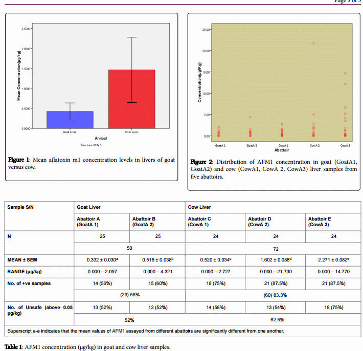
High Performance Liquid Chromatographic evidence showed the presence of aflatoxin M1 with different concentrations in 58% goat liver and 83.3% of cow liver samples (Table 1). Mean concentration of AFM1 was higher in in cow liver sample than in goat liver sample (Figure 1) (Appendix 1).
Distribution of aflatoxin M1 concentration in abattoirs
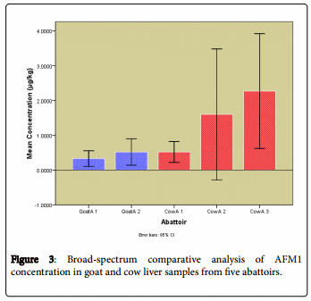
After analyzing AFM1 contamination in liver samples from the five abattoirs, a bi-variant scatter plot (Figure 2) show evidence that cow liver samples had the highest concentration of sample contamination, their maximum levels were significantly higher than that of goat liver samples. Figure 3 then shows that even the mean aflatoxin M1 levels in the cow liver samples from the three abattoirs were higher than those found in goat liver samples from two abattoirs, a broad-spectrum comparative analysis assisted this comparison.
Discussion
Conversion of aflatoxin B1 to aflatoxin M1 takes place in the liver, leading to the elevated levels of AFM1 in the organ [13]. The entrance of aflatoxins in livestock organs or tissues is mainly through their feeds, which must have been contaminated with aflatoxigenic fungi such as Aspergillus flavus or Aspergillus parasiticus either at preproduction or postproduction stages [14]. Aflatoxin M1 occurrence in livestock liver can thereby be managed by controlling level of aflatoxin B1 in feed. Analyses in this study showed unique comparative levels of AFM1 occurrence in goat (58%) and cow (83.33%) livers under study and within the abattoirs where liver sample were collected.
AFM1 level in cow liver samples (0.000-21.730 µg/kg) was significantly (p ≤ 0.05) higher than that assayed from goat liver samples (0.000-4.321 µg/kg). Also the concentrations of the toxin found in the various abattoirs were significantly different (p ≤ 0.05) from each other. The levels of AFM1 in some analyzed samples exceeded the Codex Alimentarius and European communities recommended limits, mean AFM1 concentration were 0.332 µg/kg, 0.518 µg/kg, 0.520 µg/kg, 1.602 µg/kgand 2.271 µg/kg for the five abattoirs respectively. In many countries of Europe, the low level proposed for AFM1 relates to stringent regulation of AFB1 in complementary livestock feedstuffs [15]. Studies have shown the presence of AFB1 residues in the livers of camels (0.12 µg/kg), beef calves (0.051 µg/kg), buffalo calves (0.015 µg/kg) and sheep (0.015 µg/kg) [1,2], with camels and beef liver having highest residue concentrations. Histopathological examination of liver samples with high AFB1 residues showed vacuolar degenerations, cholangitis, cirrhosis, bile duct carcinoma and hepatocellular carcinoma [16]. Thus aflatoxin residues may be implicated in massive histopathological changes to the liver tissue of animals and they had also reported histopathological changes including fatty change, cellular dissociation, necrosis, cellular infiltration, fibrosis and bile duct hiperplasia [14].
Total permissible aflatoxin level in animal feeds range from 0 to 50 parts per billion (ppb) with an average of 20 ppb [17]. Results of this study are similar to the results obtained by Atanda et al. [18], but higher than that estimated by previous studies of Zinedine et al. [19], Heshmati and Milani [20]. Though these studies were on AFM1 levels and occurrence in livestock milk not in livestock livers. However, the findings in this study lend support to those obtained in breast milk study of Coulter et al. [21]. In the study of Dashti et al. [22] fresh milk samples from Kuwaiti markets were contaminated with aflatoxin M1, with 8 samples exceeding maximum tolerant limit.

The percentage of AFM1 contamination as found in these samples were 72.95% with varying contamination levels. However, the present contamination of AFM1 in these samples was not unexpected as it may be consistent with the presence of AFB1 in feed fed to livestock, this can be seen in Udom et al. [23] Who reported mean concentration of 20.58 µg/kg and 10.08 µg/kg as well as occurrence of 91.67% and 58.33% for aflatoxin in diary feed and maintenance feed respectively, they also found that about 92% of the concentrate feed samples for dairy cattle had AFB1 concentrations exceeding the stipulated 5 µg/kg maximum limit set by the European Union (EU) [24] for dairy cattle. Most livestock brought to the abattoir in Minna are reared in free range system where uncontrolled grazing and periodic feedstock administration is involved.
Glutathione s-transferase (GST) is an enzyme that have been identified to be important in the detoxification of AFB1 and AFM1 . GST catalyzes the conjugation of the epoxides of both aflatoxins with glutathione [9]. When GST activity is high in a cell, lower concentration of toxic variations of aflatoxins is expected. In a study by Chuku and Uwakwe [25], GST activity was reported within the ranges 4.05-6.65 I.U, 2.85-5.02 I.U and 3.05-4.52 I.U in goat, cow and sheep blood respectively. This gives a possible explanation for lower AFM1 concentration in goat liver when compared to cattle liver in this study. The report of Vodela and Dalvi [26] indicated that red blood cell activities of GST decreases in the order; mouse, rats, dogs, cattle, pigs, goats and horses with the lowest activity in rabbits.
Generally, suggestion to the variations in concentration of AFM1 assumed to be dependent on the level of exposure, duration of exposure, genetic (species and breed strain), physiological (age, nutrition, and exercise) and environmental (climatic and husbandry) factors. Therefore, livestock AFB1 exposure must be reduced by good practices and controlled animal nutrition.
Conclusion
AFM1 contamination in the present investigation of livestock liver samples is alarming high and may pose a serious public health problem to animal and human health. Goat liver (52%) and cow liver (62.5%) samples were contaminated beyond the safe limit. Also cow liver samples possessed higher concentrations of AFM1 (0.000-21.730 µg/kg) than goat liver (0.000-4.321 µg/kg) samples. AFM1 contamination in the livestock liver samples in Minna, Niger state appears to be of high incidence and may pose a public health problem if stringent measures are not put in place. Awareness must be conveyed to producers, handlers, specialists and livestock husbanders.
Disclosure
The experiments comply with the current laws of research in Nigeria. The authors declare no conflict of interest.
Acknowledgements
The technical assistance of the biochemistry laboratory technologists in Federal University of Technology Minna Nigeria in sample handling is highly appreciated.
This article was originally published in Journal of Liver 2017, 6:1 DOI: 10.4172/2167-0889.1000205. This is an open-access article distributed under the terms of the Creative Commons Attribution License.
References
1. El-Shewy EA, Ashoub MA, El-Hoshy SM (1997) Toxicity of aflatoxin and ochratoxin and their residues in some animal products. Vet Hum Toxicol 13: 219-231.
2. D'Angelo A, Bellino C, Alborali GL, Biancardi A, Borrelli A, et al. (2007) Neurological signs associated with aflatoxicosis in Piedmontese calves. Vet Rec 160: 698-700.
3. Bintvihok A, Davitiyananda D (2002) Aflatoxins and their metabolites residues in Chicken tissues from 5 parts (10 provinces) of Thailand. Thai J Health Res 16: 37-50.
4. Adejumo O, Atanda, O, Raiola A, Somorin Y, Bandyopadhyay R, et al. (2013) Correlation between aflatoxin M1 content of breast milk, dietary exposure to aflatoxinB� and socioeconomic status of lactating mothers in Ogun State, Nigeria. Food Chem Toxicol 56: 171-177.
5. Iha MH, Barbosa CB, Heck AR, Trucksess MW (2014) Aflatoxin M1 and ochratoxinA in human milk in Ribeirão Preto-SP, Brazil. Food Control 40: 310-313.
6. Boudra H, Barnouin J, Dragacci S, Morgavi DP (2007) Aflatoxin M1 and ochratoxin A in raw bulk milk from French dairy herds. J Dairy Sci 90: 3197-3201.
7. Akande KE, Abubakar MM, Adegbola TA, Bogoro S (2006) Nutritional and health implications of mycotoxins in animal feeds: A Review. PJN 5: 398-403.
8. Alberto G (2014) Aflatoxin M1 Residues in Milk and their Impact on Human Health. Food Mycotoxicology 2: 1-17.
9. Neal GE, Eaton DL, Judah DJ, Verma A (1998) Metabolism and toxicity of aflatoxins M1 and B1 in human-derived in vitro systems. Toxicol Appl Pharmacol 151: 152-158.
10. Sinnhuber RO, Lee DJ, Wales JH, Landers MK, Keys AC (1974) Hepatic Carcinogenesis of Aflatoxin M1 in Rainbow Trout (Salmo gairdneri) and its Enchancement by Cyclopropene Fatty Acids. J Natl Cancer Inst 53: 1285-1288.
11. Iheanacho HE, Dutton MF, Steenkamp PA, Steenkamp L, Makun HA, et al. (2014) Real time PCR of Nor ~ 1 (aflD) gene of aflatoxin producing fungi and its correlative quantization to aflatoxin levels in South African compound feeds. J Microbiol Methods 97: 63-67.
12. AOAC (International Association of Official Analytical Chemists) (2010) Natural Toxins. Aflatoxins. In: Official Methods of Analysis, 18th edn. Gaithersburg, MD, USA 49: 4.
13. Makun HA, Dutton MF, Njobeh PB, Gbodi TA, Timothy A, et al. (2012) Aflatoxin Contamination in Foods and Feeds: A Special Focus on Africa. Trends in Vital Food and Control Engineering, Ayman +afiz Amer Eissa (Ed.) pp: 953-978.
14. Hussain Z, Khan MZ, Hassan ZU (2008) Production of aflatoxins from Aspergillus flavus and Acute aflatoxicosis in young broiler chicks. Pak J Agri Sci 45: 95-102.
15. Shipra R, Premendra DD, Subhash KK, Mukul D (2004) Detection of Aflatoxin M1 contamination in milk and infant milk products from Indian markets by ELISA. Food control 15: 287-290.
16. Al-Hizab FA, Al-Gabri, NAM, Barakat SEM (2015) Effect of Aflatoxin B1 (AFB1) Residues on the Pathology of Camel Liver. AJAVA 10: 173-178.
17. FAO/WHO (2012) Joint FAO/WHO Food standards programme codex committee on contaminants in foods. Sixth session Maastricht, The Netherlands, 26 – 30 March 2012.
18. Atanda O, Oguntubo A, Adejumo O, Ikeorah J, Akpan I (2007) Aflatoxin M1 contamination of milk and ice cream in Abeokuta and Odeda local governments of Ogun State, Nigeria. Chemosphere 68: 1455-1458.
19. Zinedine A, González-Osnaya L, Soriano JM, Moltó JC, Idrissi L, et al. (2007) Presence of aflatoxin M1 in pasteurized milk from Morocco. Int J Food Microbiol 114: 25-29.
20. Heshmati A, Milani JM (2010) Contamination of UHT milk by aflatoxin M1 in Iran. Food Control 21: 19-22.
21. Coulter JB, Lamplugh SM, Suliman GI, Omer MI, Hendrickse RG (1984) Aflatoxins in human breast milk. Ann Trop Paediatr 4: 61-66.
22. Dashti B, Al-Hamli S, Alomirah H, Al-Zenki S, Bu Abbas AB, et al. (2009) Levels of aflatoxin M1 in milk, cheese consumed in Kuwait and occurrence of total aflatoxin in local and imported animal feed. Food Control 20: 686-690.
23. Udom EI, Ezekiel CN, Fapohunda SO, Okoye ZSC, Kalu CA (2012) Incidence of Aspergillus Section Flavi and Concentration of Aflatoxin in Feed Concentrates for Cattle in Jos, Nigeria. J Veterinary Adv 2: 39-46.
24. European communities (1992) Commission Directive 92/95/EEC and Commission Directive 94/14/EC amending the annex of the Seventh Commission Directive 76/372/EEC establishing community methods of analysis for the Official control of feeding stuffs. Official Journal of European Communities 154.
25. Chuku LC, Uwakwe AA (2012) Haematological and Biochemical Studies on some Ruminants. J App Sci Environ Manage 16: 217-221.
26. Vodela JK, Dalvi RR (1997) Erythrocyte glutathione-S-transferase activity in animal species. Vet Hum Toxicol 39: 9-11002E.


