Explore
Communities in English
Advertise on Engormix

Biosynthesis and Characterization of Silver Nanoparticles from Marine Seaweed Colpomenia Sinuosa and its Antifungal Efficacy

Published: August 7, 2020
By: Dr Vishnu Kiran Manam 1 and Dr Murugesan Subbaiah 1 . / 1 Unit of Algal Biotechnology and Bio nanotechnology, PG and Research Department of Plant biology and Biotechnology, Pachaiyappa’s s College, University of Madras, Chennai, Tamilnadu, India - 600 030.
Summary

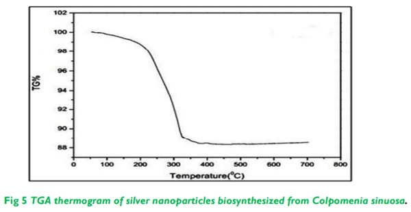
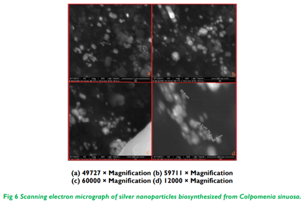
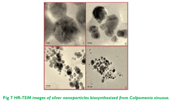
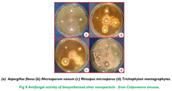
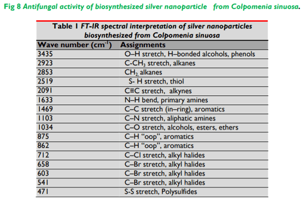
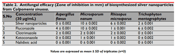
The present study illustrates the biosynthesis of colloidal silver nanoparticles (AgNPs) from marine seaweed Colpomenia sinuosa and their antifungal efficacy was determined against dermatophytic and non-dermatophytic fungi. The dermatophytic fungi used in this study are Microsporum nanum (ATCC 28951) and Trichophyton mentagrophytes (ATCC 28185) whereas the non dermatophytic fungi Aspergillus flavus (ATCC 20048) and Rhizopus microsporus (ATCC 22960). The rich content of phytochemicals, bioactive compounds and secondary metabolites in marine seaweed Colpomenia sinuosa possess the reducing/capping agents that may be environmentally acceptable and eco-friendly for the biosynthesis of silver nanoparticles. The efficacy of biosynthesized silver nanoparticles from marine macroscopic brown alga was performed using Kirby Bauer Method and the silver nanoparticles biosynthesized was characterized by UV-vis spectroscopy, Fourier Transform Infrared (FTIR) Spectroscopy, X-ray Diffraction (XRD) and Thermo gravimetric analysis. Particle size distribution and morphology were investigated by scanning electron microscope which showed silver nanoparticles in the size range of 54-85 nm. The average size of the silver nanoparticle indicated by TEM analysis was found to be 34 nm. The antifungal efficacy of silver nanoparticles at the concentration 30 µg/mL revealed greater efficacy in dermatophytic fungi while Rhizopus microsporus as non dermatophytic fungus showed better antifungal activity when compared to the standard fungal antibiotics used.

Keywords: AgNps, biosynthesis, Colpomenia sinuosa, antifungal efficacy, dermatophytes, non dermatophytes.

1. INTRODUCTION
The field of nanotechnology is one of the most active research areas in modern materials science. The biosynthesis of nanoparticles and nanomaterials as an emerging highlight has been widely attributed in the convergence of nanotechnology and biomedical sciences which opens the avenues and possibilities for wide variety of biological research and medical uses at cellular and molecular level. In the current scenario, the use of biocompatible nanoparticles in biomedical applications such as drug delivery cancer-cell diagnostics and therapeutics due to their optical properties such as surface plasmon resonance (SPR) and fluorescence has given nanotechnology a new dimension. Silver nanoparticles are one of the promising products in the nanotechnology industry. The development of consistent processes for the synthesis of silver nanomaterials is an important aspect of current nanotechnology research. One such promising process is the green synthesis which proves to be efficient without any side effects. Silver nanoparticles can be synthesized by several physical, chemical and biological methods. A possible application of silver nanoparticles is its potential as a catalyst. Silver is a safe inorganic antibacterial agent being used for centuries and is capable of killing microorganisms that cause diseases. Silver nanoparticles have been recently known to be a promising antimicrobial agent that acts for a broad range of target sites, both extracellular as well as intracellular. Biomolecules have been used for nanomaterial synthesis/functionalization and in subsequent applications for decades. Two-thirds of the world’s biomass are found in the ocean with marine species comprising around half of the total global biodiversity. Among marine organisms, marine algae have been reported to possess many health benefits to human welfare. Seaweeds are the raw material for many industrial products like agar, alginates and carrageenan; also, they continue to be widely consumed as food and medicine in Asian countries. Most edible seaweeds are known to contain significant quantities of protein, lipids, minerals, vitamins, and nutrient contents. Seaweeds are also considered to be a rich source of antioxidants. Seaweeds are marine macroalgae, a potentially renewable resource in the marine environment and are known to be an extremely rich source of bioactive compounds. Therefore, algae can be a very interesting natural resource of new metabolites with various biological activities that could be used as functional ingredients. During the last three decades, the discovery of metabolites with biological activities from marine macroalgae has increased significantly and extensive work continues on the isolation of commercially significant secondary metabolites of marine algae39. Biological activities correlate to the presence of biochemical compounds, particularly secondary metabolites. Last few decades have witnessed the rise of fungal infections especially in patients who are immunocompromised as in cancer therapy, organ, HIV infections and nosocomial infections across the world. Therefore there is an urgent need for the development of novel antifungals since the resistance towards antibiotics have emerged as a greatest threat  One of the alternatives towards resistance antibiotics is the development of silver nanoparticles. The antimicrobial properties of silver and their compounds have been known since ancient times. The recent and advanced research into metal nanoparticles has led to the use of silver nanoparticles as potent antimicrobial agents and as a catalyst due to its immense catalytic activity. The recent research in the developments of nanoparticles has led the pathway for new research in silver nanoparticles as antimicrobial agents. This present investigation illustrates green synthesis of silver nanoparticles using the extract of marine macroscopic brown seaweed Colpomenia sinuosa and characterized by UV-visible spectrophotometer. Morphological and elemental analysis was carried out by scanning electron microscopy (SEM) and transmission electron microscopy (TEM). The stability of silver nanoparticles was confirmed by Thermo gravimetric analysis. The possible biomolecules responsible for the reduction of the Ag+ ions and capping of the bio-reduced silver nanoparticles synthesized from the experimental algae Colpomenia sinuosa was identified by using Fourier Transform Infrared spectroscopy (FT-IR). The crystalline structure of the silver nanoparticles, including lattice parameters, geometry, and orientation of single crystals was studied using X-ray Diffraction (XRD) Thus, algae-mediated synthesized silver nanoparticles show more inhibition growth of dermatophytic fungi and Rhizopus microsporus in non dermatophytic fungi showed better zone of inhibition which was analyzed by agar disc diffusion method.
2. MATERIALS AND METHODS
2.1 Collection and preparation of Algal extract
Colpomenia sinuosa (Mertens ex Roth) Derbes and Soldier was collected from the intertidal regions of Leepuram, Kanyakumari District (Latitude 8°14'23.10" N, Longitude 77°20'04.02"E); South East Coast of Tamilnadu, India. Collected seaweed was washed with sea water for eliminating impurities such as sand, rocks, epiphytes and epifauna. The washed samples were preserved with 5-10% formaldehyde in sea water and transported to the laboratory in a box containing slush ice. The fumes of the formaldehyde would help to fix and preserve the seaweed material. In the laboratory, the samples were washed thoroughly in running tap water to remove salt and washed three times using distilled water which may remove metallic compounds and it was shade dried at room temperature (37 ºC) for 10 days. The dried algal materials were crushed by using mortar and pestle to get the powder form and it was stored in an airtight container. About 1 g of crushed algal powder was added with 100 ml of distilled water in 250 ml conical flask and boiled for 5-10 minutes at 60-80 °C. The crude extract was collected and stored at 4 °C for experimental use.
2.2 Bio-synthesis and characterization of silver nanoparticles
The crude extract of the experimental Colpomenia sinuosa (Mertens ex Roth) Derbes and soldier was used for the synthesis of silver nanoparticles. Silver nitrate (AgNO3-) (SD fine) was used for the synthesis of silver nanoparticles and double-distilled, deionized water was used for all the experiments. The silver nanoparticle formation was carried out by taking 500 mg of dry, shade dried powder samples of Colpomenia sinuosa in a 250 mL Erlenmeyer flask with 10-3 M aqueous AgNO3- solution and was incubated at room temperature. The pH was checked during the course of reaction and it was found to be 5.09. Nearly 95 % of bio reduction of AgNO3- ions occurred within 24 hrs at stirring condition. The biosynthesis of silver nanoparticles was characterized by UV Vis spectrophotometer (Labtron LUSB16) in the absorbance mode, and in the wavelength range between 300 to 500 nm. FTIR spectra of biosynthesized silver nanoparticles were recorded using Thermo Scientific/Nicolet iS10 spectrometer with 1 cm-1 resolution in the transmission mode from wave numbers 450 to 4000 cm-1. The Crystal structure and size of silver nanoparticles were determined using the X-ray diffractometer (Labtron LXRD-A10). Thermal stability and purity of silver nanoparticles were analyzed using Thermo Gravimetric Analysis (TGA 4000 - PerkinElmer). A scanning electron micrograph was taken using the (SEM Quanta – 400) to study the morphological characteristics of the silver nanoparticles. Further insight on morphology and the size details of the biosynthesized silver nanoparticles by the experimental algae Colpomenia sinuosa were investigated using High Resolution Transmission Electron Microscopy (HR-TEM JEOL 3010).
2.3 Fungal Susceptibility to nano silver
Antifungal activity of the biosynthesized silver nanoparticles by the experimental algae was tested against pathogenic fungi. The disc diffusion method for antifungal assay was adapted for testing silver nanoparticles. The fungal cultures were obtained from LGC Promochem India Pvt. Ltd, Peenya, and Bangalore, India. The fungal pathogens Aspergillus flavus (ATCC 20048), Rhizopus microsporus (ATCC 22960), Microsporum nanum (ATCC 28951) and Trichophyton mentagrophytes (ATCC 28185) are the fungal strains were used to study the antifungal efficacy of silver nanoparticles. The standard antifungal agents at the concentration of 30 µg/mL Fluconazole, Clotrimazole, Nalidixic acid and Ketoconazole (Hi-media) were used as zone interpretive criteria. The Mueller-Hinton agar (Hi-media) supplemented with 2% glucose and 0.5 µg/L methylene blue dye medium was used for culturing the fungal stains. The Mueller-Hinton agar is readily available and shows an acceptable batch-to-batch reproducibility while the glucose provides a stable growth of most fungi and the addition of methylene blue enhances the zone edge definition. The pH of the medium was maintained between 7.2 and 7.4 at 37|C. The inoculum is standardized to 0.5 McFarland using a densitometer and the plate was incubated at 35 °C for 24 hrs. Some strains where insufficient growth has occurred after 24 hours were necessary to be read after 48 hrs of incubation.
3. STATISTICAL ANALYSIS
The data were analyzed by using MS Excel 2007 and presented as mean ± SD of three replicates. One-way analysis of variance (ANOVA) and Tukey tests were performed by using ‘Stat plus 2009 professional’ trial version software to determine significant zone differences and means were considered as statistically significant if p < 0.05.
4. RESULTS AND DISCUSSION
4.1 Visual examination of biosynthesized silver nanoparticles
The biosynthesis of silver nanoparticles was primarily identified by color change during exposure of crude algal extract of Colpomenia sinuosa into aqueous solution of silver ions (Figure 1). The shade dried powder preparations of the experimental algae Colpomenia sinuosa were added in 10-3 M silver nitrate solution and allowed to react at 121°C for 20 minutes. The colour of the reaction solution changed to dark reddish brown. The control (without algal powder) showed no colour formation. Formation of the colour arises due to the excitation of surface plasmon vibrations where the metabolites in the algal extract act as the capping agent. The colour of the solution gradually intensified on heating which clearly indicates and confirms the formation of silver nanoparticles. After 24 hrs, there is no significant color change, indicating the saturation of the reaction of silver nanoparticle formation.
4.2 UV-visible spectroscopic analysis of biosynthesized silver nanoparticles
The absorption spectra of silver nanoparticles formed in the reaction solution were characteristic for the algae and had specific absorption maxima 428 nm for Colpomenia sinuosa (Figure 2). The broadening of the peak indicated that the silver nanoparticles synthesized from the experimental algae are poly dispersed in nature and the intensity of the band increased with the increase in reaction time. The frequency and width of the surface plasmon absorption depends largely on the size and shape of the metal nanoparticles as well as on the dielectric constant of the metal and the surrounding medium. The interaction with the biomolecules present in the aqueous part of the reaction solution by the biosynthesized silver nanoparticles from experimental algae has been indicated by UV visible spectroscopic analysis. There were no little signs of aggregation with the biosynthesized silver nano particle solution which were stable for more than six months of observation.
4.3 FTIR spectroscopic analysis of biosynthesized silver nanoparticles
The FTIR spectral measurements were carried out to identify the potential biomolecules in the crude extract of the seaweed Colpomenia sinuosa which is responsible for reducing and capping the bio reduced silver nanoparticles. Silver nanoparticles biosynthesized from experimental seaweed Colpomenia sinuosa were analyzed using FTIR spectroscopy. The local molecular environment of the organic molecules on the surface of the nanoparticles was determined by the IR spectra. FT-IR is a technique, which is used to analyze the chemical composition of many organic chemicals, semiconductor materials, gases, biological samples, inorganics, and minerals. FTIR analysis can give not only qualitative (identification) analysis of materials, but, with relevant standards, can be used for quantitative (amount) analysis. The FT-IR spectral absorbance bands of the nanoparticles of Colpomenia sinuosa were seen at 3435 cm1 (O–H stretch, H–bonded alcohols, phenols), 2923 cm-1 (C-CH3 stretch, alkanes), 2853 cm-1 (CH2, alkanes), 2519 cm-1 (S- H stretch, thiol), 2091 cm-1 (C≡C stretch, alkynes), 1633 cm-1 (N–H bend, primary amines), 1469 cm-1 (C–C stretch (in–ring), aromatics), 1103 cm-1 (C–N stretch, aliphatic amines), 1034 cm-1 (C–O stretch, alcohols, ethers), 875 cm-1 (C-H out of plane bending, aromatics), 862 cm-1 (C-H out of plane bending, aromatics), 712 cm-1 (C–Cl stretch, alkyl halides), 658 cm-1 (C–Br stretch, alkyl halides), 603 cm-1 (C–Br stretch, alkyl halides), 541 cm-1 (C–Br stretch, alkyl halides), and 471 cm-1 (S-S stretch, polysulfides) (Table 1; Figure 3). The FT-IR spectrum analysis indicates the presence of chemical bonds and the functional groups of the biomolecules responsible for the stabilization of the biosynthesized silver nanoparticles from the experimental marine brown alga Colpomenia sinuosa. The FTIR results revealed that the compounds present in the marine brown alga Colpomenia sinuosa which are aromatic, alkanes, amides or amines that may act as the capping ligand in the formation of silver nanoparticles and also possible perform the stabilization of silver nanoparticles in the aqueous medium.
4.4 X-Ray diffraction pattern (XRD)
XRD is widely used to determine the size and crystal structure of silver nanoparticles. X-ray diffractogram of the biosynthesized silver nano particle by the experimental algae Colpomenia sinuosa exhibits Bragg reflection corresponding to face centered cubic (fcc) type bulk silver. The broadened diffraction peaks around their base indicates that the silver nano particle is between nano sizes. XRD (Labtron LXRDA10) analysis of biosynthesized silver nanoparticles from Colpomenia sinuosa exhibited four distinct diffraction peaks (Figure 4). The lattice planes {1 0 0}, {1 1 0}, {1 1 1}, {2 0 0}, were identified with the corresponding Bragg’s angles of 11.58°, 32.04°, 37.89° and 46.96° respectively, which confirm the face-centered cubic structure of the silver nanoparticles. The observed peak broadening and noise were probably related to the effect of nano sized particles and the presence of various biological molecules in the reaction solution. The XRD pattern thus clearly shows that the silver nano particles are crystalline in nature.
4.5 Thermo Gravimetric Analysis (TGA) of biosynthesized silver nanoparticles
The thermogram of biosynthesized silver nanoparticles from Colpomenia sinuosa was observed and there was no major weight loss up to 220 °C and a sharp weight loss of 12 % was observed in the temperature range of 220-400 °C (Figure 5). TGA result shows that the purity of silver nanoparticles was 95% for biosynthesis of silver colloidal medium carried out by the ultra-sonication method.
4.6 Scanning Electron Microscopy (SEM) of biosynthesized silver nanoparticles
The silver nanoparticles synthesized from Colpomenia sinuosa biomass after exposure to 10-3 M aqueous silver nitrate solution for 2 hours showed the colloidal form of the particles in solution which micro precipitated on the surface of the biomass of the experimental algae. The back scattered electron image showed bright cubical and spherical area which corresponds to the silver nano particle indicating the structure of nano silver (Figure 6). The hydrogen bond and electrostatic interactions might have played a key role along with the bioorganic capping molecules of the experimental algae in the formation of the morphology and shape of silver nanoparticles. The other factor in determining the shape of the silver nano particles may be attributed to the changes in the optical and electronic properties. The stabilization of the nanoparticles by the capping agent might be due to nanoparticles not in direct contact within the aggregates. Thus the shape and morphology of the biosynthesized nanoparticles by the experimental brown alga Colpomenia sinuosa was clearly revealed by the SEM results.
4.7 Transmission Electron Microscopy (TEM of biosynthesized silver nanoparticles
The HR-TEM images of silver nanoparticles clearly revealed the formation of spherical and hexagonal shaped nanoparticles. The majority of the nanoparticles appeared spherical and only a small percentage was elongated particles that ranged in size from 5 to 50 nm The average means size of silver nanoparticles were 34 nm (Figure 7).
4.8 Antifungal efficacy of silver nanoparticles
The silver nanoparticles biosynthesized from Colpomenia sinuosa at the concentration 30 µg/mL were tested against dermatophytes and non dermatophytes. The dermatophytic fungi Microsporum nanum (10 ± 0.002 mm) and Trichophyton mentagrophytes (2 ± 0.001 mm) showed variable zone of inhibition with biosynthesized silver nanoparticles from seaweed where the fungi Microsporum nanum showed greater inhibition when compared to the fungi Trichophyton mentagrophytes which was comparatively less in the interpretive zone. The non dermatophytic fungi Rhizopus microsporus (6 ± 0.002 mm) showed intermediate zone of inhibition with biosynthesized silver nanoparticles whereas the non dermatophyte Aspergillus flavus showed no Zone formation which indicates its resistance towards biosynthesized silver nanoparticles (Figure 8). Standard antifungal agent fluconazole exhibited higher susceptibility towards dermatophytic fungi Microsporum nanum and Trichophyton mentagrophytes when compared to non dermatophytic fungi Aspergillus flavus and Rhizopus flavus which showed intermediate zone of inhibition. The antifungal agent Clotrimazole showed an intermediate zone of inhibition for both dermatophytic and non dermatophytic fungi whereas the antifungal agent Ketoconazole least zone of inhibition towards dermatophytic and non dermatophytic fungi. The Nalidixic acid antifungal showed resistance towards dermatophytic fungi and non dermatophytic fungi with no zone formation. The dermatophyte Trichophyton mentagrophytes were resistant to the antifungals Clotrimazole, Ketoconazole and Nalidixic acid. (Table 2; Figure 8).The mechanism of the inhibitory activity by the biosynthesized silver nanoparticles from marine brown alga Colpomenia sinuosa might be due to the adhesion of silver nanoparticles to the cell membrane and then penetrates in to the fungal pathogen and produce a site with little molecular weight at the centre of the fungi and then nano silver attaches to the respiratory sequence which finally causes the cell division to stop and lead to the death off the fungal cells, silver nanoparticles also release silver ions with increased antifungal efficacy. The antifungal efficacy of silver nanoparticles was determined by the type of fungal pathogen involved along with the size and surface area of the silver nano particle which may be closely associated with the formation of pits in the cell wall of the fungal58. The other possible mechanism of silver nanoparticles is that it may disrupt the membrane potential of the cell membrane of the fungal pathogen leading to the cell death.
Biosynthesis and Characterization of Silver Nanoparticles from Marine Seaweed Colpomenia Sinuosa and its Antifungal Efficacy - Image 1
 
Biosynthesis and Characterization of Silver Nanoparticles from Marine Seaweed Colpomenia Sinuosa and its Antifungal Efficacy - Image 2
 
Biosynthesis and Characterization of Silver Nanoparticles from Marine Seaweed Colpomenia Sinuosa and its Antifungal Efficacy - Image 3
 
Biosynthesis and Characterization of Silver Nanoparticles from Marine Seaweed Colpomenia Sinuosa and its Antifungal Efficacy - Image 4
 
Biosynthesis and Characterization of Silver Nanoparticles from Marine Seaweed Colpomenia Sinuosa and its Antifungal Efficacy - Image 5
 
Biosynthesis and Characterization of Silver Nanoparticles from Marine Seaweed Colpomenia Sinuosa and its Antifungal Efficacy - Image 6
 
Biosynthesis and Characterization of Silver Nanoparticles from Marine Seaweed Colpomenia Sinuosa and its Antifungal Efficacy - Image 7
 
Biosynthesis and Characterization of Silver Nanoparticles from Marine Seaweed Colpomenia Sinuosa and its Antifungal Efficacy - Image 8
 
Biosynthesis and Characterization of Silver Nanoparticles from Marine Seaweed Colpomenia Sinuosa and its Antifungal Efficacy - Image 9
 
Biosynthesis and Characterization of Silver Nanoparticles from Marine Seaweed Colpomenia Sinuosa and its Antifungal Efficacy - Image 10
5. CONCLUSION
The results concluded that the silver nanoparticles biosynthesized from marine seaweed Colpomenia sinuosa were effective against dermatophytes and intermediate effect was observed among non dermatophytic fungi especially Rhizopus microsporus. The efficacy of silver nanoparticles in this study may pave a pathway for the use of colloidal silver in the antibiotic or antifungal resistance strains especially will be helpful in treating nosocomial infections. The results also reveal that the dermatophytes are more susceptible to dermatophytes and intermediate activity was observed in the non dermatophytic fungi Rhizopus microsporus when compared to standard antifungal agents used in the study.
6. ACKNOWLEDGEMENT
I sincerely thank Mr. Raju sea diver who helped me in the collection of the sea weeds from the south east coast of tamilnadu, India. This work was carried out in part through the use of the Scanning Electron Microscopy (SEM) and Transmission Electron Microscopy (TEM) at Sophisticated Analysis Instrument Facility (SAIF), Indian Institute of Technology (IIT) – Madras, Tamilnadu, India. The Thermogravimetic analysis (TGA) was carried out at Department of chemistry, Anna University, Chennai, Tamilnadu, India. The authors thank the above institutions for their instrumentation facility for the completion of the work.
7. AUTHORS CONTRIBUTION STATEMENT
Dr Vishnu Kiran Manam designed and performed the work whereas Dr Murugesan Subbaiah suggested the relevant changes during the course of the work and the proofreading of the manuscript.
8. CONFLICT OF INTEREST
Conflict of interest declared none.
This article was originally published in International Journal of Pharma and Bio Sciences, Volume11., No 2 (Apr) 2020, pp 59-68. This is an Open Access article under the CC BY- NC-ND License (https://creativecommons.org/licenses/by-nc-nd/4.0)

1. Singh M, Manikandan S, Kumaraguru AK. Nanoparticles: A new technology with wide applications. Res. J. Nanosci. Nanotechnol. 2011; 1:1- 11. DOI: 10.3923/rjnn.2011.1.11

2. West JL Halas NJ. Engineered nanomaterials for biophotonics applications: Improving sensing, imaging, and therapeutics. Annu. Rev. Biomed. Eng. 2003; 5: 285-292. DOI: 10.1146/annurev.bioeng.5.011303.120723

3. Paciotti GF, Myer L, Weinreich D, Goia D, Pavel N, McLaughlin RE, et al. Colloidal gold: A novel nanoparticle vector for tumour directed drug delivery. Drug. Delivery. 2004; 11: 169-183. DOI: 10.1080/10717540490433895

4. Jain KK. Nanotechnology-based drug delivery for cancer. Technol. Cancer. Res. Treat. 2005; 4: 407-416. DOI: 10.1177/153303460500400408

5. Wu X, Liu H, Liu J, Haley KN, Treadway JA, Larson JP, et al. Immuno fluorescent labeling of cancer marker Her2 and other cellular targets with semiconductor quantum dots. Nat. Biotechnol. 2003; 21(1): 41-

6. DOI: 10.1038/nbt764 6. Chan WCW, Maxwell DJ, Gao X, Bailey RE, Han M, Nie S. Luminescent quantum dots for multiplexed biological detection and imaging. Curr. Opin. Biotechnol. 2002; 13 (1): 40-6. DOI: 10.1016/s0958-1669(02)00282-3

7. Sokolov K, Follen M, Aaron J, Pavlova I, Malpica A, Lotan R, et al. Real-time vital optical imaging of precancerous drugs using anti-epidermal growth factor receptor antibodies conjugated to gold nanoparticles. Cancer. Research. 2003; 63: 1999-2004. Available from; https://www.ncbi.nlm.nih.gov/pubmed/12727808

8. Alivisatos AP. The use of nanocrystals in biological detection. Nat. Biotechnol. 2004; 22 (1): 47-52. DOI: 10.1038/nbt927

9. Hirsch LR, Stafford RJ, Bankson JA, Sershen SR, Rivera B, Price RE, et al. Nano shell-mediated near-infrared thermal therapy of tumours under magnetic resonance guidance. Proc. Natl. Acad. Sci. U.S.A. 2003; 100 (23): 13549-54. DOI: 10.1073/pnas.2232479100

10. Barwal I, Ranjan P, Kateriya S, Yadav SC. Cellular oxido-reductive proteins of Chlamydomonas reinhardtii control the biosynthesis of silver nanoparticles. J.Nanobiotech. 2011; 9:56. DOI: 10.1186/1477-3155-9-56

11. Taleb A, Petit C, Pileni MP. Optical properties of selfassembled 2D and 3D super lattices of silver nanoparticles. J. Phys. Chem. B. 1998; 102 (12): 2214- 2220. DOI: 10.1021/jp972807s

12. Thirumalai AV, Prabhu D, Soniya M. Stable silver nano particle synthesizing methods and its applications. J. Bio sci. Res. 2010; 1 (4): 259-270. Available from: https://www.researchgate.net/publication/281282881_ Silver_nanoparticles_synthesis_medical_application_a nd_toxicity_effects

13. Balantrapu K, Goia D. Silver nanoparticles for printable electronics and biological applications. J. Mater. Res. 2009; 24 (9): 2828-2836. DOI: 10.1557/jmr.2009.0336

14. Tripathi RM, Saxena A, Gupta N, Kapoor H, Singh RP. High antibacterial activity of silver nanoballs against E.coli MTCC 1302, S.typhimurium MTCC 1254, B.subtilis MTCC 1133 and P.aeruginosa MTCC 2295. Dig. J. Nanomater. Bios. 2010: 5 (2): 323- 330. Available from: http://chalcogen.ro/323_Tripathi.pdf

15. Patakfalvi R, Dekany I. Preparation of silver nanoparticles in liquid crystalline systems. Colloid. Polym. Sci. 2010; 280 (5): 461- 470. DOI: 10.1007/s00396-001-0629-0

16. Rodriguez SL, Blanco MC, Lopez QMA. Electrochemical synthesis of silver nanoparticles. J. Phys. Chem. B. 2000; 104: 9683- 9688. DOI: 10.1021/jp001761r

17. Jeong SH, Yeo SY, Yi SC. The effect of filler particle size on the antibacterial properties of compounded polymer/silver fibers. J. Mater. Sci. 2005; 40: 5407- 5411. DOI: 10.1007/s10853-005-4339-8

18. Shrivastava S, Bera T, Roy A, Singh G, Ramachandrarao, Pand DD. Characterization of enhanced antibacterial effects of novel silver nanoparticles. Nanotechnology. 2007; 18 (22): 225- 103. DOI: 10.1088/0957-4484/18/22/225103

19. Bharathiraja S, Suriya J, Sekar V, Rajasekaran R. Biomimetic of silver nanoparticles by Ulva lactuca seaweed and evaluation of its antibacterial activity. Int. J. Pharm. Pharmaceut. Sci. 2012; 4(3): 139-143. Available from: https://www.researchgate.net/publication/289066797

20. Shiny PJ, Mukherjee A, Chandrasekaran, N. Marine algae mediated synthesis of the silver nanoparticles and its antibacterial efficiency. Int. J. Pharm. Pharmaceut. Sci. 2013; 5 (2): 239-241. Available from: https://shodhganga.inflibnet.ac.in/bitstream/10603/1517 97/16/16_publications.pdf

21. Mohandass C, Vijayaraj AS, Raja sabapathy R, Satheesh babu S, Rao SV, Shiva, et al. Biosynthesis of silver nanoparticles from marine seaweed Sargassum cinereum and their antibacterial activity. Ind. J. Pharm. Sci. 2013; 75 (5): 606-610. Available from: https://www.ncbi.nlm.nih.gov/pmc/articles/PMC387752 5/

22. Manal AA, Awatif AH, Khalid MO, Dalia FAE, Abdelelah AGA. Silver nanoparticles are biogenic synthesized using an orange peel extract and their use as an antibacterial agent. Intl. J. phys.ical sci. 2014; 9: 34-40. DOI: 10.5897IJPS2013.4080

23. Vishnu Kiran M, Murugesan S. Biosynthesis of silver nanoparticles from marine alga Halymenia poryphyroides and its antibacterial efficacy. Int. J. Curr. Microbiol. App. Sci. 2014; 3 (4): 96-103. Available from: https://www.ijcmas.com/Archives-17.php

24. Niemeyer CM. Nanoparticles, Proteins, and Nucleic Acids: Biotechnology Meets Materials Science. Angew. Chem. Int. Ed. 2001; 40: 4128-4158. DOI: 10.1002/1521-3773(20011119)40:22<4128::AIDANIE4128>3.0.CO;2-S.

25. Mishra VK, Temelli PF, Ooraikul Shacklock, Craigie JS. Lipids of the Red Alga, Palmaria palmate. Bot. Mari.1993; 36 (2): 169-174. DOI: 10.1515/botm.1993.36.2.169

26. Ajit KA, Meena K, Rao MM, Panda P, Mangal AK, Reddy, G et al. Marine algae: An Introduction, Food value and Medicinal uses. J. Pharm. Res.. 2011; 4 (1): 123-127.

27. Smit AJ. Medicinal and pharmaceutical uses of seaweed natural products: A review. J. App. Phycol. 2004; 16: 245–262. DOI: 10.1023/B:JAPH.0000047783.36600.ef

28. Wong KH, Cheung, PCK. Nutritional evaluation of some subtropical red and green seaweed. Part Iproximate composition, amino acid profiles and some physico-chemical properties. Food. Chem. 2000; 71: 475-482. DOI: 10.1016/s0308-8146(00)00175-8

29. Norziah MH, Ching Ch Y. Nutritional composition of edible seaweeds Gracilaria changgi. Food. Chem.. 2002; 68: 69-76. DOI: 10.1016/S0308-8146(99)00161-2

30. Ravi S, Baghel, Puja K, Reddy, CRK, Bhavanath J. Growth, pigments, and biochemical composition of marine red alga Gracilaria crassa. J. App. Phycol. 2014; 26 (5): 2143–2150. Available from: https://www.researchgate.net/publication/267042453

31. Dawes CJ, Kovach C, Friedlander M. Exposure of Gracilaria to various environmental conditions II. The effect on fatty acid composition. Bot. Mari.. 1993; 36: 289-296. DOI: 10.1515/botm.1993.36.4.283

32. Kaehler S, Kennish R. Summer and winter comparisons in the nutritional value of marine macro algae from Hong Kong. Bot. Mari. 1996; 39: 11-17. DOI: 10.1515/botm.1996.39.1-6.11

33. Sokolova EV, Barabanova AO, Homenko VA, Soloveva TF, Bogdanovich RN, Yermak IM. In vitro and ex vivo studies of antioxidant activity of carrageenans, sulfated polysaccharides from red algae. Bull. Exp. Biol. Med. 2011; 150: 426-428. DOI: 10.1007/s10517-011-1159-5

34. Chandini SK, Ganesan PV, Suresh N, Bhaskar. Seaweeds as a source of nutritionally beneficial compounds – A review. J. Food. Sci. Technol. 2008; 45: 1–13. Available from: https://www.researchgate.net/publication/298525973

35. Kladi M, Vagias C, Roussis V. Volatile halogenated metabolites from red algae. Phytochem. Rev. 2004; 3: 337-366. DOI: 10.1007/s11101-004-4155-9

36. Kashman Y, Rudi A. On the biosynthesis of marine isoprenoids. Phytochem. Rev. 2004; 3: 309-323. DOI: 10.1007/s11101-004-8062-x

37. Plaza M, Cifuentes A, Ibanez E. In the search of new functional food ingredients from algae. Trends. Food. Sci. Tech. 2008; 19: 31-39. DOI: 10.1016/j.tifs.2007.07.012

38. Holdt SL, Kraan S. Bioactive compounds in seaweed: Functional food applications and legislation. J. Appl. Phycol. 2011; 23: 543-597.

39. Faulkner DJ. Marine natural products. Nat. Prod. Rep. 2002; 19: 1-49. DOI: 10.1039/B009029H

40. Mirmirani P, Hessol NA, Maurer TA, Berger TG, Nguyen P, Khalsa A. Prevalence and predictors of skin disease in the Women’s Interagency HIV Study (WIHS). J. Am. Acad. Dermatol. 2001; 44: 785- 788. DOI: 10.1067/mjd.2001.112350

41. Kim KJ, Woo SS, Seok KM, Jong SC, Jong GK, Dong GL. Antifungal Effect of Silver Nanoparticles on Dermatophytes. J. Microbiol. Biotechnol. 2008; 18 (8): 1482–1484. Available from: http://europepmc.org/article/med/18756112

42. Nasrollahi KH, Pourshamsian P, Mansour kiaie. Antifungal activity of silver nanoparticles on some fungi. Int. J. Nano. Dim. 2011; 1 (3): 233- 239. Available from: https://www.sid.ir/en/journal/ViewPaper.aspx?ID=1943 94

43. Klasen HJ. A historical review of the use of silver in the treatment of burns. Π. Renewed interest for silver. Burns. 2000; 26: 131-138. DOI: 10.1016/s0305-4179(99)00116-3

44. Russell AD, Hugo WB. 7 Antimicrobial activity and action of silver. Prog. Med. Chem. 1994; 31: 351- 70. DOI: 10.1016/S0079-6468(08)70024-9

45. Silver S. Bacterial silver resistance: Molecular biology and uses and misuses of silver compounds. FEMS. Microbiol. Rev. 2003; 27: 341- 353. DOI: 10.1016/S0168-6445(03)00047-0

46. Baker C, Pradhan A, Pakstis L, Pochan DJ, Shah SI. Synthesis and antibacterial properties of silver nanoparticles. J. Nanosci. Nanotechnol. 2005; 5: 244- 249. DOI: 10.1166/jnn.2005.034

47. Lee BU, Yun SH, Ji JH, Bae GN. Inactivation of S. epidermidis, B. subtilis, and E. coli bacteria bioaerosols deposited on a filter utilizing airborne silver nanoparticles. J. Microbiol. Biotechnol. 2008; 18: 176- 182. Available from: https://www.ncbi.nlm.nih.gov/pubmed/18239437

48. Melaiye A, Sun Z, Hindi K, Milsted A, Ely D, Reneker DH, et al. Silver (I)-imidazole cyclophane gem-diol complexes encapsulated by electrospun tecophilic nanofibers: Formation of nanosilver particles and antimicrobial activity. J. Am. Chem. Soc. 2005; 127: 2285- 2291. DOI: 10.1021/ja040226s

49. Sondi I, Salopek SB. Silver nanoparticles as antimicrobial agents: A case study on E. coli as a model for Gram-negative bacteria. J. Colloid. Interface. Sci. 2004; 275: 177- 182. DOI: 10.1016/j.jcis.2004.02.012

50. Jiang ZJ, Liu CY, Sun LW. Catalytic properties of silver nanoparticles supported on silica spheres. J. Phys. Chem. B. 2005; 109 (5): 1730-5. 10.1021/jp046032g

51. Rajesh Kumar S, Malarkodi C, Venkat Kumar S. Synthesis and characterization of silver nanoparticles from marine brown seaweed and its antifungal efficiency against clinical fungal pathogens. Asian. J. Pharm. Clin. Res. 2017; 10 (2): 190-193. DOI: 10.22159/ajpcr.2017.v10i2.15127

52. Finegold SM, Baron EJ. “Bailey and Scotts Diagnostic Microbiology” 7th Ed., The C.V. Mosby Company, St. Louis. 1986.

53. NCCLS. Method for Antifungal Disk Diffusion Susceptibility Testing of Yeasts; Approved Guideline. NCCLS document M44-A [ISBN 1-56238-532-1]. NCCLS, 940 West Valley Road, Suite 1400, Wayne, Pennsylvania 19087-1898 USA, 2004.

54. Mukherjee P, Ahmad A, Mandal D, Senapati S, Sainkar SR., Khan MI. Fungus-mediated synthesis of silver nanoparticles and their immobilization in the mycelial matrix: A novel biological approach to nanoparticle synthesis. Nano. Lett. 2001; 1 (10): 515- 519. DOI: 10.1021/nl0155274

55. Mukherjee P, Ahmad A, Mandal D, Senapati S, Sainkar SR., Khan MI, Kumar R. Extracellular synthesis of gold nanoparticles by the fungus Fusarium oxysporum. Chem. Biochem. 2002; 3 (5): 461- 463. DOI: 10.1002/1439-7633(20020503)3:5<461::AIDCBIC461>3.0.CO;2-X

56. Gonzalo J, Serna R, Sol J, Babonneau D, Afonso CN. Morphological and interaction effects on the surface plasmon resonance of metal nanoparticles. J. Phys. Condens. Matter. 2003; 15 (42): 3001-3002. Available from; https://iopscience.iop.org/article/10.1088/0953- 8984/15/42/002/meta

57. Alt V, Bechert T, Steinruecke P, Wagener M, Seidel P, Dingeldein E, et al. An in vitro assessment of the antibacterial properties and cytotoxicity of nanoparticulate silver bone cement. Biomaterials. 2004; 25: 4383– 4391. DOI: 10.1016/j.biomaterials.2003.10.078

58. Shafaghat A. Synthesis and characterization of silver nanoparticles by the phytosynthesis method and their biological activity. Synth. React. Inorg. Metal–Org. Nano-Metal. Chem. 2015; 45: 381– 387. DOI: 10.1080/15533174.2013.819900

59. Kim J, Lee J, Kwon S, Jeong S. Preparation of biodegradable polymer/silver nanoparticles composite and its antibacterial efficacy. J. Nanosci. Nanotechnol. 2009a; 9: 1098- 1102. DOI: 10.1166/jnn.2009.c096

60. Ahmed RH, Mustafa DE. Green synthesis of silver nanoparticles mediated by traditionally used medicinal plants in Sudan. Int. Nano. Lett. 2020; 10: 1–14. DOI: 10.1007/s40089-019-00291-9

61. Anupam R, Onur B, Sudip S, Amit KM, Yilmaz MD. Green synthesis of silver nanoparticles: biomoleculenanoparticle organizations targeting antimicrobial activity. RSC. Adv. 2019; 9: 2673-2702. DOI: 10.1039/C8RA08982E

62. Anes As, Kasing A, Micky V, Devagi K, Lesley MB. Enhancement of antibacterial activity of silver nanoparticles against gram positive and gram negative using blue laser light. Intl. J. Photoene. 2019; 1-12. DOI: 10.1155/2019/2528490
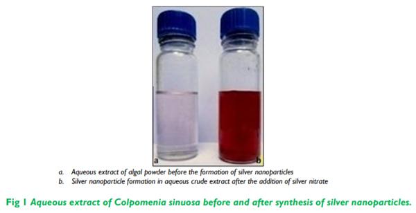
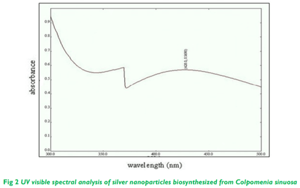
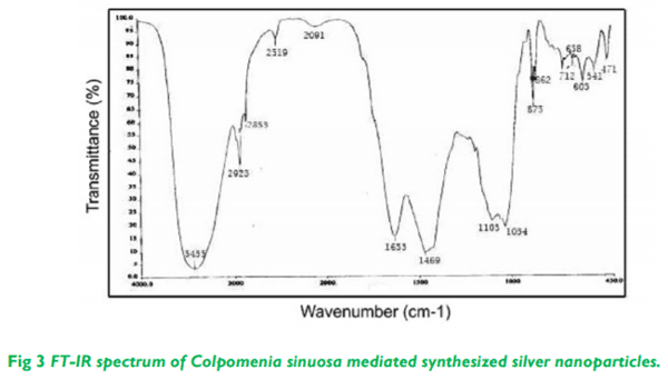
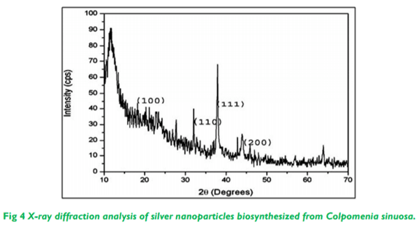
Related topics:
Authors:
DR VISHNU KIRAN MANAM
Recommend
Comment
Share
Profile picture
Would you like to discuss another topic? Create a new post to engage with experts in the community.
Join Engormix and be part of the largest agribusiness social network in the world.