Explore
Communities in English
Advertise on Engormix

Aflatoxins: Occurrence, Exposure, and Binding to Lactobacillus Species from the Gut Microbiota of Rural Ugandan Children

Published: April 15, 2020
By: Alex Paul Wacoo 1,2,3; Prudence Atukunda 4; Grace Muhoozi 5; Martin Braster 1; Marijke Wagner 1; Tim J van den Broek 6; Wilbert Sybesma 2; Ane C. Westerberg 7; Per Ole Iversen 4,8,9; and Remco Kort 1,2,10.
Summary

Author details:

1 Department of Molecular Cell Biology, Vrije Universiteit Amsterdam, 1081 HZ Amsterdam, The Netherlands; 2 Yoba for Life foundation, 1079 WB Amsterdam, The Netherlands; 3 Department of Medical Biochemistry, School of Biomedical Sciences, College of Health Sciences, Makerere University, P.O. Box 7062, Kampala, Uganda; 4 Department of Nutrition, Institute of Basic Medical Sciences, University of Oslo, 0317 Oslo, Norway; 5 Department of Human Nutrition and Home Economics, Kyambogo University, P.O. Box 1 Kampala, Uganda; 6 Department of Microbiology and Systems Biology, TNO, 3704 HE Zeist, The Netherlands; 7 Institute of Health Sciences, Kristiania University College, 0107 Oslo, Norway; 8 Division of Human Nutrition, Stellenbosch University, Tygerberg, 7505 Cape Town, South Africa; 9 Department of Hematology, Oslo University Hospital, 0318 Oslo, Norway; 10 ARTIS-Micropia, 1018 CZ Amsterdam, The Netherlands.


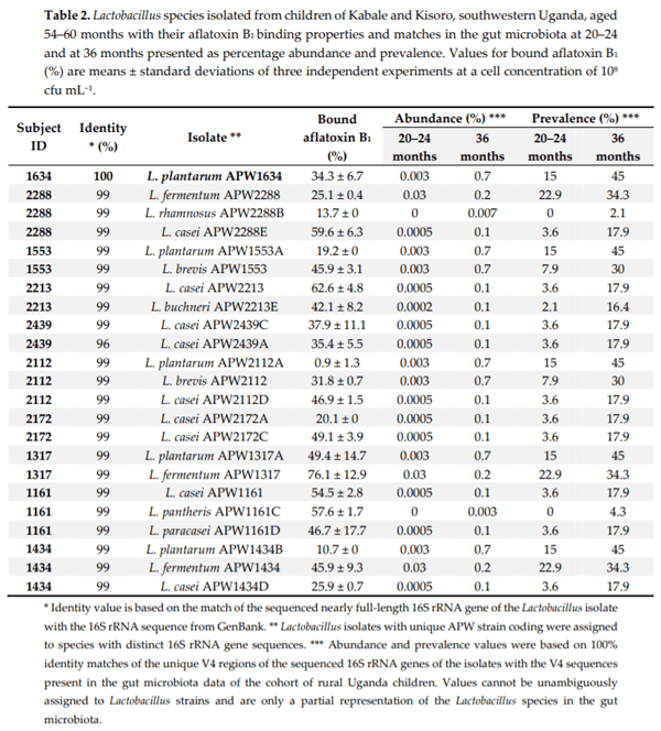
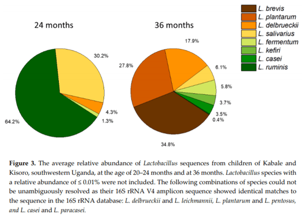
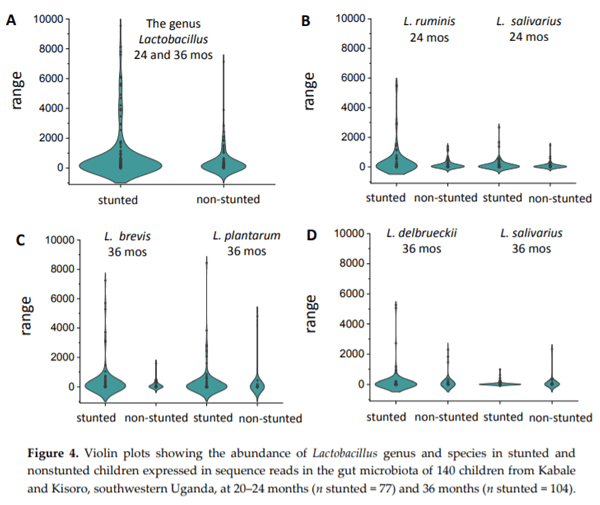
Chronic exposure of children in sub-Saharan Africa to aflatoxins has been associated with low birth weight, stunted growth, immune suppression, and liver function damage. Lactobacillus species have been shown to reduce aflatoxin contamination during the process of food fermentation. Twenty-three Lactobacillus strains were isolated from fecal samples obtained from a cohort of rural Ugandan children at the age of 54 to 60 months, typed by 16S rRNA gene sequencing, and characterized in terms of their ability to bind aflatoxin B1 in vitro. Evidence for chronic exposure of these children to aflatoxin B1 in the study area was obtained by analysis of local foods (maize flour and peanuts), followed by the identification of the breakdown product aflatoxin M1 in their urine samples. Surprisingly, Lactobacillus in the gut microbiota of 140 children from the same cohort at 24 and 36 months showed the highest positive correlation coefficient with stunting among all bacterial genera identified in the stool samples. This correlation was interpreted to be associated with dietary changes from breastfeeding to plant-based solid foods that pose an additional risk for aflatoxin contamination, on one hand, and lead to increased intake of Lactobacillus species on the other.

Keywords: Stunting; aflatoxin B1; Lactic acid bacteria; aflatoxin binding; gut microbiota

1. Introduction
The warm and humid climate conditions of sub-Saharan Africa promote the growth of fungi and associated production of mycotoxins. Approximately 25% of grains harvested annually worldwide contain mycotoxins. Ingestion of these contaminated foods can lead to disease and death [1]. Aflatoxin is the most prevalent and harmful human mycotoxin reported to date [2]. Aflatoxins are common food contaminants produced as secondary metabolites of fungi belonging to genus Aspergillus [3]. Their toxicity leads to carcinogenic and teratogenic effects as well as growth faltering, which has been confirmed in animal models, rendering aflatoxins a major food safety concern [4–6].
Four major types of aflatoxins such as B1, B2, G1, and G2 are commonly reported as contaminants of foods, including maize, ground nut, and cotton seeds. Aflatoxin B1 is the most prevalent, contributing to up to 75% of all aflatoxin contamination of foods, and it has been classified as a Group 1 carcinogen by the International Agency for Research on Cancer (IARC) in 1987 [3]. To minimize the risk of aflatoxin ingestion from contaminated foods, maximum levels for aflatoxin in nuts, grains, and oil seeds has been set up by many countries. The East Africa Community has proposed the maximum allowable level of aflatoxin as 5 µg kg−1 for aflatoxin B1 and 10 µg kg−1 for total aflatoxin [7]. Some African countries adopted codex regulatory levels, which vary between 0.5 and 15 µg kg−1 [8]. In comparison, the European Commission set the regulatory limit at 2 for B1 and 4 µg kg−1 for total aflatoxins in human foods [9].
Despite all these regulatory limits, aflatoxins are still present in dangerously high levels in groundnuts, cassava, and corn, which make up the bulk of children’s diets in Africa [10]. Approximately 74% of maize flour consumed in Kampala, Uganda, was contaminated with aflatoxins at a range from 1.8 to 268 µg kg−1 [11]. These high levels of contamination were further confirmed by Muzoora et al. who found that 72% of peanuts collected from different regions of Uganda were contaminated with aflatoxins, ranging from 1.6 to 516 µg kg−1 [12]. Due to ingestion of highly contaminated foods, Asiki et al. reported that all 100 adults and 92 children out of a total of 96 tested children had detectable levels of aflatoxin-albumin adduct [13]. This study also revealed that five babies who were exclusively breastfed tested positive for aflatoxin albumin adduct. Although a direct causal relationship has not been established, high levels of aflatoxin exposure could contribute to the high rate of stunted growth of 46% in Western Uganda [14,15].
Following ingestion of contaminated food, and reaching the upper small intestine (duodenum), aflatoxin is absorbed into the blood stream rapidly [16]. Although there is rapid absorption, aflatoxins have been found to affect the gastrointestinal tract by impairing cell growth, causing DNA damage and increasing lactate dehydrogenase activity [17]. Moreover, aflatoxins have also been reported to affect the gut microbiota. Wang et al. showed that aflatoxin Bhas the ability to alter the gut microbiota in a dose-dependent manner in rats; aflatoxin Bdid not affect gut microbiota at the phylum level, but some lactic acid bacteria were depleted [18]. Galarza-Seeber et al. also revealed that aflatoxins at a dose of one part per million (ppm) significantly decreased total lactic acid bacteria in broilers [19].
The human gut microbiota is composed of trillions of bacteria that play an important role in maintaining health [20]. The gut microbiota provides a protective barrier for the host against the proliferation of pathogenic bacteria. Gut bacteria also play a crucial role in the digestion of a wide range of foods as well as the binding and degradation of toxins [21]. Therefore, the intake of probiotics and lactic acid bacteria via fermented foods could help to reduce the uptake of aflatoxins [22]. In a study carried out on young men from Guangzhou, China, significant reduction of urinary aflatoxin was noted after administration of Lactobacillus rhamnosus LC705 and Propionibacterium freudenreichii compared to placebo [23]. A similar study showed that administration of L. casei Shirota significantly decreased the level of aflatoxin B1-lysine adduct [24]. Furthermore, a number of other studies confirmed the ability of lactic acid bacteria to bind aflatoxin B[25,26].
Despite the evidence for the toxic effects of aflatoxins, there is generally poor awareness of the risk of these toxins and a lack of proper detection methods to monitor levels in food [27]. Therefore, a portable immunosensor was developed, validated, and used to measure aflatoxin levels in maize from markets and households in Kampala [11,28], indicating that consumers, including children, are exposed to relatively high concentrations of aflatoxin. A strategy was proposed to detoxify aflatoxin in end products by fermentation with the probiotic gut isolate Lactobacillus rhamnosus yoba 2012 [29]. In this study, the exposure to aflatoxins was evaluated for 10 children (aged 54–60 months) from a cohort of 511 children. Their foods (maize flour and peanuts) were analyzed for the presence of aflatoxin Band their urine for aflatoxin M1. The aflatoxin Bbinding ability was tested of Lactobacillus species isolated from the gut microbiota of these children, and the correlation of gut Lactobacillus species with stunting for 140 children was analyzed from the same cohort at the ages of 20–24 and at 36 months.
2. Materials and Methods
2.1. Study Design and Sample Collection
In the current study, a small sample of ten children aged 54–60 months was selected on the basis of their previous growth indices at 36 months (5 stunted and 5 non-stunted children). These children took part in a randomized trial in the two districts of Kabale and Kisoro, located in the southwestern part of Uganda (Figure 1). The trial assessed the effect of an educational intervention (focusing on nutrition, hygiene, and stimulation) on their growth and development as described in detail in previous publications [30,31]. Samples of the stool and urine were taken from every child in this study. The stool was sampled using a sterile disposable stool sampling container. The stool samples from every child were put in two separate containers: one container was filled with mineral oil and kept at room temperature for the purpose of cultivation, and the other container was filled with two milliliters of 15% glycerol. The stool samples with glycerol were immediately kept on ice and transferred to the Uganda Industrial Research Institute for storage at −80 °C. Urine was sampled into a sterile, disposable plastic container and immediately stored at low temperature (0–8 °C). The ten children were widely spread across the districts of Kabale and Kisoro as indicated by subject identification number (Figure 1).
Anthropometric measurements were taken as described by Muhoozi et al. [30] and used to compute height-for-age Z–scores (HAZ) [32]. The frequently consumed regional foods (hulled and dehulled maize, and peanuts) at risk of aflatoxin contamination were identified based on a short food frequency questionnaire (Supplementary File S1). Common diet for the age group and last week’s diet of the children were evaluated. The foods were sampled and immediately stored at low temperature (0–8 °C). The in vitro measurements were carried out at the Uganda Industrial Research Institute (Kampala, Uganda) and at the Department of Molecular Cell Biology, Vrije Universiteit (Amsterdam, The Netherlands). The Lactobacillus species were isolated, and their ability to bind aflatoxin B1  was assessed. The concentration of aflatoxin B1  was determined in the food samples and aflatoxin M1 in the children’s urine.
Aflatoxins: Occurrence, Exposure, and Binding to Lactobacillus Species from the Gut Microbiota of Rural Ugandan Children - Image 1
2.2. Aflatoxin B1 in Food and Daily Intake
Hulled, dehulled maize flour, and peanut were analyzed using the ELISA Ridascreen® Aflatoxin B1 with a reported limit of detection of 1.0 µg kg−1 (R-Biopharm, Darmstadt, Germany). The enzyme immunoassay was first calibrated by the use of an aflatoxin B1 standard (0, 1, 5, 10, 20, and 50 µg l−1) in 10% (v/v) methanol as indicated in Supplementary File S2. A competitive enzyme immunoassay was used for the determination of aflatoxin B1 in cereals similar to the method described by Wacoo et al. [11]. Briefly, 5 g of homogenized maize flour was weighed into 50 mL centrifuge tubes, followed by addition of 25 mL of 70% (v/v) methanol and thorough mixing using a VWR ADC 3500 Shaker (BioSurplus, Inc, San Diego, CA, USA) for 5 minutes. In the case of peanuts, 0.4 g of sodium chloride was added to the suspension and thoroughly mixed. The suspension was then centrifuged for 10 minutes at 3500× g at room temperature. An aliquot of 50 µL of each supernatant was used for aflatoxin B1 determination using the ELISA kit.
The minimum daily aflatoxin B1 intake of children in southwestern Uganda (expressed in ng kg−1 day−1) was estimated on basis of the measured concentrations of aflatoxin B1 in maize flour and peanut sampled in each subcounty, the estimated amounts of maize and peanut consumed, and the measured body weight of the child [33]. The frequency of intake of maize flour and peanut in southwestern Uganda was obtained by a food frequency questionnaire (Supplemental File S1). The amount of maize and peanut consumed was based on the previously reported intake estimate for maize flour and products of 60 g per day for children at the age of 24 to 59 months in southwestern Uganda [34].
2.3. Aflatoxin M1 in Urine
Quantitative determination of aflatoxin M1 in urine samples was carried out using the ELISA Ridascreen® Aflatoxin Mdesigned with a limit of detection of 5 ng L−1. The ELISA kit was validated as described by the International Conference on Harmonization (ICH) (1995) [35]. The limit of detection (LOD), precision, and accuracy were determined by using concentrations of aflatoxin M1 standard (0, 125, 250, 500, 1000, and 2000 ppt) spiked in urine (Supplementary File S2). Briefly, the pH of the urine samples was first adjusted to seven. A volume of 50 µL of either standard or urine samples was pipetted and put into separate wells, followed by addition of 50 µL of enzyme conjugate and 50 µL of anti-aflatoxin Mantibody solution. The microwell plate was then mixed by shaking gently and incubated for 10 minutes at room temperature. The liquid was removed and the wells were washed three times using 250 µL of washing buffer. Then, the wells were filled with 100 µL of substrate and incubated for 5 minutes at room temperature. The reaction was stopped with 100 µL of stop solution and the absorbance taken at 450 nm wavelength using a microplate reader. The results of the standard solution were used to develop a calibration curve, and the aflatoxin Mlevels in each urine sample were determined from this curve. Urine creatinine concentrations were determined spectrophotometrically by the modified Jaffe method [36]. The aflatoxin Mconcentration from each sample was subsequently normalized to creatinine concentration in the urine. The percentage of aflatoxin, which is excreted as aflatoxin Min urine, was calculated on basis on the estimated dietary aflatoxin intake per kg body weight per day (ng), the concentration of aflatoxin Min urine (ng/mg creatinine), and the reported levels of excreted creatinine in urine per day of 15.4 mg per kg body weight per day for boys and 14.3 mg per kg body weight per day for girls [37,38].
2.4. Enumeration and Isolation of Lactic Acid Bacteria
Serial dilutions of samples were prepared in physiological saline. Total counts of lactic acid bacteria (LAB) from stool samples were determined by streaking selected serial dilutions on sterile de Man, Rogosa, Sharpe (MRS) agar (Oxoid limited, Hampshire, United Kingdom) containing 0.1% Tween 80. The plates were incubated at 37 °C for 48 h. The experiment was performed four times for every sample. After total counts of lactic acid bacteria (LAB), five colonies with distinct colony morphology were selected from each plate. The colonies were then streaked to freshly prepared MRS agar plates for identification.
2.5. Identification of Bacterial Isolates
Isolates were identified as described by Felske et al. [39]. Briefly, 16S rRNA gene fragments were amplified and sequenced using primers 8F (5’-AGAGTTTGATYMTGGCTCAG-3’) and 1512R (5’- ACGGYTACCTTGTTACGACTT-3’). The colony PCR reactions were carried out with 1 µL of each primer (10 pmol), 11 µL nuclease-free water (Promega), and 12 µL GoTaq Colorless Master Mix (Promega) in a final volume of 24 µL. To the PCR reaction mix, a small amount of a fresh colony was added using a sterile toothpick. The PCR program was set as follows: initial denaturation was carried out at 94 °C for 5 min, followed by 30 amplification cycles (30 s at 94 °C, 30 s at 55 °C, and 30 s at 72 °C), and a final extension step at 72 °C for 8 min. The PCR products were verified by electrophoresis on 1.5% (w/v) agarose gel, and sequencing was done by the Sanger sequencing method (Macrogen Inc., The Netherlands). Sequences were compared to sequences deposited in GenBank by using the (Basic Local Alignment Search Tool) BLAST algorithm, National Library of Medicine, Bethesda MD, USA [40]. All isolated strains are accessible from the strain collection of the Department of Molecular Cell Biology, Vrije Universiteit Amsterdam, The Netherlands, as well as the Department of Microbiology, School of Biomedical Sciences, College of Health Sciences, Makerere University, Uganda, in order to assure benefit sharing in accordance with the Nagoya protocol [41]. The correlation between the Lactobacillus species isolated at 54–60 months and microbiota at 20–24 and 36 months was assessed by matching the V4 amplicon sequences of our nearly full-length 16S rRNA sequences with our previously collected microbiota data (Sequence Read Archive SUB4476421) [31].
2.6. Aflatoxin B1 Binding to the Isolated Lactobacillus Species
The aflatoxin B1 binding assay was performed as described by Wacoo et al. [29]. Briefly, the isolated Lactobacillus species were cultured in de Man, Rogosa, Sharpe (MRS) broth with 0.1% (v/v) Tween 80 at 37 °C for 24 h. The cells were pelleted at 3,200 g for 10 minutes at room temperature and subsequently washed twice with physiological saline to remove excess MRS broth. The washed cell pellets were re-suspended in 2 mL of physiological saline solution. Each suspension was then serially diluted with physiological saline to obtain approximately 108 cfu mL−1. These dilutions were centrifuged, and the cell pellets re-suspended in 1.0 mg mL−1 of aflatoxin B1 followed by incubation at 37 °C for 30 minutes. After incubation, the aflatoxin B1 cell suspensions were centrifuged at 3200 g for 10 minutes at room temperature, and the residual aflatoxin B1 in the supernatant was determined using the Fluostar Omega microplate reader (BMG Labtech, Ortenberg, Germany) at an excitation of 390 nm and an emission 480 nm. Bound aflatoxin B1 was calculated by use of the formula below:
Aflatoxins: Occurrence, Exposure, and Binding to Lactobacillus Species from the Gut Microbiota of Rural Ugandan Children - Image 2
2.7. Statistical Analysis
For comparison between Lactobacillus species at 20–24 and 36 months, species were identified with BLAST of 16S rRNA amplicon sequences, and the data were presented in a pie chart of Lactobacillus species as the average percentage of the total Lactobacillus per individual. The abundances of Lactobacillus species were calculated by the percentage of all V4 16S rRNA sequence reads from the 23 isolates showing a 100% identity match in the total pool of unique sequence reads at 20–24 months and 36 months. The prevalence of Lactobacillus species was calculated as the percentage of gut microbiota compositions from the 140 children containing at least one exact match to the specific V4 sequence read from one of the 23 Lactobacillus isolates. A permutational multivariate analysis of variance (PERMANOVA) was carried on gut microbiota composition of all rural Ugandan children (n = 140) at 20–24 and 36 months and growth development scores. Analysis was performed using R version 3.3.2, with PERMANOVA as implemented in the ‘vegan’ package by Oksanen et al. using the Bray–Curtis distance measure [42]. The 16S rRNA gene sequencing data were rescaled and transformed using Wisconsin double transformation and square root transformation. The correlations between Lactobacillus species abundance in stunted (HAZ < −2.0) and nonstunted (HAZ > −2.0) children were displayed in violin plots by the use of OriginPro 2019b 9.6.5.169 (Academic).
2.8. Ethical Clearance
The study was approved by the Research Ethics committee of The AIDS Support Organization (no. TASOREC/06/15-UG-REC-009) and by the Uganda National Council for Science and Technology (no. UNCST HS 1809).
3. Results
3.1. Aflatoxin B1 Contamination in Food
In our survey on the consumption of particular foods for southwestern Uganda, beans were found to be the most frequently consumed food (Table S1). This was followed by posho/porridge (corn bread or porridge), greens (eshiga), and Irish potatoes, which were consumed at least once a day. Sweet potatoes and fermented porridge were consumed five to six times a week. Peanut, millet and sorghum porridge were taken four times a month, and dry maize with beans, cassava, and rice was eaten two to three times a month. The foods with a very high risk of aflatoxin contamination were posho/porridge, which was consumed daily, and peanuts. The intake of these foods may result in accumulation of aflatoxin in the body; thus, the ingredients of posho/porridge (maize) and peanuts were selected for further analysis.
The levels of aflatoxin B1 in hulled, dehulled maize, and peanut sampled from Kabale and Kisoro district of southwestern Uganda are shown in Figure 2. All of these food ingredients contained mean aflatoxin Blevels above the acceptable East African regulatory limit of 5 µg kg−1 for aflatoxin B1 [7]. The mean aflatoxin Bconcentration in hulled maize flour was 9.1 µg kg−1. Only 10% of the hulled maize flour samples contained undetectable levels of aflatoxin B1. Dehulled maize flour contained comparable concentrations of aflatoxin Bwith a mean of 5.3 µg kg−1, 6% higher than the East African regulatory limit of 5 µg kg−1 [7]. Approximately 50% of dehulled maize flour contained detectable levels of aflatoxin B1. Peanut samples contained, on average, higher concentrations of aflatoxin Bwith 12.8 µg kg−1. Approximately, 90% of the peanut samples contained aflatoxin Bwith levels ranging from 1.8 to 20.2 µg kg−1. More than 50% of the peanut samples contained aflatoxin Blevels higher than the East African regulatory limit of 5 µg kg−1.
Aflatoxins: Occurrence, Exposure, and Binding to Lactobacillus Species from the Gut Microbiota of Rural Ugandan Children - Image 3
3.2 Daily intake of Aflatoxin B1
Estimates for the daily intake of aflatoxin Bby the children of southwestern Uganda are shown in Table 1. The dietary aflatoxin Bintake varied from 1.12 to 88.6 ng kg−1 day−1. The overall mean dietary aflatoxin Bexposure was 50.1 ng kg−1 day−1. The mean dietary aflatoxin Bexposures to stunted and nonstunted children were 53.7 and 46.5 ng kg−1 day−1, respectively. Although there was a notable difference in the mean dietary aflatoxin Bexposure to the stunted and nonstunted children, statistically the difference was not significant (p > 0.5).
3.3. Aflatoxin M1 in Urine
The results of aflatoxin M1 analyzed from the urine samples of both nonstunted and stunted children are shown in Table 1, which also shows anthropometric parameters for the same children. The aflatoxin M1 levels in the urine samples varied from 14.8 to 168 pg mg−1 of creatinine. There was no significant difference between aflatoxin M1 levels in nonstunted children and stunted children (p > 0.05). A detectable amount of aflatoxin B1 in maize flour and peanut coincided with detectable levels of aflatoxin M1 in urine samples for all children from the different study regions. No positive correlation was observed between the minimum estimated daily intake of aflatoxin B1 and the levels of aflatoxin M1 found in urine samples.
3.4. Enumeration of Lactic Acid Bacteria
Lactic acid bacteria from the stool were counted and the results shown in Table 1. The lactic acid bacterial count in the stool samples varied from 8.9 × 10⁶ to 1.9 ×10⁸ cfu g−1. There was no significant difference between the bacterial counts between the nonstunted children (on average 7.0 × 10⁷ cfu g−1) and stunted children (on average 6.9 × 10⁷ cfu g−1). Approximately 10% of the samples contained lactic acid bacteria at the level of 106 cfu g−1. More than 50% of the samples contained lactic acid bacteria at the level of 107 cfu g−1 and 30% contained lactic acid bacterial concentrations of 108 cfu g−1.
Aflatoxins: Occurrence, Exposure, and Binding to Lactobacillus Species from the Gut Microbiota of Rural Ugandan Children - Image 4
3.5. Isolation, Identification, and Aflatoxin B1 Binding Properties of Lactobacillus Species
In this study, 23 Lactobacillus strains were isolated from fecal samples of 10 children aged 54 to 60 months (Table 2). The identification of the strains was performed based on 16S rRNA gene sequencing [31]. As nearly full-length 16S rRNA sequencing was applied, only one unambiguous identification was found on the species level of either L. casei or L. paracasei with both a percentage identity of 96% (Supplementary File S3). Though the same species of Lactobacillus was repeatedly isolated among subjects, they did not demonstrate equal binding potential to aflatoxin B1. Strains of the species of L. casei were most frequently isolated, accounting for 30% of the total Lactobacillus isolates, but it was also the most prevalent appearing in over 50% of the subjects. Both L. plantarum and L. brevis accounted for 14% of the total isolates each. However, L. plantarum was isolated in approximately 40% and L. brevis was isolated from only 20% of the subjects.
Aflatoxins: Occurrence, Exposure, and Binding to Lactobacillus Species from the Gut Microbiota of Rural Ugandan Children - Image 5
All isolated Lactobacillus strains were assessed for their ability to bind aflatoxin B1 [30]. All Lactobacillus isolates demonstrated binding of aflatoxin Bin physiological saline as shown in Table 2 at slightly variable levels. The best aflatoxin Bbinding was registered for L. fermentum APW1317 and L. casei APW2213C of 76.1% and 62.6% at 108 cfu mL−1 cell concentration. It should be noted that no systematic differences in binding ability were observed between Lactobacillus species or subjects. The amount of aflatoxin bound to the bacteria increased with a rise in cell density from 4.0 × 10⁷ to 8.1 × 10⁷ cfu mL−1. However, some strains indicated higher aflatoxin B1 binding at relatively low cell densities. This is attributed to the ability of these bacteria to coagulate and form clumps at high cell densities resulting in a smaller cell surface area for binding.
The Lactobacillus species in the gut microbiota of the children at 20–24 and 36 months accounted for 2.2% and 3.4% of the total sequence reads, respectively. All the isolated Lactobacillus species at the age of 54–60 months were found to be present at 20–24 and at 36 months, except for L. rhamnosus and L. pantheris, which could not be detected at 20–24 months. Generally, the abundance of Lactobacillus species isolated at 54–60 months, which were present at 20–24 months, varied from 0 to 0.03%. The food-derived L. fermentum was the most abundant (0.03%) and the most prevalent isolate (22.9%) found amongst the 140 children at 20–24 months (Table 2).
3.6. Distribution of Lactobacillus Species in Stool Samples
The distribution of Lactobacillus species in the stool samples of the Ugandan children is shown in Figure 3. At the age of 20–24 months, L. ruminis was the most dominant species of Lactobacillus, accounting for approximately 64.2% of the total Lactobacillus species composition of the gut microbiota. L. ruminis had an abundance of 1.3% of the total Lactobacillus species at 20–24 months. L. salivarius accounted for approximately 30.2% with abundance of 0.6% at 20–24 months. L. delbrueckii and L. fermentum accounted for 4.3% and 1% with corresponding abundance of 0.09% and 0.03%, respectively. The relative abundance of Lactobacillus species to the gut microbiota increased from 2.17% to 3.42% of all 16S rRNA sequence counts in children from 20–24 months to 36 months. Approximately four more dominant species of Lactobacillus emerged at the age of 36 months at the expense of L. salivarius and L. ruminis. There was a notable shift from autochthonous (endogenous) to allochtonous (plant derived) Lactobacillus species, most probably resulting from the change in diet from breast milk to solid food. The allochtonous Lactobacillus species L. brevis emerged at the age of 36 months and became the most dominant species with 34.8%, while the endogenous species L. salivarius and L. ruminis dropped from 64.2% and 30.2% to 6.1% and 0.4%, respectively. L. plantarum, L. delbrueckii, and L. fermentum increased from less than 0.01%, 4.3%, and 1.3% at 20–24 months to 27.8%, 17.9%, and 5.8% at 36 months, respectively. The other prominent species L. kefiri and L. casei were also found at the age of 36 months, accounting for 3.7% and 3.5%, respectively.
Aflatoxins: Occurrence, Exposure, and Binding to Lactobacillus Species from the Gut Microbiota of Rural Ugandan Children - Image 6
3.7. Lactobacillus Inversely Correlates with Growth
The permutational multivariate analysis of variance carried out on gut microbiota composition of rural Ugandan children (n = 140) with anthropometric and cognitive development scores indicated a number of significant correlations. The most pronounced variable was age (20–24 and 36 months) explaining 4.03% of the variance in the microbiota composition with p = 0.001 (Supplementary File S4). The anthropometric measures HAZ, and stunting, explained respectively 0.91% and 0.84% of the variance in the gut microbiota (p = 0.001). The genus Lactobacillus appeared as the genus with the highest correlation coefficient (0.014) for stunting among all 256 taxonomic units of bacterial genera identified in the gut microbiota. This was also evident from a representation of Lactobacillus abundance in the gut microbiota of Ugandan children at 20–24 and 36 months for stunted and nonstunted children (Figure 4A). The average number of Lactobacillus sequence reads was 801 for stunted children and 423 for nonstunted children. However, it should be noted that this difference was not significant (p > 0.05) in non-parametric tests for non-normally distributed data sets. A closer inspection on the Lactobacillus species level (Figure 4B–D) revealed that the most predominant Lactobacillus species followed this trend, except for L. salivarius at 36 months, which appeared more abundantly present in non-stunted children.
Aflatoxins: Occurrence, Exposure, and Binding to Lactobacillus Species from the Gut Microbiota of Rural Ugandan Children - Image 7
4. Discussion
A high exposure to aflatoxin-contaminated food negatively correlates with impaired growth in children [4]. The current study provides evidence that Ugandan rural children are exposed to high concentrations of aflatoxin B1 on a daily basis through consumption of contaminated food stuffs. Analysis of a set of regional maize flour samples indicated higher levels for hulled maize compared to dehulled maize. Siwela et al. were able to reduce up to 92% of aflatoxin contamination in maize through the process of dehulling [43]. Notably, the hulled maize with relatively high contamination levels is the most preferred due to the low costs. The dehulled maize is usually purchased by people categorized in the middle income class.
The estimated intake level to dietary aflatoxin in Uganda ranges from 10 to 180 ng per kg body weight per day [44]. In this study, the minimal estimated dietary aflatoxin intake levels for 80% of the children were found to be within this reported range for Uganda. The finding of 20% of the children with a minimal aflatoxin intake of less than 10 ng per kg body weight per day could result from the coincidental regional sampling of maize with a short storage time. However, lower intake values have been reported for other East African countries including Kenya (4−133 ng per kg body weight per day) [45].
The children’s exposure to aflatoxin B1 was confirmed by analysis of their urine samples for the presence of aflatoxin M1. As reported previously, approximately 1.2% to 2.2% of the dietary aflatoxin Bintake can be excreted in urine as aflatoxin M1 [46]. If two minimal intake levels below 10 ng per kg body weight are considered outliers, the average percentage of aflatoxins excreted in the urine as aflatoxin M1 equals 3.3% ± 2.8%. As in this study minimal intake levels for aflatoxin for Bwere determined, this average percentage will decrease if aflatoxin intake from other food sources such as millet, sorghum, and beans will be included in the analysis.
The ability of lactic acid bacteria including Lactobacillus to protect against food mutagens such as aflatoxins, heterocyclic amines, and phytate among others has been reported in other studies [25,26,47,48]. Physical binding to the bacterial cell wall is reported as one of the mechanisms for the mitigation of aflatoxins from the intestine [49]. In the current study Lactobacillus species were isolated, characterized and their aflatoxin Bbinding ability was tested. The Lactobacillus strains demonstrated a variable ability to bind aflatoxin B1. This variation could be attributed to the differences in structure of the proposed binding surfaces such as cell wall polysaccharides, peptidoglycan, teichoic acid, and cell wall proteins, which are known to be variable among bacterial strains of the same species [48,50].
Previous studies demonstrated that gut microbiota of newborns evolves rapidly during the first 12 months of life, remains highly dynamic up to the age of 24 of months, and becomes more stable afterwards [51]. The diet contributes significantly to this modulation [52]. At the age of 20–24 months, most of the children in our cohort of 511 children were still taking breast milk, while at the age of 36 months this was replaced by solid foods [30]. This could explain the change in composition from the autochthonous species L. salivarius and L. ruminis at 20–24 months to the allochthonous species L. plantarum, L. brevis, L. delbrueckii, L. casei, and L. fermentum at 36 months. The latter five species typically originate from plant-based foods.
A substantial part of the solid foods prepared for babies is at risk for contamination with aflatoxins [53,54]. Food contaminated with aflatoxin Bwas found to affect the gut and injures the stomach and the intestine [55]. Studies carried out in animal models have shown that aflatoxin Bpromotes intestinal damages through perturbation of the intestinal barrier and activation of cell apoptosis and cell proliferation [55]. Saran et al. hypothesized that stunted children may fail to grow due to injury as a result of recurrent infections to the gut epithelium leading to impaired gutmediated immunity, poor nutrient absorption, and poor appetite [56]. Thus, exposure to aflatoxin through solid foods from an early age could contribute to the high levels of stunted children observed in our cohort of 511 children [30].
In this study, the allochthonous Lactobacillus species isolated from the gut microbiota of Uganda children, such as L. casei, L. plantarum, L. fermentum, and L. brevis strains were shown to bind aflatoxins effectively, as also observed for these species in previous studies [57]. Therefore, it seems counterintuitive to find a significant, positive correlation for the abundance of the genus Lactobacillus and stunting, as the presence of this bacterial genus in the small intestine could possibly reduce the uptake of aflatoxins through binding. However, the abundance of Lactobacillus originating from plant-based foods may be indicative for the intake of relatively high levels of aflatoxin-contaminated foods. In contrast to the other Lactobacillus species, L. salivarius at 36 months appeared more abundant in nonstunted children. This could be related to the fact that this is a true endogenous or autochthonous Lactobacillus species in the human gut, and it has been shown to negatively correlate to Shigella-induced diarrhea in African children [58].
At this point it is not clear under which conditions binding of aflatoxin to Lactobacillus in the gut is most effective. Our binding assay has been performed in physiological saline with a neutral pH, which is different from the relatively low pH and other environmental conditions in the upper small intestine, where aflatoxins are absorbed [59]. As bacterial growth predominantly takes place in the colon, leading to concentrations up to 1011 bacteria per mL, concentrations of Lactobacillus in the duodenum may be too low (approximately 106 bacteria mL−1) to effectively remove aflatoxin through binding [60,61]. Although further studies are needed to warrant their health benefits, we propose that the Lactobacillus species isolated from Ugandan children in this study can be further developed as locally sourced probiotics [62] and are promising candidates for decontaminating of aflatoxins through fermentation of maize-containing foods prior to consumption, as recently shown for L. rhamnosus [29].
This article was originally published in Microorganisms 2020, 8, 347; doi:10.3390/microorganisms8030347. This is an Open Access article distributed under the terms and conditions of the Creative Commons Attribution (CC BY) license (http://creativecommons.org/licenses/by/4.0/).

1. Chang, W.; Xie, Q.; Zheng, A.; Zhang, S.; Chen, Z.; Wang, J.; Liu, G.; Cai, H. Effects of aflatoxins on growth performance and skeletal muscle of cherry valley meat male ducks. Anim. Nutr. 2016, 2, 186−191.

2. Feng, L.; Yang, D.; Li, H.; Tian, Y.; Zhao, T.; Zhang, R. Survey on pollution of aflatoxin b1 in foodstuffs. Parasitoses Infec. Dis. 2014, 3, 117−119.

3. Wacoo, A.P.; Wendiro, D.; Vuzi, P.C.; Hawumba, J.F. Methods for detection of aflatoxins in agricultural food crops. J. Appl. Chem. 2014, 2014, 1−15.

4. Gong, Y.Y.; Cardwell, K.; Hounsa, A.; Egal, S.; Turner, P.C.; Hall, A.J.; Wild, C.P. Dietary aflatoxin exposure and impaired growth in young children from benin and togo: Cross sectional study. Br. Med. J. 2002, 325, 20–21.

5. Mitchell, N.J.; Hsu, H.-H.; Chandyo, R.K.; Shrestha, B.; Bodhidatta, L.; Tu, Y.-K.; Gong, Y.-Y.; Egner, P.A.; Ulak, M.; Groopman, J.D. Aflatoxin exposure during the first 36 months of life was not associated with impaired growth in nepalese children: An extension of the mal-ed study. PLoS ONE 2017, 12, e0172124, 1−12.

6. Bahey, N.G.; Elaziz, H.O.A.; Gadalla, K.K.E.S. Toxic effect of aflatoxin b1 and the role of recovery on the rat cerebral cortex and hippocampus. Tissue Cell 2015, 47, 559–566.

7. East Africa Standard. Milled Maize (Corn) Products—Specification; East Africa Community: Arusha, Tanzania, 2011; pp. 1–11

8. Codex Alimentarius Commission (CAC) 2014. Codex General Standard for Contaminants and Toxins in Food and Feed. Codex Standard 193–1995. Accessed 14 January 2020 at http://www.codexalimentarius.net.

9. European Communities. Setting maximum levels for certain contaminants in foodstuffs. Commission Regulation (EC) NO. 1881/2006 of 19 December 2006. Off. J. Eur. Union 2006, 364, 5–24.

10. Matacic, C. Fungal toxins are poisoning africa’s children, says new report. Sci. News 2016, 18.

11. Wacoo, A.P.; Wendiro, D.; Nanyonga, S.; Hawumba, J.F.; Sybesma, W.; Kort, R. Feasibility of a novel onsite detection method for aflatoxin in maize flour from markets and selected households in Kampala, Uganda. Toxins 2018, 10, 1−12.

12. Muzoora, S.; Khaitsa, M.L.; Bailey, H.; Vuzi, P. Status on aflatoxin levels in groundnuts in Uganda. Pan Afr. Med. J. 2017, 27, 11.

13. Asiki, G.; Seeley, J.; Srey, C.; Baisley, K.; Lightfoot, T.; Archileo, K.; Agol, D.; Abaasa, A.; Wakeham, K.; Routledge, M.N. A pilot study to evaluate aflatoxin exposure in a rural ugandan population. Trop. Med. Int. Health 2014, 19, 592–599.

14. Kikafunda, J.; Agaba, E.; Bambona, A. Malnutrition amidst plenty: An assessment of factors responsible for persistent high levels of childhood stunting in food secure western Uganda. Afr. J. Food Agric. Nutr. Dev. 2014, 14, 2088–2113.

15. Watson, S.; Gong, Y.Y.; Routledge, M. Interventions targeting child undernutrition in developing countries may be undermined by dietary exposure to aflatoxin. Crit. Rev. Food Sci. Nutr. 2017, 57, 1963−1975.

16. Kumagai, S. Intestinal absorption and excretion of aflatoxin in rats. Toxicol. Appl. Pharmacol. 1989, 97, 88– 97.

17. Liew, W.-P.-P.; Mohd-Redzwan, S. Mycotoxin: Its impact on gut health and microbiota. Front. Cell. Infec. Microbiol. 2018, 8, 60, 1−17.

18. Wang, J.; Tang, L.; Glenn, T.C.; Wang, J.-S. Aflatoxin b1 induced compositional changes in gut microbial communities of male f344 rats. Toxicol. Sci. 2015, 150, 54–63.

19. Galarza-Seeber, R.; Latorre, J.D.; Bielke, L.R.; Kuttappan, V.A.; Wolfenden, A.D.; Hernandez-Velasco, X.; Merino-Guzman, R.; Vicente, J.L.; Donoghue, A.; Cross, D. Leaky gut and mycotoxins: Aflatoxin b1 does not increase gut permeability in broiler chickens. Front. Vet. Sci. 2016, 3, 10, 1–8.

20. Ihekweazu, F.D.; Versalovic, J. Development of the pediatric gut microbiome: Impact on health and disease. Am. J. Med. Sci. 2018, 356, 413–423.

21. Guarner, F.; Malagelada, J.-R. Gut flora in health and disease. The Lancet 2003, 361, 512–519.

22. Li, J.; Sung, C.Y.J.; Lee, N.; Ni, Y.; Pihlajamäki, J.; Panagiotou, G.; El-Nezami, H. Probiotics modulated gut microbiota suppresses hepatocellular carcinoma growth in mice. Proc. Natl. Acad. Sci. U. S. A. 2016, 113, E1306–E1315.

23. El-Nezami, H.S.; Polychronaki, N.N.; Ma, J.; Zhu, H.; Ling, W.; Salminen, E.K.; Juvonen, R.O.; Salminen, S.J.; Poussa, T.; Mykkänen, H.M. Probiotic supplementation reduces a biomarker for increased risk of liver cancer in young men from southern china. Am. J. Clin. Nutr. 2006, 83, 1199−1203.

24. Redzwan, S.M.; Mutalib, M.S.A.; Wang, J.-S.; Ahmad, Z.; Kang, M.-S.; Nasrabadi, E.N.; Jamaluddin, R. Effect of supplementation of fermented milk drink containing probiotic lactobacillus casei shirota on the concentrations of aflatoxin biomarkers among employees of universiti putra malaysia: A randomised, double-blind, cross-over, placebo-controlled study. Br. J. Nutr. 2016, 115, 39–54.

25. El-Nezami, H.; Kankaanpaa, P.; Salminen, S.; Ahokas, J. Ability of dairy strains of lactic acid bacteria to bind a common food carcinogen, aflatoxin b1. Food Chem. Toxicol. 1998, 36, 321–326.

26. Lili, Z.; Junyan, W.; Hongfei, Z.; Baoqing, Z.; Bolin, Z. Detoxification of cancerogenic compounds by lactic acid bacteria strains. Crit. Rev. Food Sci. Nutr. 2018, 58, 2727–2742.

27. Stepman, F. Scaling-up the impact of aflatoxin research in Africa. The role of social sciences. Toxins 2018, 10, 136, 1−16.

28. Wacoo, P.A.; Ocheng, M.; Wendiro, D.; Vuzi, P.C.; Hawumba, F.J. Development and characterization of an electroless plated silver/cysteine sensor platform for the electrochemical determination of aflatoxin B1. J. Sens. 2016, 2015, 1–8.

29. Wacoo, A.P.; Mukisa, I.M.; Meeme, R.; Byakika, S.; Wendiro, D.; Sybesma, W.; Kort, R. Probiotic enrichment and reduction of aflatoxins in a traditional African maize-based fermented food. Nutrients 2019, 11, 265, 1−15.

30. Muhoozi, G.K.; Atukunda, P.; Diep, L.M.; Mwadime, R.; Kaaya, A.N.; Skaare, A.B.; Willumsen, T.; Westerberg, A.C.; Iversen, P.O. Nutrition, hygiene, and stimulation education to improve growth, cognitive, language, and motor development among infants in uganda: A cluster-randomized trial. Matern. Child Nutr. 2018, 14, e12527, 1−11.

31. Atukunda, P.; Muhoozi, G.K.; van den Broek, T.J.; Kort, R.; Diep, L.M.; Kaaya, A.N.; Iversen, P.O.; Westerberg, A.C. Child development, growth and microbiota: Follow-up of a randomized education trial in Uganda. J. Glob. Health 2019, 9, 1−11.

32. WHO Multicentre Growth Reference Study Group. Who child growth standards based on length/height, weight and age. Acta Paediatr. Suppl. 2006, 450, 76, 1−11.

33. Obade, M.I.; Andang’o, P.; Obonyo, C.; Lusweti, F. Exposure of children 4 to 6 months of age to aflatoxin in Kisumu county, Kenya. Afr. J. Food Agric. Nutr. Dev. 2015, 15, 9949–9963.

34. Dary, O.; Jariseta, Z.R. Validation of dietary applications of household consumption and expenditures surveys (hces) against a 24-hour recall method in Uganda. Food Nutr. Bull. 2012, 33, S190–S198.

35. International Conference on Harmonization, Guideline. In Validation of analytical procedures: Text and methodology Q2 (R1). In Proceedings of the International Conference on Harmonization, Geneva, Switzerland, November 2005, pp. 11–12

36. Vasiliades, J. Reaction of alkaline sodium picrate with creatinine: I. Kinetics and mechanism of formation of the mono-creatinine picric acid complex. Clin. Chem. 1976, 22, 1664−1671.

37. Remer, T.; Neubert, A.; Maser-Gluth, C. Anthropometry-based reference values for 24-h urinary creatinine excretion during growth and their use in endocrine and nutritional research. Am. J. Clin. Nutr. 2002, 75, 561–569.

38. Wang, W.; Du, C.; Lin, L.; Chen, W.; Tan, L.; Shen, J.; Pearce, E.N.; Zhang, Y.; Gao, M.; Bian, J. Anthropometry-based 24-h urinary creatinine excretion reference for Chinese children. PLoS ONE 2018, 13.

39. Felske, A.; Wolterink, A.; Van Lis, R.; Akkermans, A.D. Phylogeny of the main bacterial 16S rRNA sequences in Drentsche Aa grassland soils (The Netherlands). Appl. Environ. Microbiol. 1998, 64, 871–879.

40. Altschul, S.F.; Gish, W.; Miller, W.; Myers, E.W.; Lipman, D.J. Basic local alignment search tool. J. Molecul. Biol. 1990, 215, 403–410.

41. Flach, J.; Ribeiro, C.d.S.; van der Waal, M.B.; van der Waal, R.X.; Claassen, E.; van de Burgwal, L.H. The nagoya protocol on access to genetic resources and benefit sharing: Best practices for users of lactic acid bacteria. Pharma Nutr. 2019, 100158, 1–6.

42. Oksanen, J.; Blanchet, F.; Kindt, R.; Legendre, P.; Minchin, P.; O’Hara, R.; Simpson, G.; Solymos, P.; Stevens, M.; Wagner, H. Vegan: Community ecology package version 2.3–2. Available online: https://cran.rproject.org/web/packages/vegan/index.html (accessed on 1 September 2019).

43. Siwela, A.H.; Siwela, M.; Matindi, G.; Dube, S.; Nziramasanga, N. Decontamination of aflatoxincontaminated maize by dehulling. J. Sci. Food Agric. 2005, 85, 2535–2538.

44. Okoth, S. Improving the evidence base on aflatoxin contamination and exposure in africa. CTA: 2016.

45. Liu Y, Wu F: Global burden of aflatoxin-induced hepatocellular carcinoma: a risk assessment. Environ. Health Perspect. 2010, 118, 818.

46. Sulaiman, S.H.; Jamaluddin, R.; Sabran, M.R. Association between urinary aflatoxin (AFM1) and dietary intake among adults in hulu langat district, selangor, malaysia. Nutrients 2018, 10, 460.

47. Peltonen, K.; El-Nezami, H.; Haskard, C.; Ahokas, J.; Salminen, S. Aflatoxin b1 binding by dairy strains of lactic acid bacteria and bifidobacteria. J. Dairy Sci. 2001, 84, 2152–2156.

48. Fazeli, M.R.; Hajimohammadali, M.; Moshkani, A.; Samadi, N.; Jamalifar, H.; Khoshayand, M.R.; Vaghari, E.; Pouragahi, S. Aflatoxin b1 binding capacity of autochthonous strains of lactic acid bacteria. J. Food Prot. 2009, 72, 189−192.

49. Lewis, L.; Onsongo, M.; Njapau, H.; Schurz-Rogers, H.; Luber, G.; Kieszak, S.; Nyamongo, J.; Backer, L.; Dahiye, A.M.; Misore, A. Aflatoxin contamination of commercial maize products during an outbreak of acute aflatoxicosis in eastern and central Kenya. Environ. Health Perspect. 2005, 113, 1763−1767.

50. Haskard, C.A.; El-Nezami, H.S.; Kankaanpää, P.E.; Salminen, S.; Ahokas, J.T. Surface binding of aflatoxin b1 by lactic acid bacteria. Appl. Environ. Microbiol. 2001, 67, 3086–3091.

51. Winnie-Pui-Pui Liew, Z.N.; Adilah, L.T.; Sabran, M.-R. The binding efficiency and interaction of lactobacillus casei shirota toward aflatoxin b1. Front. Microbiol. 2018, 9, 1−12.

52. Milani, C.; Duranti, S.; Bottacini, F.; Casey, E.; Turroni, F.; Mahony, J.; Belzer, C.; Palacio, S.D.; Montes, S.A.; Mancabelli, L. The first microbial colonizers of the human gut: Composition, activities, and health implications of the infant gut microbiota. Microbiol. Mol. Biol. Rev. 2017, 81, e00036–e00017.

53. Ismail, M.A.; Taligoola, H.K.; Nakamya, R. Mycobiota associated with baby food products imported into uganda with special reference to aflatoxigenic aspergilli and aflatoxins. Czech Mycol. 2008, 60, 75–89.

54. Kitya, D.; Bbosa, G.; Mulogo, E. Aflatoxin levels in common foods of south western uganda: A risk factor to hepatocellular carcinoma. Eur. J. Cancer Care 2010, 19, 516–521.

55. Liew, W.-P.-P.; Mohd-Redzwan, S.; Than, L.T.L. Gut microbiota profiling of aflatoxin b1-induced rats treated with lactobacillus casei shirota. Toxins 2019, 11, 49, 1−16.

56. Saran, S.; Gopalan, S.; Krishna, T.P. Use of fermented foods to combat stunting and failure to thrive. Nutrition 2002, 18, 393–396.

57. Ahlberg, S.H.; Joutsjoki, V.; Korhonen, H.J. Potential of lactic acid bacteria in aflatoxin risk mitigation. Int. J. Food Microbiol. 2015, 207, 87−102.

58. Lindsay, B.; Oundo, J.; Hossain, M.A.; Antonio, M.; Tamboura, B.; Walker, A.W.; Paulson, J.N.; Parkhill, J.; Omore, R.; Faruque, A.S. Microbiota that affect risk for shigellosis in children in low-income countries. Emerg. Infect. Dis. 2015, 21, 242–250.

59. Wang, R.; Jiang, L.; Zhang, M.; Zhao, L.; Hao, Y.; Guo, H.; Sang, Y.; Zhang, H.; Ren, F. The adhesion of Lactobacillus salivarius REN to a human intestinal epithelial cell line requires s-layer proteins. Sci. Rep. 2017, 7, 44029, 1−10.

60. Peles, F.; Sipos, P.; Gyori, Z.; Pfliegler, W.P.; Giacometti, F.; Serraino, A.; Pagliuca, G.; Gazzotti, T.; Pócsi, I. Adverse effects, transformation and channeling of aflatoxins into food raw materials in livestock. Front. Microbiol. 2019, 10, 2861.

61. Gratz, S.; Mykkänen, H.; Ouwehand, A.; Juvonen, R.; Salminen, S.; El-Nezami, H. Intestinal mucus alters the ability of probiotic bacteria to bind aflatoxin b1 in vitro. Appl. Environ. Microbiol. 2004, 70, 6306–6308

62. Sybesma, W.; Kort, R.; Lee, Y.-K. Locally sourced probiotics, the next opportunity for developing countries? Trends Biotechnol. 2015, 33, 197–200.

Related topics
Authors:
Remco Kort
Follow
Influencers who recommended :
Julia dvorska
Join to be able to comment.
Once you join Engormix, you will be able to participate in all content and forums.
* Required information
Would you like to discuss another topic? Create a new post to engage with experts in the community.
Create a post
Join Engormix and be part of the largest agribusiness social network in the world.
LoginRegister