Common Surgical Affections in Calves and Goat
Prevalence of Common Surgical Affections in Calves and Goat at Jhenidah Sadar
Published: February 8, 2013
By: Dr. K. M. Ehasanul Islam (Faculty of Animal Science and Veterinary Medicine, Patuakhali Science and Technology University)
INTRODUCTION
The management practices of animals and geo-climatic condition of Bangladeshare favorable for the occurrence of various diseases and disorders. The incidence of diseases varies with the species, ages, sex of the animals and season of the year (Hoaque and Samad, 1996; Saiffuzzaman, 1996; Samad, 2001). Most of the diseases are treated with medicine only; while few cases need surgical intervention in clinical veterinary practice. The importance of surgery is to save the life of an animal, to prolong the life of an animal, to hasten recovery from an injury, for elimination of a disease process, for cosmetic reasons, for correcting deformities or malformations, for the replacement of a part by an artificial one, on economic reasons or to make an animal socially acceptable, to aid in diagnosis of a suspected pathological process, for investigation in research work. Developed countries have great modern surgical facilities for treatment of both large and small animals, even for birds while such opportunities is less due to limited access of operation theater, special surgical instruments and lack of anesthetic devices required for both anesthesia induction and maintenance. In spite of obstacles, veterinary practitioners are often conducting minor surgical operations in calves and goat at field level in our country. Moreover, few surgical cases of pet animals and birds are practiced in urban area of our country. Surgical affections are classified as congenital and acquired. In calves most of the surgical affections are congenital where in goats most of the cases are acquired. A few works on surgical affections in calves and goats are done inBangladesh(Samad, 2000). This study was, therefore, undertaken with following objectives:
- To determine the prevalence of most common surgical affections in calves and goat at Jhenidah sadar.
- To identify risk factors (age, sex and breed) related to most common surgical affections in calves and goat.
MATERIALS AND METHODS
2.1 Study period and location
This study was carried out at Upazilla Livestock Office, Jhenidah sadar andZillaVeterinaryHospital, Jhenidah during the period from 10 July 2011 to 08 December 2011.
2.2 Instruments use
Razor, Shaving blade, Surgical blade, Catgut, Nylon threads, Silk threads, Artery forceps (straight and curve), Rat tooth forceps, Alli’s peritoneum holding forceps, Scissors (straight and curve), Cotton, Gauze.
2.3 Animals
Thirty seven calves and 22 goats were used for this study. All animals were brought to hospitals with the complaints of surgical conditions.
2.4 Methods of diagnosis of surgical affections
Surgical cases were diagnosed by following methods-
- Clinical history (Anamnesis)
- Clinical signs
- Physical findings: Pulse, respiration, temperature
- Inspection
- Palpation
- Exploratory surgery
2.5 Operative Technique
2.5.1 Pre-operative preparation
Physical examination was performed to determine health status of animal (pulse, respiration, temperature). Animal was kept in unfeeding condition for 6-12 hours before surgery.
Skin preparation
Preparation was conducted in the open field. Hair was removed from the surgical site by using razor with shaving blade. After shaving, skin was dried by dry cotton. Antiseptic painting was done by applying Tincture iodine (Povisep®) solution.
Sterilization of instruments
Every and each instruments were sterilized by hot water method (100°C for 30 minutes) before use.
Fluid therapy
In dehydrated animals, rehydration was done by administration of warm, sterile isotonic fluids parenteraly before and during the surgery. Checking of blood loss was controlled by ligating the potential bleeders.
2.5.2 Control and anesthesia
Xylazine hydrochloride (Rompun®, Bayer Korea, Ltd.) was administered at a dose rate of 0.01 mg/kg body weight through intramuscular route to calm down the calf. Later, 7 ml of 2% lignocaine hydrochloride (Jasocaine®, Jason Pharmaceuticals Ltd., and Dhaka, Bangladesh) was infiltrated in an inverted ‘V’ shaped manner from cranial to caudal aspect of operative site. (Klein and Firth, 1988).In goat only 2% lignocaine hydrochloride (jasocaine®, Jason Pharmaceuticals Ltd.,Dhaka,Bangladesh) had been used locally.
2.5.3 Surgical Techniques
In umbilical hernia - two elliptical incisions were made on either side of the ring. In case of a male calf, the incisions were 2-3 cm apart to prevent possible contamination of wound with urine. Following blunt dissection of the abdominal muscles, diameter of the hernial ring was measured. Then loose connective tissue and fascia were removed to create a room for incision on hernial sacs. Hernial sac was grasped and the content was pushed back to abdominal cavity. Some portion of the sac was removed and the edges of the ring were scratched and taken into apposition using the myomattress technique. Either catgut or polypropylene was used to close hernial ring. After closing the ring, skin edges were closed by horizontal mattress suture with nylon thread.
Umbilical abscess – after incision pus was removed, and the wound was packed by gauze with Tincture iodine solution. Regular dressing at alternative day was done in aseptic condition.
Atresia ani- a round incision was made on operative site, then removing of maconium from the anal opening by washing with water. Simple interrupted suture was given around the rectal opening by using silk.
Intestinal prolapse - washing of intestine with sterile saline solution, pushing of intestine within the abdominal cavity and finally closing of abdominal opening with myomattress suture technique.
Naval myiasis - maggots was removed with forceps after pouring Oil of turpentine.Tamponing of wound with gauze after soaking in tincture iodine solution.
Necrosis of eye ball - eye ball was removal after giving incision just beneath the eyelids. Finally tamponing of eyeball cavity with gauze was done.
Fracture of jaw- jaw bone was sutured with wire.
Congenital polypus- overgrowth was removed just after a simple round incision on the base of the overgrowth.
Gid disease operation, a cross incision of2.5 cm diameter was made with the surgical blade. A small incision was made over the bone to hold the periosteum with rat tooth forceps. The skull was broken with rat tooth forceps. Animal head was allowed to move when cyst is appeared. Cyst was hold with dry cotton and to pullout. After delivery of cyst, extra fluid was removed by pouring the fluid on the operative table. Stitch was removed after 8 to 10 days.
Testicular hernia- a longitudinal incision was given on the tip of the scrotum, separation of intestine from other tissue. Pushing of intestine within the abdominal cavity and finally closing of inguinal ring. Incised wound on the scrotum was closed by simple interrupted suture.
Subcutaneous cyst- a simple longitudinal incision was given on the cyst, then separation of cyst from other tissue and finally removal of cyst by holding with forceps.
Block of urethra- an elliptical incision was given on the tip of the urethral opening, separation of urethra from other tissue. Skin was sutured by using tensile strength.
Stitch abscess- evacuation of pus after loosening of suture materials. Wound was painting with tincture of iodine solution.
Hematocele -a simple longitudinal incision was given on the tip of the scrotum, and allowed to removal of blood from the scrotal cavity.
2.5.4 Post-operative management
This consisted of a course of antibiotic for 5 days. The skin stitches were removed within 10-12 days after operation. The animals were kept under supervision for a month to observe any complication if there was any.
- Streptomycin+Penicillin (Inj.-Strepto-P, Reneta pharmaceuticals ltd.Dhaka,Bangladesh.)
- Pheneramine maleate (Inj.-Astavet, Acme laboratory ltd.Dhaka,Bangladesh)
- Ketoprofen (Inj.-ketovet,Tecno drugs ltd.Dhaka,Bangladesh)
In profuse bleeding cases 5% dextrose-saline about 500-1000ml was used
Intravenously. A benzoin seal was necessary and oil of turpentine was used around the wound. The animal was kept in a clean house for few days especially until healing the wound.
REVIEW OF LITERATURE
3.1 Use of surgery in veterinary science
Hossain (1986) analyzed the 13694 case records of veterinary clinic of Bangladesh Agricultural university from 1980-1984, of which only 3484(25.4%) case were recorded as surgical and reproductive disorders. Among 2371 cases of surgical disorders, 1072 (45.2%) were wound cases, 194 (8.2%) fracture, 180 (7.6%) Gid disease, 174 (45.2%) trauma, 125 (5.3%) urilithiasis, 92 (3.9%) posthitis, 90 (3.8%) corneal opacity, 81 (3.4%) hump sore, abscess 28 (1.2%), and other conditions.
Das (1992) recorded 24.4% incidence rate of foot disease in bovine in West Bengal. They found regular overgrown hoof (9.8%) as the most common foot disorders, followed by scissors claw (5.5%), cracked hoof (4.1%), traumatic injury (1.1%), inter digital lesion (0.7%) and eruption of sole(.06%).
Das (1986) reported incidence of abscess (6.24%). Naval ill (3.6%), wart (0.61%), corneal opacity (8.58%), gangrenous mastitis (3.90%)
Mia (1967) reported the high incidence rate of urinary calculi in castrated goats of urban areas due to excessive feeding of wheat bran which is very rich in phosphate.
Hossain (1986) analyzed the Veterinary hospital records of surgical and reproductive diseases of animals in BAU campus, Mymansing.
Leipold (1983) surgical affections may be lethal, semi-lethal, or compatible with life, causing very little effect or only aesthetic effect. The affections may be caused by genetic or environmental factors or by interaction of both and are classified as lethal, sub lethal or non-lethal.
Leipold (1972) several congenital defects of appendages of calves and goat have been described as a surgical affections.
Mc Collough (1991) reported that naval area is one of the most common sites for hernia.
3.2 DIAGNOSTIC TOOLS
Kelly (1967) reported that in the investigation of an animal disease problem, the veterinarian in following traditional practice should undertake a careful and through clinical examination of the patients and the circumstances with the object of identifying the nature of affections, so that effective treatment and, where practicable, effective control measures are applied.
Samad (2000) make differential diagnosis among hernia, cyst, haematoma, tumor and abscess.
3.3 SURGICAL TECHNIQUES
Knetch (1987) stated that animals should be monitored every 15 minutes to ensure that the animal is alive and doing well. It is critical that assessment not completely rely on instrumentation to monitor the animals. Check the color of the mucus membranes, response to reflexes, heart and pulmonary functions. Instruments can be used to monitor level of oxygenation, expired carbon dioxide, electrocardiogram, electroencephalogram and temperature.
Select the correct surgical instruments for the procedure to be performed. Surgical instruments should be handled to minimize contamination for example placing on sterile drape, segregation according to function helps insure sterility for example instruments used on the skin should not be used within the abdominal cavity. Tissues should be handled gently avoiding unnecessary trauma or drying out. Only minimal dissection with appropriate instruments should be done. Blood vessels that are likely to bleed should be ligated. Avoid contamination of incisions sites.
Wounds should be closed with appropriate suture material and techniques using the right kind of needles. Non-cutting (atraumatic) taper point or round needles have no cutting edges and should be used for soft tissue like peritoneum, intestines, kidney etc. Cutting or reverse cutting needles provide a cutting edge through dense, difficult to penetrate tissues like skin.
In general absorbable sutures (e.g. Cat gut, Vicryl®, Dexon®) should be used for soft tissues. Blood vessels should be ligated with slowly absorbable (e.g. Vicryl®, Dexon®, PDS®, Maxon®) or non-absorbable sutures (e.g. Nylon, Silk). Non-absorbable sutures (e.g. Ethilon®, Prolene®, Dermalon®), surgical glue or stainless steel wound clips and staples should be used for the skin. Good surgical techniques will prevent post-surgical complications like infection, hemorrhage or even death. Proper surgical and post-surgical records should be maintained.
Non-absorbable suture materials used to close skin wounds should be removed as soon as the wound is healed (7-10 days) or within two weeks, whichever occurs first.
3.4 POST OPERATIVE CARE AND MANAGEMENT
Springer-Verlag (1976) reported that the patient after surgery should be closely watched till he completely recovers from the effect of anesthesia. The patient should be kept in clean comfortable quarters and close check on his appetite, pulse, temperature, bowl movements, conditions of the operative area should be done. Sterile pack should not be removed until do so for dressing because it may expose the incision site to possible contamination. In case, the dressing becomes wet from external contamination or wound discharges, it should be removed and fresh clean, sterile dressing is done the suture or dressing should be protected so that the patient may not able to bite or tear the wound or dressing.
Slatter (1993) in dogs, the use of Elizabethan type caller or an improvised collar of a card-board box fitted to the neck or side-stick or cradle in large animals may be helpful in protecting the site of operation from self mutilation.
Kumar (1996) the patient should be kept on easily digestible nutritive diet either orally or parentally depending upon the type of operation. In case of gastrointestinal surgery, liquid may be given orally for the first 3 to 4 days or parenteral fluids. In case oral feeding is contraindicated, the ruminal fluid or cud from healthy animal is very much beneficial to restore ruminal activity in bovine.If infection is anticipated; suitable dose of antimicrobial agents should be used. In large animals, it is necessary to keep the patient as quite as possible or he may be required to be kept on a sling or tied in such a way that he cannot lie down. In case of dehydration, severe hemorrhage or extensive surgery, the post –operative use of fluids and supportive drugs is highly beneficial. Skin suture are generally removed 8 to 10 days after complete healing has taken place.
Chemotherapeutic drugs are used as an adjunct in the treatment of surgical affections and to control or eradicate bacterial infection acquired before or during surgery. They are also used to prevent post-operative complication
Venugopalan (1967) Infra–rays and ultraviolet rays therapy, physical therapy (Fomentation, firing, diathermy, ultrasonic massage), actinotherapy (light), ultrasonic therapy used in post-operative case.
Wind and Rich (1987) Explained that administer warmed sterile isotonic fluids and keep the animals warm using hot water blankets, hot water bottle or heat lamp (avoid burns). Animals should be checked frequently preferably every 10-15 minutes, and turned from side to side until recovered. Monitor recovery from anesthesia closely and be prepared to provide respiratory support. In the pre-surgical planning phase you should have discussed with the veterinarian anticipated outcomes, for example how long it will take the animal to recover from anesthesia post-operatively, what to expect and what not to expect. Is there bleeding from the incision site? Is the color of the animal’s mucus membranes pink (good) or is it bluish (bad)? Do you anticipate lameness e.g. after orthopedic surgery or not?Monitor food and water intake after recovery from anesthesia and provide nutritional support. Is the incision site swollen? Is there discharge from the site? Swelling, discharge and discoloration of the incision site signals the need for veterinary attention. Does the animal have a fever? If the animal has a fever consult a veterinarian so treatment plan can be initiated. Administer analgesic and check for signs of discomfort or pain. The principal investigator is responsible for ensuring that post-procedural care is provided as described in the approved animal use protocol. The post-procedural monitoring and care plan should be developed in consultation with the veterinary staff. The surgical plan should include when animal is expected to return to normal behavior. If there is any question as to whether or not the animal is doing well consult a veterinarian immediately.
RESULTS AND DISCUSSION
4.1 PREVALENCE OF VARIOUS SURGICAL AFFECTOINS IN CALVES AND GOAT
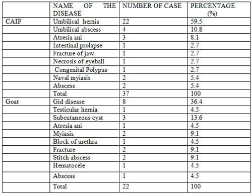
The detail result on the prevalence of surgical affections is shown in Table 1. During the study period a total 59 numbers of calves and goats were examined for surgical purpose to determine the prevalence of surgical affections in calves and goats and to find out the effect of some factors (age, sex, and breed) on that case. It was observed that most prevalence surgical affections in calves at Jhenidah sadar is umbilical hernia(59.5%, 22/37),second one is umbilical abscess(10.8%, 4/37), third one is Atresia ani(8.1%, 3/37) and number fourth is Naval myiasis and abscess (5.40%, 2/37) . In goat most common surgical affections is Gid disease (36.4%, 8/22), second one is subcutaneous cyst (13.6%, 3/22), and number third are stitch abscess, bone fracture, and vulvar myiasis (9.1%, 2/22).
Table 1. Prevalence of surgical affections in calves and goat
4.2 Effects of different variables on the occurrence of surgical affections in calves
4.2.1 Effects of age and sex
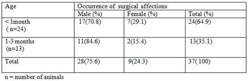
The effect of age and sex on the occurrence of surgical affections in calves is presented in Table 2. In male animals, the highest incidence (75.7%, 28/37) of the disease occurred in calves of 1-3 months old.
Like male calves, the incidence rate (24.3%, 9/37) of the disease in the female was also recorded in 1-3 months age group. The highest incidences (64.9%, 24/37) was observed in calves under 1 month of age, while the lowest incidence rate (35.1%, 13/37), was observed in 1-3 months age group.
Table 2. Effect of age and sex on the occurrence of surgical affections in calf
4.2.2 Effect of breed
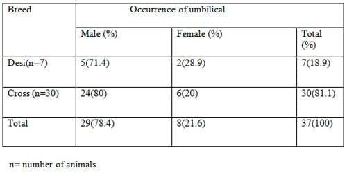
The effect of breed on the occurrence of umbilical hernia in calves is shown in Table 3. Out of 37 affected calves, 7 were desi and 30 were cross breed, the incidence of surgical affections in desi male(71.4%, 5/7 ), female(28.6%, 2/7), cross breed male(80%, 24/30), and in female(20%, 6/20).From this observation it was clear that cross bred calves(81.1%, 30/37) are more susceptible for surgical affections in compares to desi calves(18.9%, 7/37).
Table 3. Effects of breed on the occurrence of surgical affections in calves
4.3 Effects of different variables on the occurrence of surgical affections in goat
4.3.1 Effect of age and sex
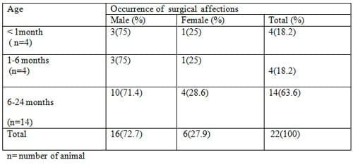
The effect of age and sex on the occurrence of surgical affections in goat is presented in Table 4. In male animals, the highest incidence (72.7%, 16/22) of the disease occurred in goat of 1-24 months of age, and the low incidence rate (27.9%, 6/22) of the disease in the female was also recorded in 1-24 months age group. In an average more surgical affections occurred in 6-24 months age (63.6%, 14/22).
Table 4. Effect of age and sex on the occurrence of surgical affections in goat
Among all surgical affections in calves umbilical hernia is available at Jhenidah sadar like inBangladesh. This prevalence agreeable with the report (Herman et al., 2000).Another report disagree with this reports (Tagi and Singh, 1998)
Calves between 1 and 3 months were most frequently affected with surgical cases. This observation about age and sex is agreeable to earlier reports (Field, 1988; Gadre et al., 1989; Rahman et al., 2001). However, Chuang et al., (2000) reported that the diseases are more prevalent in calves of below 1 month. InBangladeshdiagnosis of the affection may be delayed because animals are reared in backward system and owners are either ignorant have less interest for their management. Umbilical hernia is occurred predominantly in the male calves as compared to their female counterparts. This finding about age and sex is the agreeable with those of the report (Das and Hashim, 1996; Rahman et al., 2001) but contradictory to (Brem et al., 1985; Singh et al., 1989) who indicated females to be more susceptible to umbilical hernia than males. Higher prevalence in males may be due to large swelling at umbilical region for preputial sheath. During development of such large preputial sheath, the ventral abdominal wall may not be properly developed and leads to the formation of surgical affections before birth (Rahman et al., 2001). Naval infection in the male is also more frequent due to continuous moistening by urine.
In the present study, occurrence of umbilical hernia was significantly high in the crossbreed calves than that in the indigenous breed. The higher incidence in cross breed calves may due to preference of owners to inseminate their cows with these breeds. Pure Holstein cattle as well as the offspring ofHolsteinx indigenous cross are more likely to suffer from this congenital defect than the indigenous breed (Hayes, 1974; Kohli, 1999)
Umbilical abscess and Naval ill causes Umbilical hernia in animals. In present study abscess is more responsible than naval ill. This is higher than the study of (Swaim, 1998) the observation about others diseases is agreeable with the report of (Van Winkle, 1996)
In goats Gid disease and subcutaneous cyst is more prevalent than other surgical cases because the parasitic infection is more in Jhenidah sadar likeBangladesh(Rahman et al., 1972). The present study is agreeable with this information.
CONCLUSIONS
The following conclusions may draw from the studies on common surgical affections in calves and goat:
- Umbilical hernia in calves was fairly prevalent at Jhenidah district.
- Most Surgical affections occurred mostly in calves of 1-3 months age group, in where >1 month of age is more prone to surgical affections.
- The prevalence of surgical affections is more common in male calves than that in female calves.
- The higher incidence of surgical affections was encountered in the cross bred calves in contrast to indigenous calves.
- In Goat, Gid disease is most prevalent at Jhenidah district.
- Goats >6 months of age, and male are more susceptible to surgical affections.
REFERENCES
C. M. Lang, Springer-Verlag (1976). Animal Physiological Surgery. New York.pp. 110-115.
Das, B. R. and Hasim M.A. (1996). Studies on surgical affections in calves. Bangladesh Vet J, 30: 53-57.
Hossain, M. A. and Harman, M. A (1980). A new born calf with a supernumerary limb and atresia ani. 2: 178-179.
Kohli, R. N. (1999). Indicence of veterinary congenital defects in Iran. Indian Journal ofAnimal Science, 69 (10) : 779-780.
Kelly, W. R. (1967).Veterinary Clinical Diagnosis.3rd edn. Bailliere Tindal,London. pp.1-9.
Knetch, C. D. (1987). Hernia. In: Ruminant Surgery. 6th edn. Edited by Tyagi, R. P. S and Singh, J., CBS publishers & distributors,New Delhi. pp. 225-237.
Kumar, Amresh (1996).Veterinary Surgical Techniques.1st edn.UBS Publishers Distributors. ltd., New Dehli.pp.1-13.
Leipold, H. W., Dennis, S. M. and Hoston, K. (1972). Congenital defect of cattle: Nature cause and effect Adv Vet. Sci. Comp. Med, 16: 103-109.
Mia, A. S. (1967). Urinary calculi in farm animals and surgical treatment. Pak, J.Vet. Sci, 1 : 20-23.
Rahman, M. M., Biswas, D. and Hossian, M. A. (2001). Occurrence of umbilical hernia and comparative efficacy of different suture materials and techniques for its correction in calves. Pakistan Journal of Biological Science, 4(8) : 1026-1028.
Rahman, M. H. and Mondol, M. M. H. (1983). Helminth parasite of cattle and goats inBangladesh. Ind. Jour. Parasitology, 2: 173-174.
Samad, M. A. (2000). Clinical surgery. In: Veterinary Practitioner’s Guide. LEP publication,Dhaka. pp. 399-412.
Slatter, D. H. (1985). Text Book of small Animal Surgery, 1st edn. Edited by, W B Saunders Co.,Philadelphia, pp. 310-311.
Venugopalan (2000). Essentials of vet. Surgery. 8th edn.Oxford and IBH Publishing CO. PVT. ltd., New Delhi.pp. 330-331.
Wright, J. G. (1957). Veterinary anaesthesia. William and Wilkins Co.,Baltimore.pp. 223-224.
Wind, G. G., Rich, N. M., Urban and Schwarzenberg, (1987). Principles of Surgical Technique. The Art of Surgery. 2nd Edn.Baltimore, Munich.pp. 25-30.
Related topics
Authors:

Join to be able to comment.
Once you join Engormix, you will be able to participate in all content and forums.
* Required information
Would you like to discuss another topic? Create a new post to engage with experts in the community.
Create a post
9 de febrero de 2015
Hoaque and Samad, 1996; Saiffuzzaman, 1996; Samad, 2001"""""
Where are the references ?

4 de junio de 2013
colic treatment


You may be interested in
.jpg&w=3840&q=75)
Phileo by Lesaffre