Explore
Communities in English
Advertise on Engormix

Biosynthesis and Characterization of Silver Nanoparticles from Marine Macroscopic Red Seaweed Halymenia Porphyroides Boergesen (Crypton) and its Antifungal Efficacy Against Dermatophytic and Non-Dermatophytic Fungi

Published: October 15, 2020
By: VISHNU KIRAN MANAM*, MURUGESAN SUBBAIAH / Department of Plant Biology and Biotechnology, Unit of Algal Biotechnology and Bio-nanotechnology, Pachaiyappa’s College, University of Madras, Chennai, Tamil Nadu, India.
Summary

Objective: The current study illustrates the biosynthesis of economically scalable and energy efficient colloidal silver nanoparticles (AgNPs) from marine red seaweed Halymenia porphyroides Boergesen (Crypton) collected from Southeast coast of Tamil Nadu, India, and their antifungal efficacy against dermatophytic and non-dermatophytic fungi was evaluated.

Methods: The biosynthesis of silver nanoparticles from marine macroscopic red seaweed H. porphyroides Boergesen were synthesized by green synthesis method and characterized by UV–Vis spectroscopy, Fourier transform infrared (FT-IR) spectroscopy, X-ray diffraction, thermogravimetric analysis (TGA), scanning electron microscope (SEM), and transmission electron microscopy (TEM). The efficacy of silver nanoparticles against dermatophytic and non-dermatophytic fungi was performed by disk diffusion method.

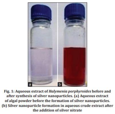
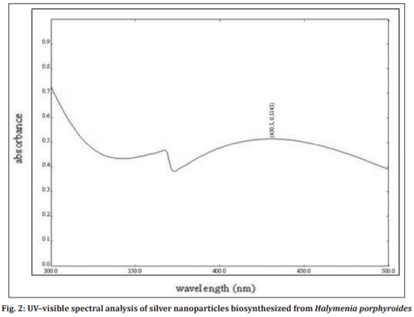
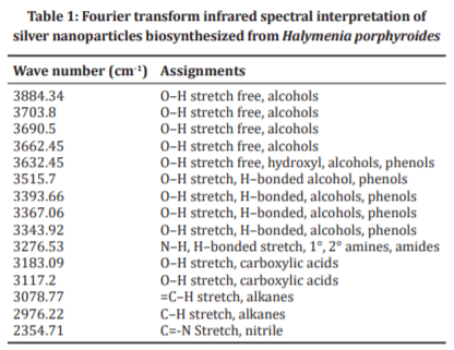
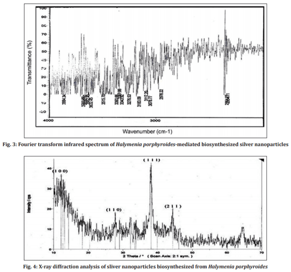
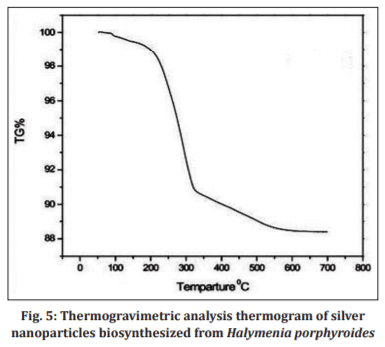
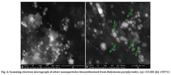
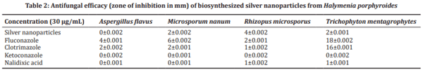
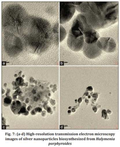
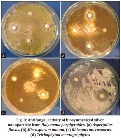
Results: The presence of silver nanoparticles with an average size between 34.3 and 80 nm and exhibiting face-centered cubic structure was confirmed. SEM revealed the morphology of the nanoparticles as spherical and TEM exhibited the nanoparticle distribution. The FT-IR spectra confirmed the presence of potential biomolecules in the seaweed crude extract which is responsible for reducing and capping the bioreduced silver nanoparticles. The UV absorption spectra at 430.5 nm revealed the characteristic spectra of the silver nanoparticles. The purity and the thermal stability of silver nanoparticles were revealed by TGA. Silver nanoparticles showed significant efficacy against dermatophytes and Rhizopus microsporus among nondermatophytes.

Conclusion: Intermediate efficacy was observed against dermatophytes and among non-dermatophytic fungi R. microsporus exhibited better efficacy, whereas Aspergillus flavus were resistant to the biosynthesized silver nanoparticle.

Keywords: AgNPs, Biosynthesis, Halymenia porphyroides, Antifungal efficacy, Dermatophytes, non-dermatophytes.

INTRODUCTION
Nanotechnology and nanoscience are the study of production, manipulation, and application of materials ranging in size from less than a micron to that of individual atoms [1]. The major key aspect of nanotechnology involves in the development of rapid and reliable experimental protocols such as biological and chemical processes for the synthesis of nanomaterials over wide range of chemical compositions, sizes, high monodispersity, as well as large-scale production [2]. Due to the potential for achieving specific processes and selectivity, especially in biological and pharmaceutical industries, nanomaterials and nanoparticles are considered as an important and increasing scientific research [3-8]. The increasing research and growth in the science of nanotechnology during the past few decades has opened up new avenues of fundamental and applied frontiers in the fields of materials science and engineering, such as bionanotechnology [9], quantum dots [10], surface-enhanced Raman scattering [11], and nanobiotechnology [12]. Silver nanoparticles (AgNPs), which have a high specific surface area, have been studied widely because of their unique physicochemical characteristics including catalytic activity, optical properties, electronic properties, antimicrobial activity, and magnetic properties [13-15]. Silver nanoparticles have been synthesized by various synthetic methods involving physical, chemical [16], and biochemical techniques [17] as well as biological methods using “green chemistry” protocols [18] and find their applications in food chemistry [19], biomedicine [20], and agriculture [21]. Biological/green synthesis of silver nanoparticles using microorganism [22-24], enzyme [25], plant or plant extract [26], and seaweed extract [27] has been in practice as alternatives to chemical and physical methods since they have been considered as economically stable and ecofriendly. The presence of high organic and inorganic biochemical constituents [28], phytochemicals [29], and bioactive compounds [30] in seaweeds may act as the capping/reducing agents in the biosynthesis of nanoparticles. The rise in fungal infections, especially the nosocomial infections and in patients with immunocompromised condition in cases such as cancer therapy, organ, and in HIV infections, has alarmed the researchers for an alternative treatment toward fungal pathogens [31]. One such alternative and need for novel antifungal agents are silver nanoparticles since the fungal resistance toward antibiotics has been emerging as a major threat worldwide [32,33]. Nanoparticles can be considered as potential antifungal agents [34] despite the marginal attention have been documented on the effect of silver nanoparticles as antifungal agents [35-38]. However, certain studies have been performed on the dermatophytic fungi with regard to nanoparticles [39,40]. This present investigation illustrates green synthesis of silver nanoparticles using the extract of marine macroscopic red seaweed Halymenia porphyroides and characterized by UV–visible spectrophotometer. Morphological and elemental analysis was carried out by scanning electron microscopy (SEM) and transmission electron microscopy (TEM). The stability of silver nanoparticles was confirmed by TGA. The possible biomolecules responsible for the reduction of the Ag+ ions and capping of the bioreduced silver nanoparticles synthesized from the experimental algae H. porphyroides were identified using Fourier-transform infrared (FT-IR) spectroscopy. The crystalline structure of the silver nanoparticles, including lattice parameters, geometry, and orientation of single crystals, was studied using X-ray diffraction (XRD). Thus, algae-mediated synthesized silver nanoparticles showed intermediate zone of inhibition toward dermatophytic fungi and Rhizopus microsporus in non-dermatophytic fungi which showed better zone of inhibition, whereas Aspergillus flavus showed nil zone of inhibition which was analyzed by agar disk diffusion method.
METHODS
Collection and preparation of red seaweed extract
The marine red seaweed H. porphyroides Boergesen (Crypton) was collected on summer season from 2.5 m rapid intertidal regions of the Gulf of Mannar – Mandapam (latitude 9°17”N, longitude 79°11”E), Ramanathapuram district, Southeast Coast of Tamil Nadu, South India. Collected seaweed was washed with seawater for eliminating impurities such as sand, rocks, epiphytes, and epifauna. The washed samples were preserved with 5–10% formaldehyde in seawater and transported to the laboratory in a box containing slush ice. The fumes of the formaldehyde would help to fix and preserve the seaweed material. In the laboratory, the samples were washed thoroughly in running tap water to remove salt and washed 3 times using distilled water which may remove metallic compounds and it was shade dried at room temperature (37°C) for 10 days. The dried seaweed materials were crushed using mortar and pestle to get the powder form and it was stored in an air-tight container. About 1 g of crushed seaweed powder was added with 100 ml of distilled water in 250 ml conical flask and boiled for 5–10 min at 60–80°C. The crude extract was then collected and stored at 4°C for experimental use [41].
Biosynthesis and characterization of silver nanoparticles
The crude extract of the experimental marine red seaweed H. porphyroides Boergesen (Crypton) was used for the synthesis of silver nanoparticles. Silver nitrate (AgNO3-) (SD fine) was used for the synthesis of silver nanoparticles and double-distilled, deionized water was used for all the experiments. The silver nanoparticle formation was carried out by taking 500 mg of dry, shade dried powder samples of H. porphyroides in a 250 mL Erlenmeyer flask with 10-3 M aqueous AgNO3- solution and was incubated at room temperature. The pH was checked during the course of reaction and it was found to be 5.09. Nearly 95% of bioreduction of AgNO3- ions occurred within 24 h at stirring condition. The biosynthesis of silver nanoparticles was characterized by UV–Vis spectrophotometer (Labtron LUS-B16) in the absorbance mode and in the wavelength range between 300 to 500 nm. FT-IR spectra of biosynthesized silver nanoparticles were recorded using Thermo Scientific/Nicolet iS10 spectrometer with 1 cm-1 resolution in the transmission mode from wavenumbers 450–4000 cm-1. The crystal structure and size of silver nanoparticles were determined using the X-ray diffractometer (Labtron LXRD-A10). Thermal stability and purity of silver nanoparticles were analyzed using TGA 4000 – PerkinElmer. A scanning electron micrograph was taken using the SEM Quanta – 400 to study the morphological characteristics of the silver nanoparticles. Further insight into morphology and the size details of the biosynthesized silver nanoparticles by the experimental algae H. porphyroides were investigated using high-resolution TEM (HR-TEM JEOL 3010).
Fungal susceptibility to nanosilver
Antifungal activity of the biosynthesized silver nanoparticles by the experimental algae H. porphyroides was tested against pathogenic fungi. The disk diffusion method for antifungal assay was adapted for testing silver nanoparticles [42]. The fungal cultures were obtained from LGC Promochem India Pvt. Ltd., Peenya, Bengaluru, India. The fungal pathogens A. flavus (ATCC 20048), R. microsporus (ATCC 22960), Microsporum nanum (ATCC 28951), and Trichophyton mentagrophytes (ATCC 28185) are the fungal strains which were used to study the antifungal efficacy of silver nanoparticles. The standard antifungal agents at the concentration of 30 µg/mL fluconazole, clotrimazole, nalidixic acid, and ketoconazole (HiMedia) were used as zone interpretive criteria. The Mueller-Hinton agar (HiMedia) supplemented with 2% glucose and 0.5 µg/L methylene blue dye medium was used for culturing the fungal stains. The Mueller-Hinton agar is readily available and shows an acceptable batch-to-batch reproducibility while the glucose provides a stable growth of most fungi and the addition of methylene blue enhances the zone edge definition [43]. The pH of the medium was maintained between 7.2 and 7.4 at 37°C. The inoculum is standardized to 0.5 McFarland using a densitometer and the plate was incubated at 35°C for 24 h. Some strains where insufficient growth has occurred after 24 h were necessary to be read after 48 h of incubation.
Statistical analysis
The data were analyzed using MS Excel 2007 and presented as mean  ±  SD of three replicates. One-way analysis of variance and Tukey tests were performed using “Stat plus 2009 professional” trial version software to determine significant zone differences and means were considered as statistically significant if p<0.05.
RESULTS AND DISCUSSION
Visual examination
The biosynthesis of silver nanoparticles was primarily identified by color change during exposure of crude seaweed extract of H. porphyroides into aqueous solution of silver ions which is shown in Fig. 1. The shade dried powder preparations of the experimental seaweed H. porphyroides were added in 10-3 M silver nitrate solution and allowed to react at 121°C for 20 min. The colour of the reaction solution changed to dark reddish. The control (without seaweed powder) showed no colour formation. Formation of the colour arises due to the excitation of surface plasmon vibrations where the metabolites in the seaweed extract act as the capping agent. The colour of the solution gradually intensified on heating which clearly indicates and confirms the formation of silver nanoparticles. After 24 h, there is no significant color change, indicating the saturation of the reaction of silver nanoparticle formation.
UV–visible spectroscopic analysis
The silver nanoparticles synthesized by marine red seaweed H. porphyroides were analyzed using UV–visible spectrophotometer (Labtron LUS-B16). The absorption spectra of silver nanoparticles formed in the reaction solution were characteristic of the nanoparticle from the seaweed and had specific absorption maxima at 430.5 nm which is shown in Fig. 2. The characteristic absorption peaks of the silver nanoparticles are in the range between 410 and 440 nm, which confirms the synthesis and formation of silver nanoparticles [44,45].
Biosynthesis and Characterization of Silver Nanoparticles from Marine Macroscopic Red Seaweed Halymenia Porphyroides Boergesen (Crypton) and its Antifungal Efficacy Against Dermatophytic and Non-Dermatophytic Fungi - Image 1
 
Biosynthesis and Characterization of Silver Nanoparticles from Marine Macroscopic Red Seaweed Halymenia Porphyroides Boergesen (Crypton) and its Antifungal Efficacy Against Dermatophytic and Non-Dermatophytic Fungi - Image 2
 
Biosynthesis and Characterization of Silver Nanoparticles from Marine Macroscopic Red Seaweed Halymenia Porphyroides Boergesen (Crypton) and its Antifungal Efficacy Against Dermatophytic and Non-Dermatophytic Fungi - Image 3
The frequency and width of the surface plasmon absorption depend on the size and shape of the metal nanoparticles as well as on the dielectric constant of the metal itself and the surrounding medium [46]. The broad peaks of the absorption spectra indicate the presence of biosynthesized silver nanoparticles from the experimental seaweed H. porphyroides with large size distribution and are polydispersed. The interaction with the biomolecules present in the aqueous part of the reaction solution by the biosynthesized silver nanoparticles from experimental seaweed H. porphyroides has been indicated by UV–visible spectroscopic analysis. There were no little signs of aggregation with the biosynthesized silver nanoparticles solution which were stable for more than 6 months of observation.
FT-IR spectroscopic analysis
The FT-IR spectral measurements were carried out to identify the potential biomolecules in the crude extract of the seaweed H. porphyroides which is responsible for reducing and capping the bioreduced silver nanoparticles. Silver nanoparticles biosynthesized from experimental seaweed H. porphyroides were analyzed using FT-IR spectroscopy which is shown in Fig. 3 and Table 1. The local molecular environment of the organic molecules on the surface of the nanoparticles was determined by the IR spectra. FT-IR spectroscopy is a technique which is used to analyze the chemical composition of many organic chemicals, semiconductor materials, gases, biological samples, inorganics, and minerals. FT-IR analysis can give not only qualitative (identification) analysis of materials but, with relevant standards, also can be used for quantitative (amount) analysis. The FT-IR spectrum analysis of silver nanoparticles biosynthesized from red seaweed manifests an absorption peak at 3884.34 cm-1 (O–H stretch free, alcohols), 3703.80 cm-1 (O–H stretch free, alcohols), 3690.50 cm-1 (O–H stretch free, alcohols), 3662.45 cm-1 (O–H stretch free, alcohols), 3632.45 cm-1 (O–H stretch free, hydroxyl, alcohols, phenols), 3515.70 cm-1 (O–H stretch, H–bonded alcohol, phenols), 3393.66 cm-1 (O–H stretch, H–bonded, alcohols, phenols), 3367.06 cm-1 (O–H stretch, H–bonded, alcohols, phenols), 3343.92 cm-1 (O–H stretch, H–bonded, alcohols, phenols), 3276.53 cm-1 (N–H,H–bonded stretch, primary, secondary amines, amides), 3183.09 cm-1 (O–H stretch, carboxylic acids), 3117.20 cm-1 (O–H stretch, carboxylic acids), 3078.77 cm-1 (=C–H stretch, alkanes), 2976.22 cm-1 (C–H stretch, alkanes) [47], and 2354.71 cm-1 (C=N stretch, nitrile) [48]. The results revealed that the capping ligand of the silver nanoparticles may be an aromatic compound or alkanes or amines [49]. The biological molecules such as secondary metabolites could possibly play a major role in the synthesis and stabilization of the metal nanoparticles [50,51]. The Vander Waals forces between nitrogen and oxygen atoms as well as the release of protein molecules in biocompound of the experimental seaweed H. porphyroides may be responsible for the biosynthesis and stabilization of silver nanoparticles in an aqueous solution. These results obtained are in good agreement with the literature [52,53].
XRD pattern
XRD is a widely used to determine the size and crystal structure of silver nanoparticles. X-ray diffractogram of the biosynthesized silver nanoparticles by the experimental seaweed H. porphyroides exhibits Bragg reflection corresponding to face-centered cubic (fcc) type bulk silver. The broadened diffraction peaks around their base indicate that the silver nanoparticles are between nanosizes. XRD (Labtron LXRD-A10) analysis of biosynthesized silver nanoparticles from H. porphyroides exhibited four distinct diffraction peaks is shown in Fig. 4. The XRD diffraction pattern of biosynthesized silver nanoparticles from H. porphyroides showed four distinct diffraction peaks at 12.13°, 28.77°, 39.87°, and 46.11° with 2θ values corresponding to the lattice planes {1 0 0}, {1 1 0}, {1 1 1}, and {2 1 1} which are indexed as crystalline silver fcc phase [54]. The observed peak broadening and noise was probably related to the effect of nanosized particles and the presence of various crystalline biological macromolecules in the experimental seaweed extract. The results are in agreement with the findings of earlier investigations as reported by Paneerselvam et al. (2012) [55] Chandra and Kumar (2015) [56]. The diffraction patterns of silver nanoparticles biosynthesized from the experimental seaweed H. porphyroides indicate the uniqueness of the crystalline structure, phase purity, degree of crystallinity, and the unit cell parameters. The presence of some unassigned peaks in the X-ray diffractogram of the biosynthesized silver nanoparticles from the experimental seaweed H. porphyroides may be due to the biomolecules and proteins in the seaweed extract which acts as stabilizing agents [57]. The XRD pattern thus clearly shows that the silver nanoparticles are crystalline in nature.
TGA
The biosynthesized silver nanoparticles from the experimental seaweed H. porphyroides were subjected to TGA 4000 – PerkinElmer which is shown in Fig. 5. The thermal stability, purity, and humidity of the particles were determined using TGA [58]. The TGA relies on a high degree precision in three measurements: Weight, temperature, and temperature change. It can be applied to silver nanoparticles to determine changes in weight in relation to the changes in temperature [59]. Thermogravimetric analysis is commonly employed in research and testing to determine the characteristics of materials such as degradation temperature, absorbed moisture content of nanoparticles and polymers [60]. The silver nanoparticles biosynthesized from H. porphyroides showed a weight loss in a two-step process, the first step indicated a weight loss of 9% at the temperature up to 340°C which corresponds to the decomposition of bioorganic compounds present in the nanoparticle powder [61] and the second step involved weight loss of 3% at the temperature ranging from 340 to 650°C which corresponds to the decomposition of amines [62]. Thermal decomposition of the Ag+1 bioorganic complex at high temperature (200°C) results in an Ag atom and organic molecules [63]. The crystallite growth of silver nanoparticles biosynthesized from the experimental seaweed H. porphyroides appears to be consistent with “Ostwald ripening” where the stability gradient diffuses from the surfaces of small particles to the surfaces of larger particles in the Ag+1-bioorganic solution [64]. The TGA results of the biosynthesized silver nanoparticles, from experimental seaweed H. porphyroides, show 95% purity and their stabilization was observed at 200°C which are in agreement with the findings of the earlier investigations reported by Forough and Farhadi (2010) [65] and Amjad et al. (2012) [66]. The 95% purity of the silver colloidal medium from the experimental seaweed H. porphyroides was determined by an ultrasonication method which eliminates and separates silver nanoparticles from bioorganic complexes [67].
SEM
The morphology and shape of these silver nanoparticles were carried out using SEM Quanta – 400, as shown in Fig. 6. The silver nanoparticles biosynthesized from H. porphyroides biomass after exposure to 10-3 M aqueous silver nitrate solution for 2 h showed the colloidal form of the particles in solution which microprecipitated on the surface of the biomass of the experimental seaweed. In the present study, the SEM analysis of silver nanoparticles, besides being present in colloidal form in solution, was also microprecipitated on the surface of the biomass, which were clearly indicating that the nanoparticles formed by the reduction of Ag+ ions are bound to the surface of the cells. The silver nanoparticles biosynthesized from H. porphyroides formed were predominated in cubical and spherical structures with uniform shape as reported by Chandran et al. (2006) [68]. The brighter cubical and spherical area of the back scattered electron image corresponds to the silver nanoparticle indicating the cubic and spherical structure of silver. It is known that the shape of the metal nanoparticles has considerable changes their optical and electronic properties [69]. SEM pictures of silver nanoparticles from the experimental seaweed H. porphyroides showed that they were intact after the reaction and immobilization. The experimental results showed that the diameter of prepared silver nanoparticles in the solution was about 34.3–80.5 nm for H. porphyroides. Silver nanoparticles have a strong tendency to agglomerate which reduces the surface to volume ratio and thereby produce the catalytic effect. Therefore, a stabilizing agent is often used to prevent agglomeration. However, the agent is adsorbed on the surface of the nanoparticles, shielding them from the oxidant and reluctant and thereby inhibiting the catalysis [70].
Biosynthesis and Characterization of Silver Nanoparticles from Marine Macroscopic Red Seaweed Halymenia Porphyroides Boergesen (Crypton) and its Antifungal Efficacy Against Dermatophytic and Non-Dermatophytic Fungi - Image 4
 
Biosynthesis and Characterization of Silver Nanoparticles from Marine Macroscopic Red Seaweed Halymenia Porphyroides Boergesen (Crypton) and its Antifungal Efficacy Against Dermatophytic and Non-Dermatophytic Fungi - Image 5
 
Biosynthesis and Characterization of Silver Nanoparticles from Marine Macroscopic Red Seaweed Halymenia Porphyroides Boergesen (Crypton) and its Antifungal Efficacy Against Dermatophytic and Non-Dermatophytic Fungi - Image 6
TEM
The HR-TEM is the technique used to determine the size and particle distribution of silver nanoparticles [71]. The size and distribution of biosynthesized silver nanoparticles were observed by taking the micrograph from drop-coated films from silver complex solution with the extract of the experimental seaweed H. porphyroides. The TEM (HR-TEM JEOL 3010) images showed the formation of spherical shaped silver nanoparticles and are found in aggregates ranging from 5 to 50 nm, as shown in Fig. 7. The average mean size of silver nanoparticles from H. porphyroides was 32 nm. Similar results were recorded by Murugesan et al. (2011) [72] using red alga Gracilaria edulis extract as a reducing and capping agent. The shape of the metal nanoparticles has considerably changed their optical and electronic properties [73]. The HR-TEM images do not confirm the presence of capping agents on the silver nanoparticles, but under careful observation, it is noted that the silver nanoparticles are surrounded by a faint thin layer of other material, which may be the capping organic material from the experimental seaweed H. porphyroides. The silver nanoparticles of the TEM images are not in physical contact but are separated from each other by a uniform inter-particle distance. The results of HR-TEM findings indicate that the seaweed mediated silver nanoparticle shape varies as cubical and spherical which are in agreement with earlier investigations reported by Raghunandan et al. (2009) [74] and Noruzi et al. (2011) [75].
Antifungal efficacy of silver nanoparticles
The silver nanoparticles biosynthesized from H. porphyroides at the concentration 30 µg/mL were tested against dermatophytes and nondermatophytes. The dermatophytic fungi M. nanum (2±0.002 mm) and T. mentagrophytes (2±0.001 mm) showed intermediate zone of inhibition with biosynthesized silver nanoparticles from seaweed H. porphyroides. The non-dermatophytic fungi R. microsporus (4±0.002 mm) showed greater zone of inhibition with biosynthesized silver nanoparticles, whereas the non-dermatophyte A. flavus showed no zone formation which indicates its resistance toward biosynthesized silver nanoparticles (Fig. 8). Standard antifungal agent fluconazole exhibited higher resistance toward dermatophytic fungi M. nanum and T. mentagrophytes when compared to non-dermatophytic fungi A. flavus and R. microsporus which showed intermediate zone of inhibition. The antifungal agent clotrimazole showed an intermediate zone of inhibition for both dermatophytic and non-dermatophytic fungi and strong zone of inhibition was observed with T. mentagrophytes, whereas the antifungal agent ketoconazole showed least zone of inhibition toward dermatophytic and non-dermatophytic fungi. The nalidixic acid antifungal showed resistance toward dermatophytic fungi and non-dermatophytic fungi with least zone formation. The dermatophyte T. mentagrophytes were more susceptible to the antifungals clotrimazole and fluconazole (Fig. 8 and Table 2). The adhesion and penetration of silver nanoparticles to the cell membrane of the fungal pathogen thereby producing a with less molecular weight at the center of the fungi resulting in the attachment of nanosilver to the respiratory sequence and consequently stopping the cell division leading to the death of fungal cells might be the mechanism involved in the efficacy pathway of the silver nanoparticles [76]. The size and the surface are of the silver nanoparticles largely determine the formation of pits in the fungal cell wall [77]. Disrupting the membrane potential of the cell membrane of the fungal pathogen, leading to the cell death, might be the other possible mechanism [78-81]. The antimicrobial and antifungal properties of biosynthesized sliver nanoparticles from marine seaweed extract were confirmed from the current study on antifungal efficacy [82,83].
Biosynthesis and Characterization of Silver Nanoparticles from Marine Macroscopic Red Seaweed Halymenia Porphyroides Boergesen (Crypton) and its Antifungal Efficacy Against Dermatophytic and Non-Dermatophytic Fungi - Image 7
 
Biosynthesis and Characterization of Silver Nanoparticles from Marine Macroscopic Red Seaweed Halymenia Porphyroides Boergesen (Crypton) and its Antifungal Efficacy Against Dermatophytic and Non-Dermatophytic Fungi - Image 8
 
Biosynthesis and Characterization of Silver Nanoparticles from Marine Macroscopic Red Seaweed Halymenia Porphyroides Boergesen (Crypton) and its Antifungal Efficacy Against Dermatophytic and Non-Dermatophytic Fungi - Image 9
CONCLUSION
The results from the present investigation have concluded that the silver nanoparticles biosynthesized from marine seaweed H. porphyroides showed intermediate effect against dermatophytes, whereas among non-dermatophytic fungi, especially R. microsporus, exhibited better efficacy and on the other non-dermatophyte A. flavus were resistant to the biosynthesized silver nanoparticles. As elaborated earlier not much work has been documented in the efficacy of silver nanoparticles toward fungal pathogens, whereas this current study may pave a pathway for the use of colloidal silver in the antibiotic or antifungal resistance strains, especially will be helpful in treating immunocompromised patients and in nosocomial infections. The results also reveal that the dermatophytes are susceptible to silver nanoparticles with intermediate, whereas the non-dermatophytic fungi R. microsporus exhibited much better zone of inhibition toward the algal-meditated silver nanoparticles when compared to standard antifungal agents used in the study. This study forms the basis of the exoskeleton for more research with silver nanoparticles toward fungal pathogens and drug-resistant strains.

ACKNOWLEDGMENT
I sincerely thank Mr. Raju sea diver who helped me in the collection of the seaweeds from the Southeast coast of Tamil Nadu, India. This work was carried out in part through the use of the SEM and TEM at Sophisticated Analysis Instrument Facility, Indian Institute of Technology – Madras, Tamil Nadu, India. The TGA was carried out at the Department of Chemistry, Anna University, Chennai, Tamil Nadu, India. The authors thank the above institutions for their instrumentation facility for the completion of the work.

AUTHORS’ CONTRIBUTIONS
Dr. Vishnu Kiran Manam designed and performed the work, whereas Dr. Murugesan Subbaiah suggested the relevant changes during the course of the work and the proofreading of the manuscript.

CONFLICTS OF INTEREST
The authors declare that they have no conflicts of interest.
AUTHORS’ FUNDING
The research work performed by authors by self-financial scheme and no financial assistance received from any funding agencies.
 
This article was originally published in Asian Journal of Pharmaceutical and Clinical Research, Vol 13, Issue 8, 2020, 174-181. DOI: http://dx.doi.org/10.22159/ajpcr.2020.v13i8.38262. This is an Open Access article under the CC BY license (http://creativecommons.org/licenses/by/4.0/)

1. Prashant M, Nisha KR, Sudesh KY. Biosynthesis of nanoparticles: Technological concepts and future applications. J Nanopart Res 2008;10:507-17.

2. Saifuddin N, Wong CW, Nur Yasumira AA. Rapid biosynthesis of silver nanoparticles using culture supernatant of Bacteria with microwave irradiation. E J Chem 2009;6:61-70.

3. Alivisatos P. The use of nanocrystals in biological detection. Nat Biotechnol 2004;22:47-52.

4. Love JC, Estroff LA, Kriebel JK, Nuzzo RG, Whitesides GM. Self-assembled monolayers of thiolates on metals as a form of nanotechnology. Chem Rev 2005;105:1103-69.

5. Mirkin CA, Taton TA. Semiconductors meet biology. Nature 2000;405:626-7.

6. Nie S, Xing Y, Kim GJ, Simons JW. Nanotechnology applications in cancer. Annu Rev Biomed Eng 2007;9:257-88.

7. Wang MD, Shin DM, Simons JW, Nie S. Nanotechnology for targeted cancer therapy. Expert Rev Anticancer Ther 2007;7:833-7.

8. Daniel MC, Astruc D. Gold nanoparticles: Assembly, supramolecular chemistry, quantum-size-related properties, and applications toward biology, catalysis, and nanotechnology. Chem Rev 2004;104:293-346.

9. Goodsell DS. Bionanotechnology: Lessonsfrom Nature. New Jersey: Willey-Less; 2004. p. 1-8.

10. Chan WC, Nie S. Quantum dot bioconjugates for ultrasensitive nonisotopic detection. Science 1998;281:2016-8.

11. Tian Z, Ren B. Adsorption and reaction at electrochemical interfaces as probed by surface-enhanced Raman spectroscopy. Annu Rev Phys Chem 2004;55:197-229.

12. Klefenz H. Nanobiotechnology: From molecules to systems. Eng Life Sci 2004;4:211-8.

13. Kowshik M, Ashtaputre S, Kharrazi S. Extracellular synthesis of silver nanoparticles by a silver-tolerant yeast strain MKY3. Nanotechnology 2003;14:95-100.

14. Souza GI, Marcato PD, Duran N, Esposito E. Utilization of Fusarium oxysporum in the Biosynthesis of Silver Nanoparticles and its Antibacterial Activities. Brazil: Presented at 10th National Meeting of Environmental Microbiology; 2004. p. 25.

15. Duran N, Marcato PD, Alves OL, Souza G. Mechanistic aspects of biosynthesis of silver nanoparticles by several Fusarium oxysporum strains. J Nanotechnol 2005;3:8.

16. Sinha S, Pan I, Chanda P, Sen SK. Nanoparticle’s fabrication using ambient biological resources. J Appl Biosci 2009;19:1113-30.

17. Huang J, Li Q, Sun D, Lu Y, Su Y, Yang X, et al. Biosynthesis of silver and gold nano particles by novel sundried Cinnamomum camphora leaf. Nanotechnology 2007;18:105104-14.

18. Kiran MV, Murugesan S. Biosynthesis and characterization of silver nanoparticles from marine macroscopic brown seaweed Colpomenia sinuosa (Mertens ex Roth) Derbes and Solier. J Adv Chem Sci 2020;6:663-6.

19. Li H, Li F, Wang L, Shengv J, Xin Z, Zhao L. Effect of nanopacking on preservation quality of Chinese jujube (Ziziphus jujuba Mill. Var. inermis (Bunge) Rehd). Food Chem 2009;114:547-52.

20. Chaloupka K, Malam Y, Seifalian AM. Nanosilver as a new generation of nanoproduct in biomedical applications. Trends Biotechnol 2010;28:580-8.

21. Park HJ, Sung HK, Kim HJ, Choi SH. A new composition of nanosized silica-silver for control of various plant diseases. J Plant Pathol 2006;22:295-302.

22. Klaus T, Joerger R, Olsson E, Granqvist CG. Silver-based crystalline nanoparticles, microbially fabricated. Proc Natl Acad Sci U S A 1999;96:13611-4.

23. Konishi Y, Ohno K, Saitoh N, Nomura T, Nagamine S, Hishida H, et al. Bioreductive deposition of platinum nanoparticles on the bacterium Shewanella algae. J Biotechnol 2007;128:648-53.

24. Nair B, Pradeep T. Coalescense of nanoclusters and formation of submicron crystallites assisted by Lactobacillus strains. Cryst Growth Des 2002;2:293-8.

25. Willner I, Baron R, Willner B. Growing metal nanoparticles by enzymes. Adv Mater 2006;18:1109-20.

26. Shankar SS, Rai A, Ahmad A, Sastry M. Rapid synthesis of Au, Ag, and bimetallic Au core Ag shell nanoparticles using Neem (Azadirachta indica) leaf broth. J Coll Interf Sci 2004;275:496-502.

27. Kiran MV, Murugesan S. Biosynthesis and characterization of silver nanoparticles from marine macroscopic red seaweed Halymenia porphyroides Boergesen (Crypton). J Nanosci Tech 2020;6:886-90.

28. Kiran MV, Murugesan S. Biochemical investigation of marine seaweeds Colpomenia sinuosa and Halymenia porphyroides collected along south east coast of Tamil Nadu, India. Eur J Biomed Pharm Sci 2020;7:414-7.

29. Kiran MV, Murugesan S. Phytochemical, amino acid, fatty acid and Vitamin investigation of marine seaweeds Colpomenia sinuosa and Halymenia porphyroides collected along south east coast of Tamil Nadu, India. World J Pharm Res 2020;9:1088-102.

30. Kumar KA, Kumar SR, Rengasamy R. Review on bioactive potential in seaweeds (Marine Macroalgae): A special emphasis on bioactivity of seaweeds against plant pathogens. Asian J Plant Sci 2020;9:227-40.

31. Rajasulochana P, Dhamotharan R, Murugakoothan P, Murugesan S, Krishnamoorthy P. Biosynthesis and characterization of gold nanoparticles using the alga Kappaphycus alvarezii. Int J Nanosci 2010;9:511-9.

32. Swaminathan S, Murugesan S, Damodarkumar S, Dhamotharan R, Bhuvaneswari S. Synthesis and characterization of gold nano particles from alga Acanthophora specifera (VAHL) Boergesen. Int J Nanosci Nanotech 2011;2:85-94.

33. Dhamotharan R, Punitha D, Murugesan S, Subha TS. Brown algalbiomass mediated biosynthesis of gold nanoparticles. Int J Nanosci Nanotech 2010;1:37-44.

34. Pana CA, Milan K, Renata V, Robert P, Jana S, Vladimir K, et al. Antifungal activity of silver nanoparticles against Candida spp. Biomaterials 2009;30:6333-40.

35. El-Rafie MH, Mohamed AA, Shaheen TI, Hebeish A. Antimicrobial effect of silver nanoparticles produced by fungal process on cotton fabrics. Carbohydr Polym 2010;80:779-82.

36. Gogoi SK, Gopinath P, Paul A, Ramesh A, Ghosh SS, Chattopadhyay A. Green fluorescent protein-expressing Escherichi coli as a model system for investigating the antimicrobial activities of silver nanoparticles. Langmuir 2006;22:9322-8.

37. Roe D, Karandikar B, Bonn-Savage N, Gibbins B, Roullet JB. Antimicrobial surface functionalization of plastic catheters by silver nanoparticles. J Antimicrob Chem 2008;61:869-76.

38. Zeng F, Hou C, Wu SZ, Liu XX, Tong Z, Yu SN. Silver nanoparticles directly formed on natural macroporous matrix and their anti-microbial activities. Nanotechnology 2007;18:1-8.

39. Kim KJ, Sung WS, Moon SK, Choi JS, Kim JG, Lee DG. Antifungal effect of silver nanoparticles on dermatophytes. J Microbiol Biotech 2008;18:1482-4.

40. Monali G, Jayendra K, Avinash I, Aniket G, Mahendra R. Fungusmediated synthesis of silver nanoparticles and their activity against pathogenic fungi in combination with fluconazole. Nanomed Nanotech Biol Med 2009;5:382-6.

41. Finegold SM, Baron EJ. Bailey and Scotts Diagnostic Microbiology. 7th ed. St. Louis: The C.V. Mosby Company; 1986.

42. NCCLS. Method for Antifungal Disk Diffusion Susceptibility Testing of Yeasts; Approved Guideline. Pennsylvania: NCCLS; 2004. p. 19087.

43. Rajesh KS, Malrakodi C, Venkat KS. Synthesis and characterization of silver nanoparticles from marine brown seaweeds and its antifungal efficiency against clinical fungal pathogens. Asian J Pharm Clin Res 2017;10:190-3.

44. Henglein A. Physicochemical properties of small metal properties in solution: Micrelectrode reactions, chemisorption’s, composite metal particles and the atom to metal transistion. J Phys Chem 1993;97:5457-71.

45. Kumar P, Senthamilselvi S, Prabha AL, Kumar KP, Kumar RS, Govindaraju M. Synthesis of silver nano particles from Sargassum tenerrimum and screening phytochemicals on its antibacterial activity. Nano Biomed Eng 2012;4:12-6.

46. Sastry M, Patil V,Sainkar SR. Electrostatically controlled diffusion of carboxylic acid derivatized silver colloidal particles in thermally evaporated fatty amine films. J Phys Chem B 1998;102:1404-10.

47. Silverstein RM, Webster FX. Spectrometric Identification of Organic Compounds. New York: John Wiley & Sons; 1998.

48. O’Coinceanainn MO, Astill C, Schumm S. Potentiometric FTIR and NMR studies of the complexation of metals with theaflavin. Dalton Trans 2003;5:801-7.

49. Inbakandan D, Venkatesan R, Khan A. Biosynthesis of gold nanoparticles utilizing marine sponge Acanthella elongata (Dendy, 1905). Colloids Surf B Biointerfaces 2010;81:634-9.

50. Sathyavathi R, Krishna MB, Rao SR, Saritha R, Rao DN. Biosynthesis of silver nano particles using Coriandrum sativum leaf extract and their application in nonlinear optics. Adv Sci Lett 2010;3:1-6.

51. Rashmi S, Preeti V, Sadhna P. Enzymatic formation of gold nanoparticles using Phanerochaete chrysosprium. Adv Chem Eng Sci 2011;1:154-62.

52. Chen Q, Liu G, Chen G, Mi T, Tai J. Green synthesis of silver nanoparticles with glucose for conductivity enhancement of conductive ink. Bioresources 2016;12:608-21.

53. Zavoi S, Fetea F, Ranga F, Pop R, Baciu R, Socaciu C. Comparative fingerprint and extraction yield of medicinal herb phenolics with hepatoprotective potential, as determined by UV-Vis and FT-MIR spectroscopy. Not Bot Horti Agrobot Cluj Napoca 2011;39:82-9.

54. Leff DV, Brandt L, Heath JR. Synthesis and characterization of hydrophobic, organically soluble gold nanocrystals functionalized with primary amines. Langmuir 1996;12:4723-30.

55. Paneerselvam C, Ponarulselvam S, Murugan K, Kalimuthu K, Thangamani S. Synthesis of silver nanoparticles using leaves of Catharanthus roseus Linn. G. Donn and their antiplasmodial activities. Asian Pac J Trop Biomed 2012;2:574-80.

56. Chandra DA, Kumar SS.Mycogenic silver nanoparticle biosynthesis and its pesticide degradation potentials. Int J Tech Enhanc Emer Eng Res 2015;3:108-13.

57. Daizy P. Biosynthesis of Au, Ag and Au-Ag nanoparticles using edible mushroom extract. Spectrochimic Acta Part A 2009;73:374-81.

58. Bajpai SK, Mohan YM. Recent Advances in Nanoscience and Technology. Dubai: Bentham Science Publishers; 2009.

59. Assael ML, Gialou K. Measurement of the thermal conductivity of stainless steel AISI 304 L up to 550 K. Int J Therm Phys 2003;24:1145-53.

60. Ananth AN, Umapathy S, Sophia J, Mathavan T, Mangalaraj D. On the optical and thermal properties of in situ/ex situ reduced Ag NP’s/ PVA composites and its role as a simple SPR-based protein sensor. Appl Nanosci 2011;1:87-96.

61. Mbhele ZH, Salemane MG, van Sittert DG, Nedeljkovic JM, Djokovic V, Luyt AS. Fabrication and characterization of silverpolyvinyl alcohol nano composites. Chem Mater 2003;15:5019-24.

62. Xu B, Qian L, Liu X, Song C, Yan Z. Synthesis and characterization of magnesium substituted aluminophosphate molecular sieves with AEL structure. J Nat Gas Chem 2004;13:231-7.

63. Yu-Yuan S, Sun B, Zhou Z, Yong-Tao W, Mei-Fang Z. Size-controlled and large-scale synthesis of organic-soluble Ag nanocrystals in water and their formation mechanism. Prog Nat Sci Mat Int 2011;21:447-54.

64. Smith A. Particle Growth in Suspensions. London: Academic Press; 1983. p. 3-15.

65. Forough M, Farhadi K. Biological and green synthesis of silver nanoparticles. Turk J Eng Environ Sci 2010;34:281-7.

66. Amjad H, Jamaroz K, Muhammad FK. Synthesis and characterization of antimicrobial polymer containing silver nano particles. Pak Oral Dent J 2012;32:539-49.

67. Dhandapani P, Supraja N. Extracellular synthesis of silver nano particles by marine thermophilic Bacteria. Int J Pharm Biol Arch 2012;3:1418-23.

68. Chandran SP, Chaudhary M, Pasricha R, Ahmad A, Sastry M. Synthesis of gold and silver nano particles using aloe vera plant extract. J Biotechnol Prog 2006;22:577-83.

69. Crawford BJ, Burke RD. TEM and SEM methods. Cell Biol 2004;74:411-41.

70. Jiang ZJ, Liu CY, Sun LW. Catalytic properties of silver nanoparticles supported on silica spheres. Am Chem Soc 2004;62:4459-63.

71. Ahmad A, Mukerjee P, Senapati S, Mandal D, Khan MI, Kumar R. Extracellular biosynthesis of silver nano particles using the fungus Fusarium oxysporum. Colloids Surf B 2003;28:313-8.

72. Murugesan S, Elumalai M, Dhamotharan R. Green synthesis of silver nanoparticles from marine alga Gracilaria edulis S.G (Gmelin) P.C. Silva. Biosci Biotech Res Commun 2011;4:105-10.

73. Shipway AN, Katz E, Willer I. Nano particle arrays on surfaces for electronic, optical and sensor applications. Chem Phys Chem 2000;1:18-52.

74. Raghunandan D, Basavaraja S, Mahesh B, Balaji S, Manjunath SY, Venkataraman A. Biosynthesis of stable poly shaped gold nano particles from microwave-exposed aqueous extracellular anti-malignant guava (Psidium guajava) leaf extract. Nano Biotechnol 2009;5:34-41.

75. Noruzi M, Zare D, Khoshnevisan K, Davoodi D. Rapid green synthesis of gold nano particles using rosa hybrid petal extract at room temperature. Spectrochimica Acta Part A 2011;79:1461-5.

76. Alt V, Bechert T, Steinruecke P, Wagener M, Seidel P, Dingeldein E, et al. An in vitro assessment of the antibacterial properties and cytotoxicity of nanoparticulate silver bone cement. Biomaterials 2004;25:4383-91.

77. Shafaghat A. Synthesis and characterization of silver nanoparticles by the phytosynthesis method and their biological activity. Synth React Inorg Metal Org Nano Metal Chem 2015;45:381-7.

78. Kim J, Lee J, Kwon S, Jeong S. Preparation of biodegradable polymer/ silver nanoparticles composite and its antibacterial efficacy. J Nanosci Nanotechnol 2009;9:1098-102.

79. Ahmed RH, Mustafa DE. Green synthesis of silver nanoparticles mediated by traditionally used medicinal plants in Sudan. Int Nano Lett 2020;10:1-14.

80. Anupam R, Onur B, Sudip S, Amit KM, Yilmaz MD. Green synthesis of silver nanoparticles: Biomolecule-nanoparticle organizations targeting antimicrobial activity. RSC Adv 2019;9:2673-702.

81. Anes AS, Kasing A, Micky V, Devagi K, Lesley MB. Enhancement of antibacterial activity of silver nanoparticles against gram positive and gram negative using blue laser light. Int J Photoene 2019;2019:1-12.

82. Sunita P, Rajeshwari S, Rajiv P, Rajendran V, Seenivasan R. Green synthesis of silver nanoparticle from leaf extract of Aegle marmelos and evaluation of its antibacterial activity. Int J Pharm Pharm Sci 2015;7:169-73.

83. Vivek VA, Subhash N, Shakilabanu A, Gino AK. Characterization and biological evaluation of silver nanoparticles synthesized by aqueous root extract of Desmodium gangeticum for its antioxidant, antimicrobial and cytotoxicity. Int J Pharm Pharm Sci 2014;7 Suppl 1:182-6.

Related topics
Authors:
DR VISHNU KIRAN MANAM
Recommend
Comment
Share
Profile picture
Would you like to discuss another topic? Create a new post to engage with experts in the community.
Join Engormix and be part of the largest agribusiness social network in the world.