Fosfomycin after oral administration to broiler chickens
Residual profile of fosfomycin after oral administration to broiler chickens
Published: October 20, 2011
By: N Mestorino*, M Daniele, A Moncada Cárdenas, M Dadé, JO Errecalde - Cátedra de Farmacología. Facultad de Ciencias Veterinarias. Universidad Nacional de La Plata, La Plata, Buenos Aires, Argentina. INCAM S.A., Cañuelas, Buenos Aires.
Summary
To ensure delivery of safe foods to consumers, withdrawal times for each new formulation must be set according to the maximum residual limits established by regulatory agencies. This study was performed in broiler chickens treated PO with a fosfomycin formulation at 10 mg/kg body weight in water during 5 days. Thirty chickens were treated and slaughtered at different times until 8 days after treatment; samples of muscle, skin/fat, liver, and kidney were collected. The fosfomycin concentrations in tissues were quantified by microbiological method with Micrococcus luteus ATCC 9341 as microorganism control.
Key Words: Fosfomycin, Chickens, Tissues, Residues.
Introduction
Consumer safety is based on a series of measures including maximum residue limits and acceptable daily intakes as the most important ones. Acceptable daily intakes are determined in laboratory animals and extrapolated to humans through the application of safety factors. Thus, knowing how much of a particular food a human consumes daily, it is determined which is the maximum amount of a given chemical that may be present in food under complete safety conditions. Fosfomycin (L-cis-1 ,2-epoxypropyl-phosphonic acid) is a bactericidal antibiotic with a broad spectrum of activity that inhibits the synthesis of bacterial cell wall. It effectiveness has been proven in vitro against Gram-positive and Gram-negative bacteria (Kahan et al., 1974). It is used in chickens for the treatment of infections caused by Escherichia coli and Salmonella spp. (Fernández et al., 1998, 2001, 2002, Prescott, 2000). Pharmacokinetic studies in broiler chickens have shown that fosfomycin sodium salt is a product very soluble in water, with low protein binding, and low molecular weight, which hinders its diffusion within tissues such as muscle or fat, reaching a high concentration in the kidney. Aramayona et al. (1997) showed that fosfomycin levels in chicken tissues 24 hours after completing the treatment were below the detection limit. A Maximum Residue Limit for fosfomycin in edible tissues has not been established in the European Community, however The Japan Food Chemical Research Foundation has set a maximum residue limit (MRL) for bovine tissues of 0.5 ppm. Though in the veterinary pharmaceutical industry there are several formulations based on fosfomycin, there are few pharmacokinetic studies and residual profiles performed in animals intended for human consumption. Thus, the objective of this study was to establish a restriction period so that tissues from chickens treated with a fosfomycin-based formulation developed by Cevasa S.A., according to manufacturer's instructions, are at acceptable residual levels for consumption according to The Japan Food Chemical Research Foundation guidelines.
Materials & Methods
A group of 30 animals were conventionally kept and fed, with access to balanced food and water "ad libitum". Food was controlled throughout the trial period, to be certain there were no substances with antimicrobial power.
Formulation was administered orally in drinking water for five consecutive days, following the directions of the laboratory. The animals were treated with a formulation developed by Cevasa based on 25% of calcium fosfomycin at a ratio of 0.6 g/L of water.
Once the treatment was completed, we proceeded with the slaughter of 6 animals per group at days 1, 2, 4, 6 and 8 days after last drug administration. Samples of muscle, skin/fat, liver, kidney and feathers were obtained. Samples were washed with saline solution and placed in nylon bags perfectly labeled to be stored at -20°C until extraction and assay.
Fosfomycin concentrations in samples obtained were determined by microbiological method (cylinder plate technique) using Sarcina lutea ATCC 9341 as standardized control strain at 50% of transmittance. We used plates of 25 x 25 charged with 120 mL of culture medium inoculated in monolayer. We put 49 stainless steel cylinders in each plate, using a previously established design. Standards (100 uL) were seeded in duplicate and each problem sample (100 uL) was seeded in quadruplicate.
The waiting period (withdrawal time, WT) was calculated using statistical program WT1.4 of EMEA.
Results & Discussion
The microbiological method was linear at concentrations between 0.125 and 1 µg/mL. We obtained a quantification limit of 0.125 µg/mL or µg/g in all tissues tested, and a detection limit of 0.0625 µg/mL or µg/g. Percentage recovery in muscle was 71.50 ± 5.48% with a variation coefficient of 7.67%; in skin/fat it was 75.55 ± 7.77% with a variation coefficient of 10.26%; in liver it was 82.69 ± 15.83% with a variation coefficient of 19.32% and in kidney it was 74.38 ± 6.21% with a variation coefficient of 8.11%.
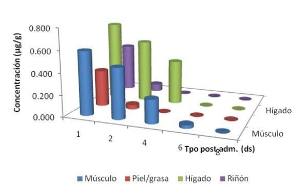
In muscle, fosfomycin was detected at concentrations above the MRL established by The Japan Food Chemical Research Foundation for bovine tissues (0.5 ppm) in 5 out of 6 samples obtained at day 1 post-administration. At 2 days post-administration, fosfomycin was quantified in 2 out of 6 treated animals, while the rest of the time the levels were below the LOD (0.0625 µg/g).). The concentrations in skin/fat were 0.337 ± 0.15 µg/g in the first post-administration sample to fall below the LOQ on the second day post-administration. The liver was the tissue where the highest concentrations were found, and they continued above established MRL until 2 days post-administration, falling below it 4 days after treatment ended. This finding generally agrees with a previous study done by our research group, but with fosfomycin sodium. In kidney, concentrations were 0.447 ± 0.169 on the first day post-treatment ends, falling into the LOD from day 2 and thereafter no more was detected (Fig. 20).
As regards to calculation of waiting time using the WT 1.4 program, this could only be performed for muscle and liver tissues, in which concentrations were determined at least in three sampling points (1, 2 and 4 days after completion of treatment), while in fat and kidney we only measured levels at two points (1 and 2 days after treatment ended). Because the program applies the method of linear regression on logotransformed data, at least we need 3 points.
Finally, in Fig. 1 we present average concentrations determined in each tissue tested (N = 6) and waiting times set for muscle (6.62 days) and liver (5 days).
Fig. 1. Fosfomycin average tissual profile in chicken edible tissues following 25% calcium fosfomycin administration at a ratio of 0.6 g/L of water during 5 days
Conclusion
As a final conclusion, this study shows that after administration of 25% calcium fosfomycin (Cevasa S.A.) at a ratio of 0.6 g/L in drinking water during 5 days, there must be a waiting period of 7 days before sending the animals for human consumption.
Bibliography
Aramayona J, Bregante M, Solans C, Rueda S, Fraile LJ, García MA. 1997. Pharmacokinetics of fosfomycin in chickens after a single intravenous dose and tissue levels following chronic oral administration. Veterinary Research 28:51-588.
Fernández A, Payuelo R, Gómez J, Ramos J, Loste A, Marca M. 1998. Efficacy of phosphomycin in the control of Escherichia coli infection of broiler chickens. Research in Veterinary Science, 65:201-204.
Fernández A, Lara C, Loste A, Calvo S, Marca M. 2001. Control of Salmonella enteritidis phage type 4 experimental infection by fosfomycin in newly hatched chicks. Comparative Immunology. Microbiology and Infectious Diseases 24:207-216.
Fernández A, Lara C, Loste A, Marca M. 2002. Efficacy of calcium fosfomycin for the treatment of experimental infections of broiler chickens with Escherichia coli O78: k80. Veterinary Research Communications 26:427-436.
Japan Food Chemical Research Foundation. URL:http://www.m5.ws001.squarestart.ne.jp/
foundation/agrdtl.php?a_inq=71900. Acceso: 01/04/2011.
foundation/agrdtl.php?a_inq=71900. Acceso: 01/04/2011.
Kahan FM, Kahan JS, Cassidy PJ, Kropp H. 1974. The mechanism of action of fosfomycin (phosphonomycin). Ann N Y Acad. Sci. 235:364-86.
Prescott JF. 2000. Peptide antibiotics: polymixins, glycopeptides, streptogramins and bacitracin. pp. 190. In Antimicrobial Therapy in Veterinary Medicine, 3rd edn. Eds Prescott J, Baggot D, Walter R, Iowa State University Press, Ames, IA.
Content from the event:
Related topics:
Recommend
Comment
Share

Would you like to discuss another topic? Create a new post to engage with experts in the community.


You may be interested in
.jpg&w=3840&q=75)