Big liver and spleen disease or hepatitis splenomegaly syndrome (HSS) is clinically characterized by increased mortality and decreased egg production in both broiler breeder hens and egg-type chickens, usually ranging from 30 to 72 wk of age. On gross examination, blood-tinged fluid can frequently be observed in the coelomic cavity, and liver and spleen are typically both enlarged (19).
The causative agent of HSS is the avian hepatitis E virus (HEV), which is a nonenveloped, single-stranded RNA virus with an icosahedral capsid symmetry that belongs to the family Hepeviridae (5). HEV has been identified in several animal species (25), and based on host tropism and genetic relatedness, strains genetically characterized, thus far, can be classified into two genera: genus Orthohepevirus (all mammalian and avian HEV isolates) and genus Piscihepevirus (cutthroat trout virus [23]). Currently, four avian HEV genotypes have been described in chicken flocks worldwide. Genotype 1 has been identified in Australia and Korea, genotype 2 is present in North America, genotype 3 is present in Europe and China, and more recently, a novel putative genotype 4 has been detected in Hungary and Taiwan (2,11,14,18,23). A significant proportion of chicken flocks worldwide is seropositive for avian HEV, even though seropositive flocks do not necessarily suffer from HSS (19). Often, the identification of avian HEV is considered a minor causative agent in diagnostic investigations and frequently regarded as insignificant when another poultry disease has been diagnosed in the same chicken. However, HEV-associated disease has been experimentally confirmed (4).
Spotty liver syndrome, or vibrionic hepatitis, is characterized by an acute drop in weekly egg production of 10 to 25%, accumulative mortality up to 10%, and loose feces in predominantly free-range laying hens under peak lay (8). Postmortem features usually consist of an enlarged liver, with multifocal grey and white 1-to-2-mm foci. A proportion of birds may also have fibrinous perihepatitis and excess clear abdominal or pericardial fluid or a combination of them. Microscopic lesions consist of random multifocal necrotic hepatitis. Vibrio-like organisms, later identified as Campylobacter jejuni, were suggested as the likely cause (21). Despite its initial association with the disease, C. jejuni has not been conclusively linked to the development of the focal lesions characteristic of spotty liver disease, as the presence of Campylobacter spp. in the liver alone has been shown not sufficient to cause the disease (13). The etiology of spotty liver syndrome has not been established; currently, there is insufficient evidence to describe risk factors or a specific control measure.
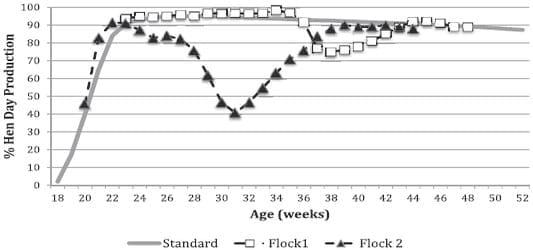
Fig. 1. Egg production from two flocks affected with hepatitis E virus. Flock 1 represents the least-affected flock and corresponds to case 1 in Table 1. Flock 2, corresponding to case 6 in Table 1, was the most severely affected flock (41.7% egg production at 31 wk of age) and remained below expectations for months.
The present report describes the detection and characterization of avian HEV in 10 organic laying hen flocks displaying clinical signs similar to spotty liver syndrome between September 2012 and March 2014 and documents the clinical, pathologic, and microbiologic findings.
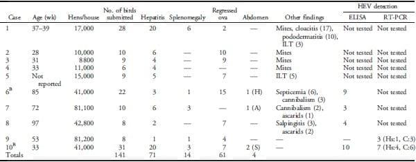
Table 1. Age and hen distribution in affected houses and pathologic and diagnostic findings.A
 A
AThe number under the gross observations and in parenthesis represents the number of birds. HEV 5 hepatitis E virus; RT-PCR 5 reverse transcriptase–PCR; ILT 5 infectious laryngotracheitis; H 5 hemorrhage; A 5 ascites, clear fluid without blood; S 5 serosanguineous exudate; Hs 5 helicase; C 5 capsid.
BTwo different (consecutive) flocks housed in the same barn.
Materials and methods.
Case history. Between September 2012 and March 2014, 28-to-97- wk-old organic laying hens experienced a 20%–40% decreased in egg production (Fig. 1) and an increase in mortality of up to 1%. Clinical signs lasted 3–5 wk. The outbreaks occurred in 10 separated flocks, housed in nine different houses (Table 1), and chickens were obtained from two independent companies. Houses on these farms were operated on all-in-all-out principles. Nonorganic flocks, housed adjacent to affected flocks, were not involved.
Pathology. A total of 141 organic layer chickens were submitted on 12 different occasions to the Avian Health and Food Safety Laboratory, Washington State University, (Puyallup, WA) for diagnostic workup. Birds submitted alive were euthanatized with carbon dioxide (1), and blood samples were taken. Postmortem examinations were performed for all submitted birds.
Samples of liver, spleen, oviduct, ovary, esophagus, crop, proventriculus, gizzard, small and large intestine, pancreas, trachea, lung, heart, kidney, skeletal muscle, and brain were collected and fixed in 10% neutral buffered formalin for a minimum of 24 hr. All tissues were dehydrated in graded alcohols, cleared in xylene, embedded in paraffin, sectioned at 4 mm, stained with hematoxylin and eosin, and examined under a light microscope. In addition, selected sections of liver were subjected to special stains for further characterization, including Gram stained by modified Brown-Hopps (6), Congo red staining (15), periodic acid Schiff (PAS) (7), Ziehl-Neelsen stain (17), and silver stain (22).
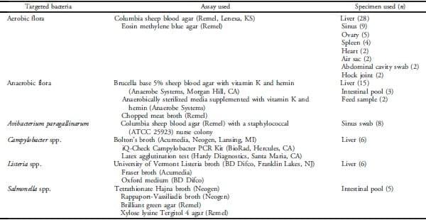
Bacteriology. Specimens from submitted birds were collected and subjected to different bacterial culture approaches (Table 2).
Table 2. Approaches to determine presence of bacterial organisms.A
 A
ACulture conditions represent standard conditions and are not listed.
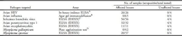
Serology. Targeted pathogens, assays used, and the number of serum samples tested are summarized in Table 3. Samples were tested for antibodies against avian HEV by an in-house indirect ELISA described elsewhere (4). Briefly, a purified truncated recombinant open reading frame 2 capsid protein of the prototype strain of avian HEV expressed in Escherichia coli was coated onto 96-well plates (Nunc; Thermo Fisher Scientific, Agawam, MA). A horseradish peroxidase–conjugated goat anti-chicken immunoglobulin Y (IgY) fragment crystallizable (Gallus Immunotech, Fergus, Ontario, Canada) was used as the secondary antibody. The optical density (OD) values were measured at 450 nm. Three internal controls, a positive control, a negative control, and a cutoff control, were included on each plate. The results were calculated as antibody index (sample OD and cut-off serum mean OD), with results ,0.9, 0.9–1.1, and $1.1 defined as negative, inconclusive, and positive, respectively.
Virology. Ten livers were collected and sent overnight to the Veterinary Diagnostic Laboratory at Iowa State University, where they were tested for avian HEV RNA by nested reverse transcriptase– (RT-) PCR, as previously described (9). Briefly, RNA extractions on liver homogenates were performed by using the QIAampH Viral RNA Mini Kit (Qiagen, Valencia, CA). Extracts were subsequently used for detection of the partial helicase and capsid genes of avian HEV, as described (24) in nested RT-PCR reactions. Briefly, for helicase gene detection, the external primer set 59-TGTTATYACACCCACCAARACGYTG- 39 and 59-CCTCRTGGACCGTWATCGACCC-39 and the internal primer set 59-GCCACGGCTRTTACACCYCAYGT-39 and 59-GACCCRGGRTTCGACTGCTT-39 were used. For the capsid gene, the external primer set 59-TCGCCYGGTAAYACWAATGC- 39 and 59-GCGTTSCCSACAGGYCGGCC-39 and the internal primer set 59-ACWAATGCYAGGGTCACCCG-39 and 59-ATGTACTGRCC RCTSGCCGC-39 were used. PCR products with the expected size were examined on a 1% agarose gel excised and purified with the QIAquick Gel Extraction Kit (Qiagen).
Table 3. Pathogens targeted, assays used, and number of samples used for different serology assays.
 A
ABilliam et al., Ref. 4.
BSigma Aldrich Inc., St. Louis, MO: agarose base; National Veterinary Service Laboratories, Ames, IA: antigen and controls.
CIDEXX Laboratories Inc., Westbrook, ME.
DCharles River Labs, Wilmington, MA.
Sequencing of the RT-PCR product of positive avian HEV RNA samples was performed directly on both strands at the Iowa State University DNA Facility, Ames, Iowa (Applied Biosystems 3730xl DNA Analyzer). Sequences were aligned with published data by using the Basic Local Alignment Search Tool at the National Center for Biotechnology Information (http://www.ncbi.nlm.nih.gov/). Sequences were compiled by using Lasergene software and the Clustal V alignment algorithm (DNAStar, Madison, WI). For sequence analysis, the sequences of the helicase and the capsid genes were compared with each other and with sequences of other avian HEV isolates representing genotypes 1 (GenBank accession nos. AM943647 and JN597006), 2 (GenBank accession nos. AY535004, EF206691, and EU919187), 3 (GenBank accession nos. AM943646 and GU954430), and 4 (GenBank accession nos. JN997392 and KF511797).
Toxicology.
Two feed samples were sent to Alltech (Winchester, KY) for comprehensive 37+ mycotoxin analysis.
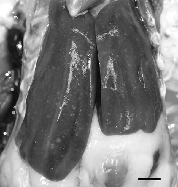
Fig. 2. Characteristic 1-to-2-mm, whitish, pinpoint, multifocal liver lesions. Ruler 5 1 cm.
Results.
The necropsy findings are summarized in Table 1. The most common macroscopic lesions were multifocal white pinpoint foci (Fig. 2) on the liver surface without visible liver enlargement. Regressing ova were also common among submitted birds. Only 9.9% (14/141) of all birds had a slightly enlarged spleen. Fluid in the abdominal cavity, with or without blood, was only observed in 2.8% (4/141) of the examined birds.
Histology revealed multifocal necrotizing hepatitis that ranged from mild to severe. Occasionally, low numbers of heterophils were identified in some of the affected areas (Fig. 3a), while other areas were infiltrated mainly by lymphocytes (Fig. 3b). Multinucleated giant cells were noted occasionally. Hepatocytes did not have increased cytoplasmic deposition of fat. Liver sections stained with Congo red and viewed with polarized light did not show apple-green birefringence associated with amyloid. No organisms were noted by Gram, PAS, acid fast, or silver stains.
No significant aerobic bacteria were isolated. Campylobacter sp. was detected by PCR in one of the six livers tested. All six livers were negative for Campylobacter by culture. Clostridium sp. was isolated from 8/15 livers, both intestinal pools, and both feed samples. Clostridium species identified by Rap ID Ana II System (Remel, Lenexa, KS) included Clostridium perfringens (three livers and one intestinal pool), Clostridium bifermentans (two livers), and Clostridium tetani (one liver). The Clostridium species could not be determined in two livers, one intestinal pool, and the feed samples. All 26 serum samples from birds from affected flocks were antibody positive for avian HEV virus. On the other hand, four samples that were from two nonaffected flocks, housed next to an affected flock, were negative for avian HEV antibodies. All serum samples, from affected and nonaffected flocks, were seropositive to infectious bronchitis virus, avian paramyxovirus type 1, and avian encephalomyelitis. Twenty of the 57 serum samples from affected flocks were positive for Mycoplasma synoviae. All the serum samples tested were negative for antibodies against avian influenza and Mycoplasma gallisepticum.
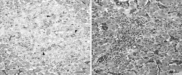
Fig. 3. Histopathology of the liver reveals multifocal moderate-to-severe necrotizing hepatitis with infiltration of heterophils (arrowheads; a) or lymphocytes (b). Hematoxylin and eosin staining. Ruler 5 2.5 mm.
Fig. 4. Sequencing results show that the identified avian HEV strain presented 100% nucleotide identity for the 361-bp fragment of the helicase gene with the U.S. prototype (GenBank accession no.AY535004).
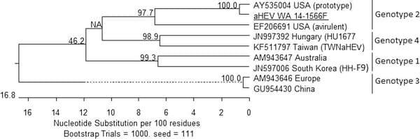
Of the 10 liver samples analyzed by RT-PCR, 50% (5/10) were positive for avian HEV helicase gene RNA (386 bp), and 90% (9/10) were positive for the avian HEV capsid gene RNA (242 bp). Two avian HEV RNA-positive samples were further sequenced. Both genes belonged to genotype 2, which is the only genotype found in the United States. The sequence of the 361-bp fragment of the helicase gene (Fig. 4) showed 100% nucleotide identity with the U.S. prototype. The sequence of the 262-bp fragment of the capsid gene (Fig. 5) showed 99.6% nucleotide identity with an avirulent U.S. avian HEV strain and 89.5% nucleotide identity with the U.S. prototype strain. All sequences reported in this study were deposited in the GenBank database under accession nos. KM262769 through KM262771.
Among all mycotoxins tested, only fumonisins were detected, and the identified levels, 4,440 ppb, were below the low risk for mycotoxicosis for layers (4,440 ppb).
Discussion.
The present report describes the detection and characterization of avian HEV in organic layer flocks experiencing clinical disease. Increased mortality and a drop in egg production were the main clinical signs. The clinical history and gross lesions in affected birds were atypical for avian HEV infection. The drop in egg production (up to 40%) was higher than the 20% reported in the literature (19).
Gross changes in the cases included in this study were similar to spotty liver syndrome described in free-range layers from Europe, Australia, and the United States (8,10,16). Only 2.8% (4/141) of the examined birds had fluid, with or without blood, around the liver or in the abdomen. Histologically, multifocal necrotic hepatitis was the most consistent finding, and contribution of amyloid to this lesion was ruled out by special stains. Previous studies (20) demonstrated large amounts of avian HEV antigen in clinically affected birds; however, often the presence of antigen demonstrated by immunohistochemistry was not associated with pathologic lesions. Although immunohistochemistry testing in the affected livers could be beneficial to demonstrate the antigen in the liver, it was not attempted in this study due to the lack of access to a reliable working method.
Significant bacterial agents or mycotoxins were not recovered or identified in any of the affected birds. Therefore, a diagnosis of avian HEV was made after demonstration of avian HEV RNA in the livers by nested RT-PCR. The sequence in the affected flocks involved avian HEV strain shared nucleotide identity of 100% in the helicase gene and of 99.6% in the capsid gene with other genotype 2 isolates. Genotype 2 is the only genotype found in the United States thus far (3,9,12). Furthermore, all 26 serum samples collected from four affected houses were positive for anti-HEV antibodies, while sera from birds from two nonaffected houses adjacent to an affected house were seronegative for avian HEV IgY. The high prevalence (100%) of antibodies against HEV in affected flocks is consistent with exposure to the virus in those flocks. The lack of antibodies in nonaffected flocks may be due to the absence of the virus in those houses or to the small sample size (only four birds tested). Although the prevalence of avian HEV antibodies has been estimated to be 71% in chicken flocks, in general, only 30% of the chickens in the United States have antibodies against avian HEV (19).
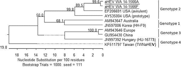
Fig. 5. Sequencing results show that the identified avian HEV strain presented 99.6% nucleotide identity for the 262-bp fragment of the capsid gene.
The source of the HEV infection on the investigated farms remains unknown, but all the affected flocks were located in close proximity, and a common local source of infection is likely. In general, poultry houses are cleaned thoroughly between flocks; however, the survivability of this fecal-oral–transmitted virus in organic houses that cannot be cleaned or disinfected (i.e., yard), may be extended compared with regular nonorganic houses. While standard caged birds do not have access to the floor or feces, organic flocks do. Finally, the role of wildlife in spreading the disease cannot be ignored. At the end of 2014 and early 2015, highly pathogenic avian influenza (HPAI) was detected in the state of Washington. Because of the high risk of infection with HPAI, organic flocks did not have access to the outdoors during this time, and the consecutive flocks were not affected with avian HEV.
In the affected flocks described in the current study, avian HEV infection was characterized by multifocal necrosis of the liver without hepatomegaly. Multiple factors, such as host genotype, genetics of virus, timing and dose of infection and interaction with other management factors, preexisting immunity, and other agents, may explain the variation in clinical effects (4,20). Diagnosis of avian HEV cannot be made based on clinical signs or lesions alone but, rather, should be based on the demonstration of specific viral RNA in samples from flocks.
References.
1. American Veterinary Medical Association. AVMA guidelines for the euthanasia of animals: 2013 edition. American Veterinary Medical Association, Schaumburg, IL) 2013.
2. Ba´nyai, K., A ´ . G. To´th, E ´ . Ivanics, R. Gla´vits, K. Szentpa´li-Gavalle´r, and A ´ . Da´n. Putative novel genotype of avian hepatitis E virus, Hungary, 2010. Emerg. Infect. Dis. 18:1365–1368. 2012.
3. Bilic, I., B. Jaskulska, A. Basic, C. J. Morrow, and M. Hess. Sequence analysis and comparison of avian hepatitis E viruses from Australia and Europe indicate the existence of different genotypes. J. Gen. Virol. 90:863–873. 2009.
4. Billam, P., T. LeRoith, R. S. Pudupakam, F. W. Pierson, R. B. Duncan, and X. J. Meng. Comparative pathogenesis in specific-pathogenfree chickens of two strains of avian hepatitis E virus recovered from a chicken with Hepatitis–Splenomegaly syndrome and from a clinically healthy chicken. Vet. Microbiol. 139:353–261. 2009.
5. Cao, D. J., and X. J. Meng. Molecular biology and replication of hepatitis E virus. Emerg. Microbes Infect. 1:e17. 2012.
6. Carson, F. L., and C. Hladik. Brown-Hopps modification of the Gram strain. In: Histotechnology: a self instructional text, 3rd ed. F. L. Carson and C. Hladik, eds. American Society for Clinical Pathology Press, Hong Kong, China. pp. 231–233. 2009.
7. Carson, F. L., and C. Hladik. Periodic acid Schiff. In: Histotechnology: a self instructional text, 3rd ed. American Society for Clinical Pathology Press, Hong Kong, China. pp. 119–123. 2009.
8. Crawshaw, T., and R. Irvine. Spotty liver syndrome in poultry in Great Britain. Vet. Rec. 170:317–318. 2012.
9. Gerber, P. F., D. W. Trampel, and T. Opriessnig. Identification and characterization of avian hepatitis E virus in 2013 outbreaks of hepatitis-splenomegaly syndrome in two US layer operations. Avian Pathol. 43:357–363. 2014.
10. Grimes, T., and R. Reece. Spotty liver disease—an emerging disease in free-range egg layers in Australia. In: Proc. 60th Western Poultry Disease Conference, Sacramento, CA. p. 53–56. 2011.
11. Hsu, I. W. Y., and H. J. Tsai. Avian hepatitis E virus in chickens, Taiwan, 2013. Emerg. Infect. Dis. 20:149–151. 2014.
12. Huang, F. F., G. Haqshenas, H. L. Shivaprasad, D. K. Guenette, P. R. Woolcock, C. T. Larsen, F. W. Pierson, F. Elvinger, T. E. Toth, and X. J. Meng. Heterogeneity and seroprevalence of a newly identified avian hepatitis E virus from chickens in the United States. J. Clin. Microbiol. 40:4197–4202. 2002.
13. Jennings, J. L., L. C. Sait, C. A. Perrett, C. Foster, L. K. Williams, T. J. Humphrey, and T. A. Cogan. Campylobacter jejuni is associated with, but not sufficient to cause vibrionic hepatitis in chickens. Vet. Microbiol. 149:193–199. 2011.
14. Kwon, H. M., H. W. Sung, and X. J. Meng. Serological prevalence, genetic identification, and characterization of the first strains of avian hepatitis E virus from chickens in Korea. Virus Genes 45:237–245. 2012.
15. Lillie, R. D. Benhold’s Congo red amyloid stain. In: Histopathologic technic and practical histochemistry, R. D. Lillie, ed. The Blakinston Company, Inc., New York. p. 293. 1954.
16. Linares, J. A. Interesting cases from the poultry laboratory. In: Proc. 57th Western Poultry Disease Conference, Puerto Vallarta, Mexico. p. 266. 2008.
17. Luna, L. G. Ziehl-Neelsen for acid fast bacteria. In: Manual of histologic staining methods of the Armed Forces Institute of Pathology, 3rd ed. L. G. Luna, ed. McGraw-Hill Publications, p. 220. 1968.
18. Marek, A., I. Bilic, I. Prokofieva, and M. Hess. Phylogenetic analysis of avian hepatitis E virus samples from European and Australian chicken flocks supports the existence of a different genus within the Hepeviridae comprising at least three different genotypes. Vet. Microbiol. 145:54–61. 2010.
19. Meng, X. J., and H. L. Shivaprasad. Avian hepatitis E virus infections. In: Diseases of poultry, 13th ed. D. E. Swayne, J. R. Glisson, L. R. McDougald, L. Nolan, D. L. Suarez, and V. L. Nair, eds. John Wiley & Sons, Inc., Ames, IA. pp. 494–512. 2013.
20. Morrow, C. J., G. Samu, E. Matrai, A. Klausz, A. M. Wood, S. Richter, B. Jaskulska, and M. Hess. Avian hepatitis E virus infection and possible associated clinical disease in broiler breeder flocks in Hungary. Avian Pathol. 37:527–535. 2008.
21. Peckham, M. C. Avian vibrionic hepatitis. Avian Dis. 2:348–358. 1958.
22. Sheehan, D. C., and B. B. Hrapchak. Grocott’s ammoniacal silver stain for fungi. In: Theory and practice of histotechnology, 2nd ed. D. C. Sheehan and B. B. Hrapchak, eds. C. V. Mosby Co., St. Louis, MO. p. 246. 1980.
23. Smith, D. B., P. Simmonds, International Committee on Taxonomy of Viruses Hepeviridae Study Group, S. Jameel, S. U. Emerson, T. J. Harrison, X. J. Meng, H. Okamoto, W. H. M. van der Poel, and M. A. Purdy. Consensus proposals for classification of the family Hepeviridae. J. Gen. Virol. 95:2223–2232. 2014.
24. Sun, Z. F., C. T. Larsen, F. F. Huang, P. Billam, F. W. Pierson, T. E. Toth, and X. J. Meng. Generation and infectivity titration of an infectious stock of avian Hepatitis E Virus (HEV) in chickens and cross-species infection of turkeys with avian HEV. J. Clin. Microbiol. 42:2658–2662. 2004.
25. Yugo, D. M., and X. J. Meng. Hepatitis E Virus: Foodborne, Waterborne and Zoonotic Transmission. Int. J. Environ. Res. Public Health 33:4507–4533. 2013.


.jpg&w=3840&q=75)