Introduction
Egyptian aquaculture represents a unique model of cohabitating freshwater fishes that live together in confined space permitting continuous interaction between different aquatic species and their environment. Polyculture system currently adopted by the Egyptian earthen pond aquaculture has increased the potential of transspecies infections with different pathogens spatially distributed within pond water, cohabitating fish body surfaces or those located on the same fish host (Eissa et al., 2008). External bacteria such as F. columnare (Amin et al., 1988) and protozoan infections like M. tilapiae (Faisal and Shalaby, 1987; Eissa et al., 2006) are the most eminent byproduct of such trans-species interaction in the Egyptian polyculture system.
Flavobacterium columnare is the causative agent of the Columnaris disease, a dermotropic disease that affect a wide range of cultured and wild ranging freshwater fishes around the globe (Austin and Austin, 2007) . F. columnare is a Gram negative very long rod (0.3 - 0.7 μm width and 5 - 15 μm length) which belongs to the family Flavobacteriacea, a group of yellow pigmented bacteria (Austin and Austin, 2007).
The taxonomy of the bacterium has been changed several times over the years on the basis of morpho - chemical criteria. The old taxonomical names of the pathogen included, Chondrococcus columnaris, Cytophaga columnaris, and Flexibacter columnaris (Bernardet and Grimont, 1989). The most recent name "Flavobacterium columnare" was approved by the microbiological community after molecular typing of the worldwide archived strains (Bernardet et al., 1996).
The ubiquitous nature of the pathogen in freshwater habitats and the ability for fish to contract the disease after environmental insults puts F. columnare among the most prevalent pathogens in cultured, ornamental, and wild fish populations (Shotts and Starliper, 1999). F. columnare was isolated from a wide range of coldwater, temperate and tropical fishes. It has been isolated from several fish species throughout the world, including the economically important species such as Nile tilapia (Oreochromis niloticus) (Amin et al., 1988), common carp (Cyprinus carpio) (Bootsma and Clerx, 1976), channel catfish (Ictalurus punctatus) (Bader et al., 2003), goldfish (Carassius auratus), eels (Anguilla japonica, Anguilla Anguilla) (Anderson and Conroy, 1969; Wakabayashi, 1970); flounder, Paralichthys olivaceous (Baxa et al. 1986) and Salmonine species such as rainbow trout (Oncorhynchus mykiss); brown trout (Salmo trutta); brook trout (Salvelinus fontinalis) (Bullock et al., 1986).
Columnaris is a contagious disease that can be transmitted horizontally through direct contact, skin wounds as well as through orofecal route (Bullock et al., 1986; Welker et al., 2005). Due to the ubiquitous nature of the F. columnare in the freshwater, an injury to the skin or gills of fish with elevation of water temperature may quickly initiate the columnaris infection. It has been noted that F. columnare forms microcysts composed of huge number of the organism inside the water with very long existence cycle that can reach up to 1 year or more (Pacha and Porter, 1968; Bullock et al., 1986). These microcysts are good source of infection to the surrounding fishes (Bullock et al., 1986). The period between exposure to F. columnare and the outbreak of clinical disease varies greatly, depending upon the virulence of the strain and the ambient water temperature (Holt et al., 1975; Morrison et al., 1981).
It has been documented that temperature rise above 20 ºC enhances the bacterial pathogenicity and virulence by the following mechanisms: increases the bacterial growth rate by an average of 30 %; increases the adhesion capacity of the bacterium to the fish tissues (Decostere et al., 1999); Chondrotin AC lyase, an enzyme produced by F. columnare (Griffin, 1992) which degrades polysaccharides, particularly those found in cartilaginous connective tissue (Teska, 1993) reaches its peak of activity at temperatures above 25 ºC . The sharp decrease in dissolved oxygen due to temperature rise above 25 ºC jeopardizes the immune system of fish by increasing the potentials of the ubiquitous bacterial invasion (Amend, 1983; Bullock et al., 1986; Suomalainen et al., 2005).
F. columnare infection can be clinically diagnosed through its characteristic clinical picture in the affected fish. In scaly fishes, the infection initially appears as milky veiled erosions on the dorsal aspect of the fish´s body which may progress to extensive ulcers (Amend, 1983). In scaly-less fishes, the signs start with simple ulcers which predominately end with extensive saddle back like ulcers exposing the underlying musculatures (Morrison et al., 1981). Fin and gill rot is another sign of the progressive infection in both scaly and scaly-less fishes (Bullock et al., 1986; Latremouille, 2003).
Morphological and biochemical characteristics of the pathogen are unique. The presence of hay stalk like piles (columns) of the pathogen in the unstained wet mounts is presumptive for the pathogen. Production of certain extracellular products and pigments by F. columnare were utilized by Griffin (1992) to produce a key screen (Griffin Screen) for the cultural and biochemical identification of the pathogen. The production of the yellow flexirubin pigment (identified by the chromoshift of the colonies from yellow to pink after the addition of 3 % KOH) (Griffin, 1992); production of the extracellular galactosamine glycan (identified by the binding of the colonies to Congo red dye) (Johnson and Chilton, 1966; McMurdy,1969); production of gelatinase enzyme (Bertolini and Rohovec, 1992); production of Chondrotin AC lyase, which degrades polysaccharides (Teska, 1993); growth in the presence of 0.5 % NaCl (Bernardet and Gimont, 1989) and growth in the presence of polymyxin B and neomycin sulfate (Bullock et al., 1986) are all suggestive for F. columnare (Griffin, 1992).
Polymerase chain reaction (PCR) based techniques employing species-specific primers have been used for the detection of F. columnare (Toyama et al., 1996; Wakabayashi et al., 1999; Darwish et al., 2004). The first six hundred nucleotides in the 5´ terminus of the 16S rDNA contain enough genetic information to permit precise alignment of bacterial sequences to the major lines of descent; this terminus has been hypothesized as the area of concern for molecular analysis (Liesack et al., 1997). F. columnare has been identified with species specific PCR primers to a portion of the 16S ribosomal RNA gene (Bader et al., 2003; Darwish et al., 2004). This technique can differentiate F. columnare from other species of flavobacteria but it does not demarcate subspecies strains of the same bacteria. Adopting wide spectrum PCR to 16S rDNA pursued by random fragment length polymorphism (RFLP) analysis then amplicon sequencing was able to delineate several subspecies including F. columnare, F. psychrophilum and F. johnsoniae (Tiirola et al., 2002). Most recently, Zhang, and Arias, 2010 used RT-PCR to identify and molecularly characterize three genes from a shotgun genomic library of F. columnare virulent strain ALG-00-530 which were further selected for differential expression analysis based on sequence similarity to assumed virulence genes from related species.
Fishes are liable to F. columnare infection following certain levels of stress. Abrupt variations in water temperature are likely to induce and speed up the progression of this disease. High mortalities among cold, temperate, tropical fishes were globally noticed to be associated with high surge of temperature during late spring, summer and fall seasons (Hilger et al., 1991). Temperatures above 16 ºC in cold water and 25 ºC in temperate water could be a triggering factor in initiation of the infection (Holt et al., 1975; Suomalainen et al., 2005). The severity of columnaris is also affected by other environmental factors; for example, the impact of the disease may increase under conditions of low dissolved oxygen or high concentrations of ammonia (Amend, 1983; Bullock et al. 1986; Suomalainen et al., 2005). Mortality rates can be extremely high, with 60 to 90% mortality common (Bullock et al., 1986). Large numbers of F. columnare have been isolated from water during outbreaks and good survival of the pathogen take places over a wide range of water pH and hardness (Amend, 1983; Bullock et al., 1986). Increased pond rearing density is another critical stress factor likely to induce an outbreak of columnaris (Wedemeyer, 1974).
Parasites and bacteria are permanent inhabitants of freshwater fish farms. The concurrent occurrence of some external parasites together with some ubiquitous bacteria represents a kind of symbiotic relationship (Rintamäki-kinnunen and Valtonen, 1997; Eissa, 2005; Xu et al., 2007). Rintamäki-kinnunen and Valtonen, 1997 reported that 30 % of fishes with parasitic infection had a coincident Flavobacterial infection. However, interactions between parasites and bacterial disease are poorly understood (Pylkkö et al., 2006; Xu et al., 2007). In the current study M. tilapiae was concurrently reported together with F. columnare from the same sites of infection. M. tilapiae is a myxosporean protozoon that affect African cichlids inducing number of external lesions such as corneal opacity due to the presence of the plasmodial spores of the protozoon in the cornea, frontal skin ulcer and head cysts which might progress to form what is called "hole in the head like lesions" (Faisal and Shalaby, 1987; Eissa et al., 2006). Taxonomical Identification of the M. tilapiae spore retrieved from the Egyptian Nile tilapias was briefly described by many of the Egyptian and African authors (Faisal and Shalaby, 1987; Reed et al., 2002; Eissa et al., 2006).
The current study discusses the concurrent occurrence of M. tilapiae and F. columnare during an episode of mass mortalities among the earthen pond reared Nile tilapia during the early summer season.
Materials and Methods
Fish
In the early summer of 2009, 50 Nile tilapia (75 gm in average) and 25 Nile catfish (250 gm in average) were collected from a polyculture earthen pond at a semi- intensive fish farm in Sharkiya Province, mid-zone Nile Delta, Egypt. The pond was suffering from an event of sudden mortalities associated with sharp surge of water temperature from 25 ºC to 30 ºC. Tilapias and catfish brought alive to the Fish Diseases and Management Laboratory (FDML) at Faculty of Veterinary Medicine, Cairo University. Fish were kept in well-aerated, temperature adjusted water aquaria until examined.
Sampling and sample processing
Tilapias were euthanized with an overdose of MS 222 (Tricaine methane sulfonate, Finquel- Argent Chemical Laboratories, Washington) and visually inspected for any abnormalities before examination is adopted. Lesions were photographed and documented using Sony digital camera (Japan).
Microscopical smears were made from scraps of the skin lesions of both Nile tilapias and Nile catfish. Methylene blue and Giemsa stained fresh smears were examined using regular light microscope as well as dissecting microscope. Photos for the retrieved protozoon were taken and sketched then used for taxonomical identification.
Isolation and biochemical identification
Bacteriological swabs from skin lesions of Nile tilapia and Nile catfish were cultured into tryptic soy broth (TSB) (Remel, Lenexa, KS, USA) and onto Hsu-Shotts agar plates (Bullock et al., 1986). Inoculated broth and agar plates were incubated at 25 ºC, 30 ºC, 37 ºC and checked daily for up to 5 days.
Bacterial isolates were presumptively identified using both cultural characteristics and conventional biochemical tests recommended by Griffin (1992) (Table 1). Biochemical tests include catalase with 3% hydrogen peroxide solution, adding potassium hydroxide 3 % onto the bacterial colonies on Hsu-Shotts agar plates, Congo red binding (by flooding 24 hr cultures with 5 ml of Congo red dye), gelatin hydrolysis, growth at TSB, growth at 25 ºC, 30 ºC, 37 ºC , growth at 0.5 % and 1 % salt (NaCl).
Molecular Identification
Chromosomal DNA was extracted from 100 μl of the bacterial suspension (a single colony of each of the isolated bacteria suspended in 100 μl of sterile saline) using DNeasy tissue extraction kit (QIAGEN, Valencia, CA) according to the manufacturer´s instructions. The extracted DNA was amplified using specific primer set specifically for F. columnare (Darwish et al., 2004). Two specific primers, Forward (598- CAGTGGTGAAATCTGGT-614) and Reverse (1260- GCTCCTACTTGCGTAGT- 276) were efficiently used. The negative controls consisted of a PCR mixture with molecular grade water (negative QC control) and another with Pseudomonas florescence DNA template (negative QA control). Positive control (FC+) consisted of a PCR mixture with DNA extracted from known F. columnare (ATCC 49512).
Thermal cycling and amplification procedures were done according to the method described by Darwish et al. (2004). Thermal cycling was done in a thermal cycler (Bio-Rad, Hercules, CA) using 100 ng of template DNA, 20 mM dNTPs, 0.2 mM of each primer (Forward and Reverse), 1X reaction buffer and 2.5 U of Taq DNA polymerase (Invitrogen, Carlsbad, CA ) in 100 ml reaction mixture. The amplification procedure consisted of 30 cycles of amplification. Each cycle consisted of three steps, denaturation of the chromosomal DNA, primer annealing to the bacterial isolate DNA template and primer extension, as follows: 94 ºC for 0.5 min, 45 ºC for 0.5 min and 72 ºC for 2 min. The 30 amplification cycles were preceded by initial denaturation at 94 ºC for 10 min and the last cycle was pursued by 8 min primer extension period at 72 ºC after which the final mixture was held at 4 ºC. The PCR products were eletrophoresed in 1% agarose gel (Invitrogen), stained with ethidium bromide, visualized with ultraviolet light, and photographed using a Kodak EDAS system (Eastman Kodak, Rochester, NY). Isolates were considered positive when a 675 bp band was detected.

Water quality assessment
Water temperature and pH were measured using a waterproof digital combo pH meter and thermometer (HI 98127 (pHep 4) - Hanna instruments Inc., RI, USA). Dissolved oxygen (DO) concentration were measured using a digital dissolved oxygen meter (HI 9142 - Hanna instruments Inc., RI, USA). Total ammonia nitrogen (TAN mg/L) was determined following the method described by Chattopadhyay (1998).
Results
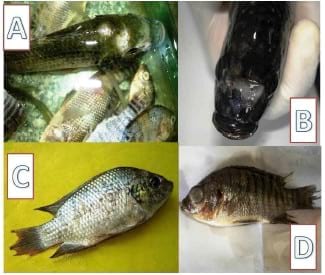
Clinical examination of the collected Nile tilapia revealed the presence of number of external clinical abnormalities including, apical gill lamellar necrosis, marked skin ulcerations at the frontal head region (Figure 1-A); typical hole in the head like lesion with marked liquefaction of the underlying frontal tissues (Figure 1-B); marked signs of fin rot (Figure 1-C) with / without corneal opacity (Figure 1-D). Examination of the cohabitating Nile catfish revealed the presence of typical signs of saddle back like ulcer on the dorsal part of the fish trunk region (Figure 2).
Clinical records also revealed that more than 75 % (38 out 50 fish) of the examined Nile tilapias were associated with head cysts that usually progress to frontal ulcers then penetrate deeper in the skull to form hole- in- the- head like lesion (Figure 1). Microscopical examination of unstained wet mounts made from these lesions revealed the presence of piles "hay stalks" of very long bacteria presumptive for F. columnare. Giemsa stained smears from the head lesions revealed the presence of very long rods (8- 15 μm). Further, Gram stained smears from the same lesions revealed the presence of large number of very long gram negative rods scattered between mucus, gills, skin and muscle tissue debris.


Methylene blue and Giemsa stained impressions made from skin, muscles debris of the head cysts revealed the presence of a myxosporean plasmodial spores (Figure 7) that presumptively identified as M. tilapiae with spore body oblong to oval with anterior and posterior ends bluntly rounded, 14.0-15.5 (15.0 ± 0.39) μm in length. Widest region of spore observed towards centre of spore body, 12.0-12.6 (12.3 ± 0.27) μm in width. Characteristic two spherical / pyriform polar capsules of equal size located in anterior portion of the spore that encompasses four / six coils of polar filaments.


Nile catfish mortalities were nearly close to that of their cohabitating Nile tilapia (18 out of 25 fish - 72 %). Clinically, catfishes typically presented the characteristic saddle back like ulcer on the dorsal part of the fish trunk region with the underlying musculature layer exposed in many of the cases (Figure 2). Microscopical examination of unstained wet mounts made from the lesions revealed the presence of piles "hay stalks" of very long bacteria presumptive for F. columnare. Gram stained smears from the same skin ulcers and underlying musculatures revealed the presence of long gram negative rods (8- 15 μm) very similar to those found in the smears of Nile tilapia head lesions. No external protozoa or worms were found in any of the smears made from the above mentioned lesions.
Bacterial isolates retrieved from both Nile tilapia and Nile catfish were presumptively identified as F. columnare using both cultural characteristics and conventional biochemical tests (Figure 3, Figure 4, Figure 5, and Table 1). Consistent with F. columnare standard ATCC 49512 strain, both Nile tilapia and Nile catfish isolates produced a 675 bp band when molecularly tested by means of PCR (Figure 6).
Water quality parameters of the affected pond including temperature, pH, total ammonia nitrogen (TAN) and dissolved oxygen were determined and compared to the average normal values that were regularly measured by authorities for the same pond prior to the event of mass mortalities. Abrupt increase in temperature from (25 ºC to 30 ºC), pH from (6.5 to 8.8), total ammonia (TAN) from (0.3 to 3 PPM) and sharp decrease in dissolved oxygen from (6 to 3 PPM) were the environmental stimulus that initiated and boosted the infection (Table 2).
Discussion
The continuous global eruptions of pandemics among farm animals and poultry, necessitates the search for safe alternatives as what is called "Blue Revolution" or aquaculture (Scholtissek and Naylor, 1988). Despite the fact that aquaculture is the savior for the humanity from the uprising shortage of animal proteins. Yet, it is one of the human activities that possess potential negative effects on aquatic species due to the increased potential of pathogens spread (Groombridge, 1992). The worldwide reports of F. columnare isolation from wide varieties of aquaculture facilities including cold, temperate and tropical areas support the aforementioned fact (Bullock et al., 1986).
An acute episode of mass mortalities among polycultured earthen pond reared Nile tilapia and catfish have occurred during the early summer of 2009. The event of the mortalities was the motive for several investigations adopted during a field trip to an aquaculture station in Sharkiya province. Records indicated that mortalities among the two farmed species were close to 75 % of the entire earthen pond population. These results were similar to that of Eissa et al., 2006 who reported a peak of mortalities among the earthen pond raised Nile tilapias from the same aquaculture station during the summer of 2006. This seasonal eruption of mortalities among such polycultured fish populations from the same aquaculture stations is highly presumptive for the endemic existence of one or multiple infectious agents. Our laboratory investigations revealed the isolation of F. columnare, a long gram negative bacterium together with M. tilapiae (myxosporean spore) from the same lesions. Such diagnosis was achieved using number of laboratory tools including bacteriological isolation on selective media, biochemical characterization, and molecular confirmation.

The variant degrees of external lesions (erosions, ulcers and fin rot) among the examined tilapias during the episode of mass mortalities coincides with those detailed in many of the literatures published through the past few decades (Morrison et al.,1981; Amend et al., 1983; Bullock et al., 1986; Amin et al., 1988; Latremouille, 2003). Our apical lamellar gill lesions were in complete accordance with those previously reported by several authors for similar natural columnaris infections in earthen pond raised Nile tilapia populations from Nile Delta, Egypt (Amin et al., 1988) as well as those from other species like carp, rainbow trout, brown trout, eels, and channel catfish from different geographical areas around the globe (Bootsma and Clerx, 1976; Amend et al., 1983; Bullock et al., 1986). The nature and severity of the typical saddle back like ulcers seen in our examined Nile catfishes were previously recorded in other types of catfishes including channel catfish during summer season outbreaks in diverse geographical areas around the globe (Hawke and Thune, 1992; Thomas-Jinu and Goodwin, 2004).
Hole-in-the-head like lesion was found in many of the examined Nile tilapias. M. tilapiae, a species specific myxosporean spore that affects African cichlids including Nile tilapia was found concurrently with F. columnare in such lesions. The two diverse infectious agents were incriminated as the cause of the sequence of the pathology associated with the induction of hole in the head lesions.
Eissa et al., 2006 have retrieved the active plasmodial spores of M. tilapiae from the pathology associated with hole in the head like lesions of 80 % of the earthen pond reared Nile tilapias at the same farm from which our current samples were collected during the summer of 2006. The detection of M. tilapiae vegetative plasmodial stages in the head lesions of the same type of fish collected from the same aquaculture facility during the same season (summer 2009) confirms the endemic existence of the myxosporean spore in the earthen ponds of that facility. This assumption is highly strengthened by the conclusion of Eissa et al., 2006 who explained that the endemic nature of the spore in such facility has derived from the active distribution of the tubificid oligocheat worm which act as an intermediate host for the myxobolus species in the mud of the ponds and that´s why the spore were retrieved again after 3 years of occurrence from the same pond.
In accordance with Egyptian agricultural standard regulations, the aquaculture facility under investigation usually use freshwater from streams that is mixed with agricultural drainage water (Eissa et al., 2009). This type of water usually brings up many of the biological agents including the intermediate host (tubificid oligocheat) from theagricultural lands into the aquaculture facility water. In addition, the affected fish farm is a transient stop for some migratory birdscrossing northern Egypt (Eissa et al., 2008) which could be lodged with the active stages of such myxsporean parasite inside theirintestinal tracts. Such dynamic process could further facilitate the transmission, establishment and spread of the active spores to the aquaculture facility ponds through the continuous shedding of their droppings.
The sudden environmental changes complicated with faulty management during the early hot summer were highly incriminated as predisposing factors for initiating the concurrent F. columnare (Wakabayashi, 1991) and M. tilapiae infection in the polyculture earthen pond raised tilapias. The abrupt rise in water temperaturefrom 25 ºC to 30 ºC is known to enhance the bacterial pathogenicity and virulence by increasing the bacterial growth rate; increases the adhesion capacity of the bacterium to the fish tissues (Decostere et al., 1999); activates the Chondrotin AC lyase (Griffin, 1992) which degrades polysaccharides, particularly those found in cartilaginous connective tissue (Teska, 1993). The sharp decrease in dissolved oxygen (from 6 to 3 ppm) due to temperature rise above 25 ºC has jeopardized the immune system of fish by increasing the potentials of the ubiquitous bacterial and parasitic invasions (Amend, 1983; Bullock et al. 1986; Suomalainen et al., 2005). Further, the increased rearing capacity at the investigated aquaculture facility was responsible for the expected increase in organic matter load, sharp increase in ammonia levels (from 0.3 to 3 ppm) with consequent rise in water pH (from 6.5 to 8. 8) and increased physical contact chances between different fishes are the most possible triggering factors for initiation, establishment and spread of infection (Sniezko, 1974; Holt et al., 1975; Morrison et al., 1981; Bullock et al., 1986; Suomalainen et al., 2005).
Egyptian agricultural drainage water often contains organochlorine and organophosphate pesticides (Hassanein et al., 1999; Eissa et al., 2009). Such chemicals are known to cause immunosuprresion among exposed fishes (Dunier et al., 1991; Dunier and Swicki, 1993; Dunier, 1996; Cuesta et al., 2008). Moreover, a large number of industrial facilities including textile, ceramic, glass, electrical cable, battery and metallurgical factories surround the geographical zone of this aquaculture facility (Eissa et al., 2009). These facilities constitute potential sources of heavy metals (Lead, cadmium and mercury), which might reach fish either through direct drainage or atmospheric deposition ending up with permanent damage to the fish immune system components (epidermal layer of the skin, anterior kidney and spleen) (Dunier ,1996; Sweet and Zelikoff, 2001). The aforementioned complex of environmental, management, population dynamics (Wakabayashi, 1991)and other chemical pollution factors are those enemies that primarily destroyed fish innate and specific immune barriers and opened portals of entry for the naturally existing infectious agents (Anderson, 1990; Dunier and Swicki, 1993; Ross, 2002) including the ubiquitous F. columnare (MacFarlane et al., 1986) .
Concurrent parasitic / bacterial infections are the real scenarios for infections in the natural environments where water bodies are naturally inhabited by the ubiquitous bacteria and parasites. Sun et al., 2009 explained an endosymbiotic relationship between F. columnare and the parasitic ciliate Ichthyophthirius multifiliis in which the bacteria adheres to the parasite through association with the cilia. The case is not only restricted to the endosymbiont relationship between the parasite and bacteria, but also extended to a synergistic type of relationship in which the parasite enhances the bacterial invasion to the fish skin with consequent bacterial disease development (Kabata, 1985; Plumb, 1997). In our case, the detection of M. tilapiae plasmodial vegetative stages in large numbers at the head lesions (frontal head erosions, ulcers and hole- in- the- head lesions) together with F. columnare is highly suggestive of a synergistic mechanism for induction of the head pathology and mortalities. This assumption could be supported by Bandilla et al., 2006 who concluded that Argulus coregoni parasitic infection increased the susceptibility of Rainbow trout to F. columnare infection. M. tilapiae easily penetrates the skin of scalyless parts of the fish body such as frontal area at the head. The myxsporean parasite secretes number of proteolytic enzymes (chemotrypsin like enzymes / proteases) that were able to digest the entire skin, hypodermis and underlying musculature with an ultimate progress to what is called hole-in - the head like lesions (Eissa et al., 2006). Such extensive damage to the fish skin has initiated further invasion with the ubiquitous F. columnare. This explanation coincides with those previously published by Kabata, 1985; Buchmann and Bresciani, 1997; Plumb, 1997 for similar cases of concurrent parasitic / bacterial infections.
The endemic nature of M. tilapiae in the investigated polyculture pond and the seasonal resurgence of mortalities in summer of 2006 - 2009 with concomitant isolation of F. columnare from the affected fishes might give clue to a complicated long term case of concurrent infection. M. tilapiae spores can live in the tubificid oligocheat worm (intermediate host) and ponds mud for more than a year (Faisal and Shalaby, 1987; Eissa et al., 2006). Similarly, F. columnare microcysts can stay viable in the ponds water or mud for more than a year (Bullock et al., 1986). This long period of existence for both infectious agents in the ponds environment will always be a vulnerable source of seasonal or environmental stress related mass mortalities.
The polyculture system makes use of the benefits of interspecies maximum food utilization (natural and artificial) to maximize the fish production cycle yield. However, the rearing of different fish species in a confined space (earthen pond in our case) could be an ideal media for interspecies infectious diseases transmission or even evolution of new pathogens´ virulence mechanisms by which it could infect a cohabitating fish species that never been infected by such pathogens. In our investigated polyculture earthen pond, Nile catfish has contracted the F. columnare infection from their cohabitating Nile tilapias or from the long term source of infection (bacterial water microcysts). We also suggest that F. columnare infection in Nile catfish was favored by the scaly-less nature and some skin injuries that rendered catfish liable for the cohabiting bacterial invasion. The synergistic increase in the secretion of proteases, chemotrypsin like enzymes by M. tilapiae (Eissa et al., 2006) and Chondrotin AC lyase, adhesion factor by F. columnare (Griffin, 1992; Teska, 1993; Decostere et al., 1999) resulted in extensive skin and gill damage with consequent increase in mortalities due to osmoregulatory failure.
The taxonomical measures of the M. tilapiae reported in the current study coincide with those reported by Eissa et al. (2006); Faisal and Shalaby (1987) and Reed et al. (2002). Cultural and biochemical conventional test results for the retrieved Nile tilapia and Nile catfish bacterial isolates were in complete accordance with those came in Griffin screen for F. columnare (Griffin, 1992). The banding patterns of both isolates on the agarose gel electrophoresis were completely consistent with that of ATCC 49512 standard positive F. columnare isolate (Darwish et al., 2004).
In conclusion, the current study reports on the seasonal resurgence of mass mortalities due to F. columnare infection in the polycultured Nile tilapia and Nile catfish during the early summer. Further, the endemic existence of the myxosporean spore M. tilapiae concurrently with some environmental stresses (abrupt temperature rise, polluted pond water) and faulty management (high ammonia, high pH and low dissolved oxygen) are the potential initiating factors for such outbreaks.
Acknowledgments
We are grateful to Dr. Nashwa Ezzeldeen, Professor of Microbiology at the Faculty of Veterinary Medicine, Cairo University and Dr. Ossama Saleh, Researcher at the Central Laboratory of Aquaculture Research, Abbasa for their endless support in sample collection, processing and microbiological assays performed during the entire study.
References
Abolarin M.O. (1974): Myxobolus tilapiae sp. nov. (Protozoa: Myxosporida) from three species of freshwater tilapia in Nigeria. Journal of West African Science Association, 19, 109-114.
Amend D.F. (1983): Columnaris (Flexibacter columnaris) disease of freshwater fishes and a brief review of other flexibacterial disease of fish. Pages 139-151 in D.P.
Anderson, M.M. Dorson, and P. Dubourget, editors. Antigens of fish pathogens. Collection Foundation Marcel Merieux, Lyon, France. Amin N.E., Abdallah I.S., Faisal M., Easa M. El-S., Alaway T., Alyan S.A. (1988): Columnaris infection among cultured Nile tilapia Oreochromis niloticus. Antonie van Leeuwenhoek 54: 509-520.
Anderson D. (1990): Immunological indicators: effect of environmental stress on immune protection and disease outbreaks. American Fisheries Society Symposium, 8, 38 - 50. Anderson J. I.W., Conroy D.A. (1969): The pathogenic myxobacteria with special reference to fish disease. Journal of Applied Bacteriology, 32, 30-39.
Austin B., Austin D.A. (2007): Bacterial Fish Pathogens: Diseases of Farmed and Wild Fish. Chichester: Praxis Publishing Ltd. Bader J.A., Shoemaker C.A., Klesius P.H. (2003): Rapid detection of columnaris disease in channel catfish (Ictalurus punctatus) with a new species-specifi 16-S rRNA gene-based PCR primer for Flavobacterium columnare. Journal of Microbiological Methods, 52 (2), 209-220.
Bandilla M., Valtonen E.T., Suomalainen L-R., Aphalo P.J., Hakalahti T. (2006): A link between ectoparasite infection and susceptibility to bacterial disease in rainbow trout. International Journal of Parasitology, 36, 987-991.
Baxa D.V., Kawai K., Kusuda R. (1986): Characteristics of gliding bacteria isolated from diseased cultured flounder, Paralichthys olivaceous. Fish Pathology, 21,251- 58.
Bernardet J.F., Grimont P.A.D. (1989): Deoxyribonucleic acid relatedness and phenotypic characterization of Flexibacter columnaris sp. nov., nom. rev., Flexibacter psychrofilus sp. nov., nom. ver., and Flexibacter maritimus. International Journal of Systemic Bacteriology, 39 (3), 346-354.
Bernardet J.F., Segers P., Vancanneyt M., Berthe M., Kerters K., Vandamme P. (1996): Cutting a Gordian Knot: Emended classification and description of the genus Flavobacterium, emended description of the family Flavobacteriaceae, and proposal of Flavobacterium hydatis nom. nov. (basonym, Cytophaga aquatilis Strohl and Tait 1978). International Journal of Systemic Bacteriology, 46 (1), 128-148.
Bertolini J.M., Rohovec J.S.(1992): Electrophoretic detection of proteases from different Flavobacterium columnare strains and assessment of their variability. Diseases of Aquatic Organisms, 12, 121-128.
Bootsma R., Clerx J.P.M. (1976): Columnaris disease of cultured carp (Cyprinus carpio L); Characterization of the causative agent. Aquaculture, 7, 731-384. Buchmann K., Bresciani J. (1997): Parasitic infection in pond-reared rainbow trout Oncorhynchus mykiss in Denmark. Diseases of Aquatic Organisms, 28, 125-138.
Bullock G.L. (1972): Studies on Selected Myxobacteria Pathogenic for Fishes and on Bacterial Gill Disease in Hatchery-reared Salmonids. Washington: U.S. Fish Wildlife Service Technical Paper # 60
Bullock G.L., Hsu T.C., Shotts E.B. (1986): Columnaris disease of salmonids. U.S. Fish and Wildlife Service, Fish Disease Leaflet, 72, 9 pp.
Chattopadhyay G.N. (1998): Chemical Analysis of Fish Pond Soil and Water. Daya Publishing House. New Delhi, India.
This article was originally published on IBC Interdisciplinary Bio Central Journal in June 04, 2010. Engormix.com thanks the author and the journal for this contribution.